Focus on Carcinoembryonic Antigen (CEA)/CD66e/CEACAM5

Carcinoembryonic antigen CEA, is also known as CD66e or CEACAM5,as a key molecule in the field of biomedical research, occupies a unique position in tumor-related studies.
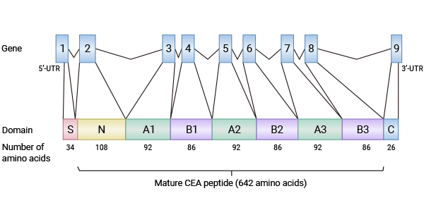
The Structure of CEA
CEA is a member of the immunoglobulin superfamily (IgSF), and its encoding gene is CEACAM5, which plays a crucial role in human physiological and pathological processes. CEA is anchored to the cell membrane via glycosylphosphatidylinositol (GPI), a characteristic that endows it with two forms of existence: membrane protein and secreted protein, laying the foundation for its participation in various physiological activities. When the membrane anchoring region is cleaved by phospholipase C and phospholipase D, CEA will enter the circulatory system, and this process is of great significance in the development of tumors.
At the molecular structure level, CEA is composed of 7 immunoglobulin-like domains, including 1 variable (IgV)-like domain (N domain) and 6 IgC-like domains (A1, B1, A2, B2, A3, B3). Before and after these domains, there are a signal peptide (1 - 34AA) and a precursor peptide (686 - 702AA), respectively, and the precursor peptide will be removed during the maturation process. This structural feature of CEA, especially its abundant immunoglobulin-like domains, provides a structural basis for its exertion of multiple biological functions. The presence of these domains enables CEA to interact specifically with other molecules and participate in important processes such as cell adhesion and signal transduction.
The CEA Family
The CEA gene family is large and complex. According to the similarity of sequences and functions, it can be divided into three categories: CEA-related cell adhesion molecules (CEACAM), pregnancy-specific glycoproteins (PSG), and pseudogenes. Among them, the CEACAM category contains 12 gene members, and the protein sequence similarity among these members ranges from 17% to 88%. Moreover, the extracellular regions of all members show a high degree of glycosylation modification. The difference in glycosylation modification is particularly significant in cancer cells. The molecular weight of CEA in normal cells is approximately 72kDa, while in cancer cell lines and samples from cancer patients, its molecular weight can be as high as 180 to 200kDa. This change affects the functional characteristics of CEA and plays an important role in the occurrence and development of tumors.
Cell-Cell Interaction
As a cell adhesion molecule, CEA is anchored to the cell membrane via GPI and can connect the epithelial cell membrane and adjacent cell groups. CEA co-localizes with integrin α5β1, which enhances the binding ability of cells to fibronectin and promotes the interaction between cells and the extracellular matrix. At the same time, CEA can also exert its cell adhesion function through antiparallel CEA-CEA homotypic interactions and CEA-CEACAM1, CEA-CEACAM6 heterotypic interactions, maintaining stable connections between cells and playing an indispensable role in the construction and maintenance of tissue morphology.
Immune Response
The high expression of CEA in tumor tissues is relatively common, and this tumor-specific glycosylated CEA can interact with DC-SIGN on dendritic cells, inhibiting the specific immune response of dendritic cells related to tumor progression. This mechanism allows tumor cells to escape the surveillance and attack of the immune system, creating favorable conditions for the growth and spread of tumors. In the process of tumor immune escape, this function of CEA has become a major challenge in tumor treatment, and it also prompts researchers to deeply study how to break this immune suppression and enhance the body's immune response to tumor cells.
Anoikis
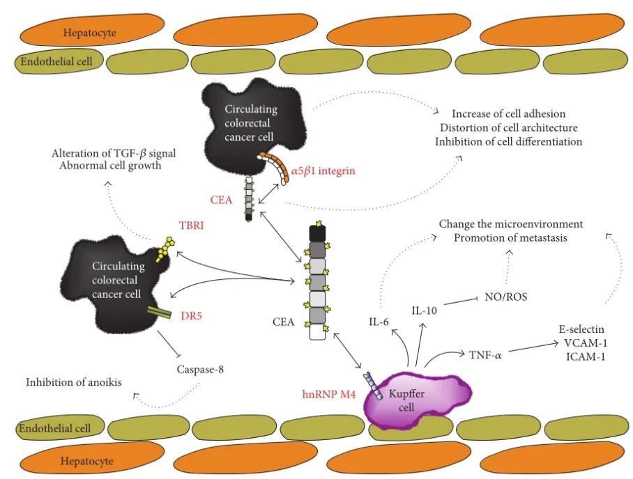
Anoikis is a phenomenon of cell death caused by cells detaching from the extracellular matrix, which is of great significance for maintaining tissue homeostasis. However, during the metastasis of tumor cells, they can resist anoikis through various mechanisms. CEA plays a key role in this process. It can activate the integrin signaling pathway, disrupt tissue structure, and inhibit cell differentiation and anoikis. CEA on the surface of circulating tumor cells can directly bind to DR5, inhibiting the downstream death signal transmission mediated by DR5, enabling cancer cells to evade anoikis. In addition, CEA can also interact with the TGF-β type I receptor (TβRI), altering the downstream TGF-β signaling pathway and promoting the proliferation of tumor cells, further driving the development and metastasis of tumors.
Cancer Cell Metastasis
CEA also plays an important role in tumor metastasis. Taking colorectal cancer as an example, when the level of CEA released by colorectal cancer cells rises, it will trigger the activation of Kupffer cells in the liver. CEA binds to the hnRNP M4 molecule on the surface of Kupffer cells, inducing Kupffer cells to overexpress cytokines and changing the liver microenvironment, allowing colorectal cancer cells to survive in the liver and promoting metastasis. Moreover, other terminally differentiated macrophages (such as alveolar macrophages) and the surface of certain cancer cells also express hnRNP M4, and the binding of CEA to it may play a similar role in the lung metastasis of tumors and other processes. Although the specific mechanism by which CEA affects cancer cell metastasis has not been fully clarified, this discovery provides a new direction for the study of tumor metastasis mechanisms and the formulation of tumor treatment strategies.
Conclusion
The structure and function of carcinoembryonic antigen CEA are closely related, and it has important impacts on multiple key links such as the occurrence, development, metastasis, and immune escape of tumors. With the continuous in-depth research, it is expected that in the future, based on a deep understanding of CEA, more accurate and effective tumor diagnostic methods and treatment strategies can be developed, bringing new hope for conquering tumor diseases.
Product Information
| Gatalog Num | Product Name | Product Parameters | Price |
| S0B2091P | CEA(CD66e) Recombinant Rabbit mAb,PBS Only (SDT-098-54) | Host : Rabbit | Inquiry |
| S0B2091 | CEA(CD66e) Recombinant Rabbit mAb (SDT-098-54) | Host : Rabbit | $880 |
| S0B3054 | CEA Recombinant Rabbit mAb (SDT-098-41) | Host : Rabbit | $80 |
| S0B3055 | CEA Recombinant Rabbit mAb (SDT-098-54) | Host : Rabbit | $80 |
| S0B3056 | CEA Recombinant Rabbit mAb (SDT-098-61) | Host : Rabbit | $80 |
| UA010376 | CEACAM-5/CD66e His Tag Protein, Cynomolgus | Host : Cynomolgus | $520 |
| Expression System : HEK293 | |||
| Conjugation : Unconjugated | |||
| UA010368 | CEACAM-5/CD66e Fc Chimera Protein, Human | Host : Human | $520 |
| Expression System : HEK293 | |||
| Conjugation : Unconjugated | |||
| UA010288 | CEACAM-5/CD66e His Tag Protein, Human | Host : Human | $520 |
| Expression System : HEK293 | |||
| Conjugation : Unconjugated | |||
| UA010746 | Biotinylated CEACAM-5/CD66e His&Avi Tag Protein, Human | Host : Human | $560 |
| Expression System : HEK293 | |||
| Conjugation : Biotin | |||
| UA010697 | Biotinylated CEACAM-5/CD66e Fc&Avi Tag Protein, Human | Host : Human | $490 |
| Expression System : HEK293 | |||
| Conjugation : Biotin | |||
| UA010475 | FITC-Labeled CEACAM-5/CD66e Fc Chimera Protein, Human | Host : Human | $490 |
| Expression System : HEK293 | |||
| Conjugation : FITC | |||
| UA010474 | FITC-Labeled CEACAM-5/CD66e His Tag Protein, Human | Host : Human | $490 |
| Expression System : HEK293 | |||
| Conjugation : FITC |


