Fig1. The fluorescent intensity from antibody-probe protein complex internalization into cell is specific for B7-H3 on Hela cell.
pH-sensitive IgG labeling reagents Max(Green)
pH-sensitive IgG labeling reagents Max(Green)
Price:
Regular price
$880 USD
Regular price
Sale price
$880 USD
Unit price
per
For shipping services or bulk orders, you may request a quotation.
Secure checkout with

View full details
Product Details
Product Details
Product Specification
| Molecular Weight | 31kDa |
| Storage Buffer | PBS, PH7.4 with 5% trehalose is added as protectants |
| Reconstitution |
1. Before opening the tube cap, centrifuge the sample tube at 5000g for 3-5min at room temperature to ensure the lyophilized sample to settie down at the bottom of the tube. 2. Dissolved lyophilized protein sample in sterile water based on the recommended volume 10uL(10ug package). 3. After adding sterile water, cover the lid and mix them bygently tapping the tube for 5-10 times. Note: Do not vortex or vigorously pipette sample.
|
| Stability & Storage | · 12 months from date of receipt, lyophilized powder stored at -20 to -80℃. |
Background
Antibody internalization labeling probe. It can bind to candidated antibodies to produce soluble complex that only fluoresce in acidic environments for the study of antibody internalization.
The pH-sensitive Labeling probe binds to your primary antibodies via Fc binding protein to a pH-dependent fluorescent molecular. This fluorescent complex reporter will increase intensity as the pH of its surroundings becomes more acidic, as evident when exposed to the environment inside a cell. The internalized antibodies are detected by measuring fluorescence intensity of the cells.
This product can becan be used for human IgG1, IgG2, IgG3, IgG4, rabbit IgG, mouse IgG1, IgG2a, IgG2b and IgG3 and can be detected with Flow cytometry FITC or AF488 filter.
Guidelines
Avoid vigorous vortex after reconstitution.
Avoid repeated freeze-thaw cycles.
Picture
Picture
Bioactivity
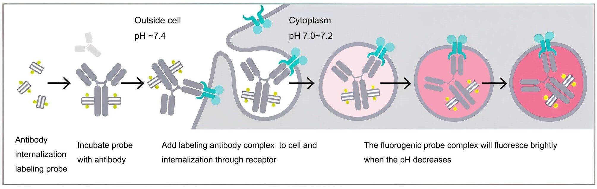
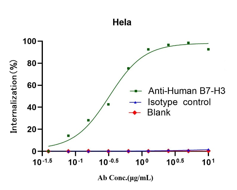

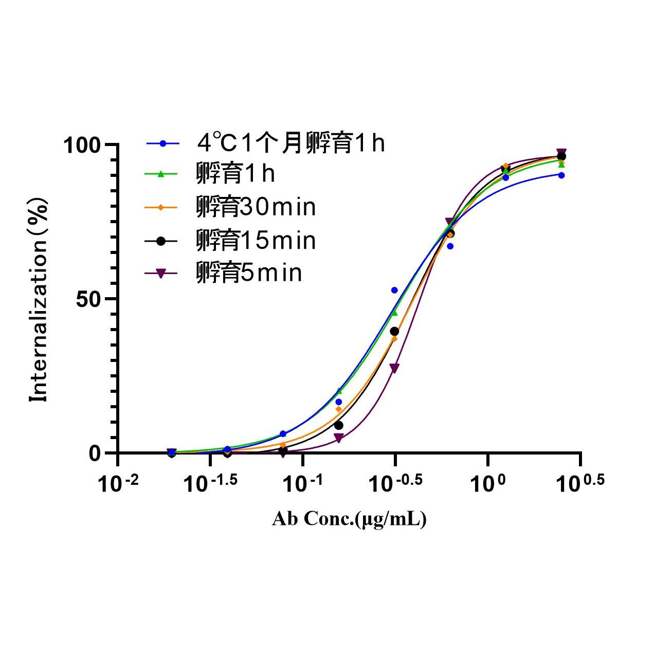
Fig2. Comparison of antibody for B7-H3 internalization effects between UA070122 and UA070080 on Hela cells.
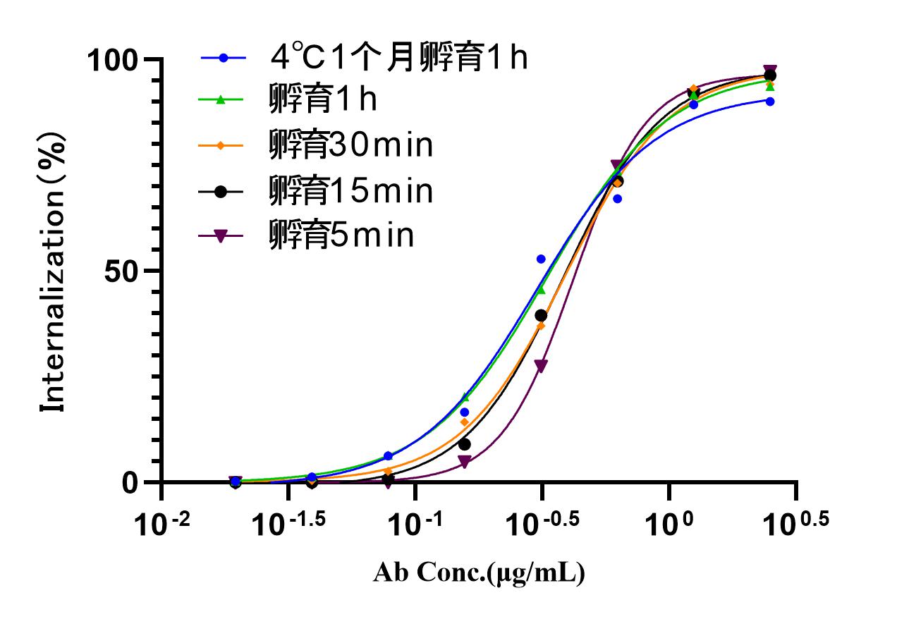
The influence of long-term stability and incubation time