Immunoprecipitation (IP/CoIP) kit
Immunoprecipitation (IP/CoIP) kit
Price:
Regular price
$438 USD
Regular price
Sale price
$438 USD
Unit price
per
For shipping services or bulk orders, you may request a quotation.
Secure checkout with

View full details
Product Details
Product Details
Product Specification
| Description |
Introduction: Immune (co) precipitation (IP/CoIP) is based on the specific affinity of antibodies and proteins. By capturing antibodies that specifically bind to proteins or antigens, target proteins can be captured and enriched from complex samples, and the interacting proteins or other biological macromolecules can be determined.
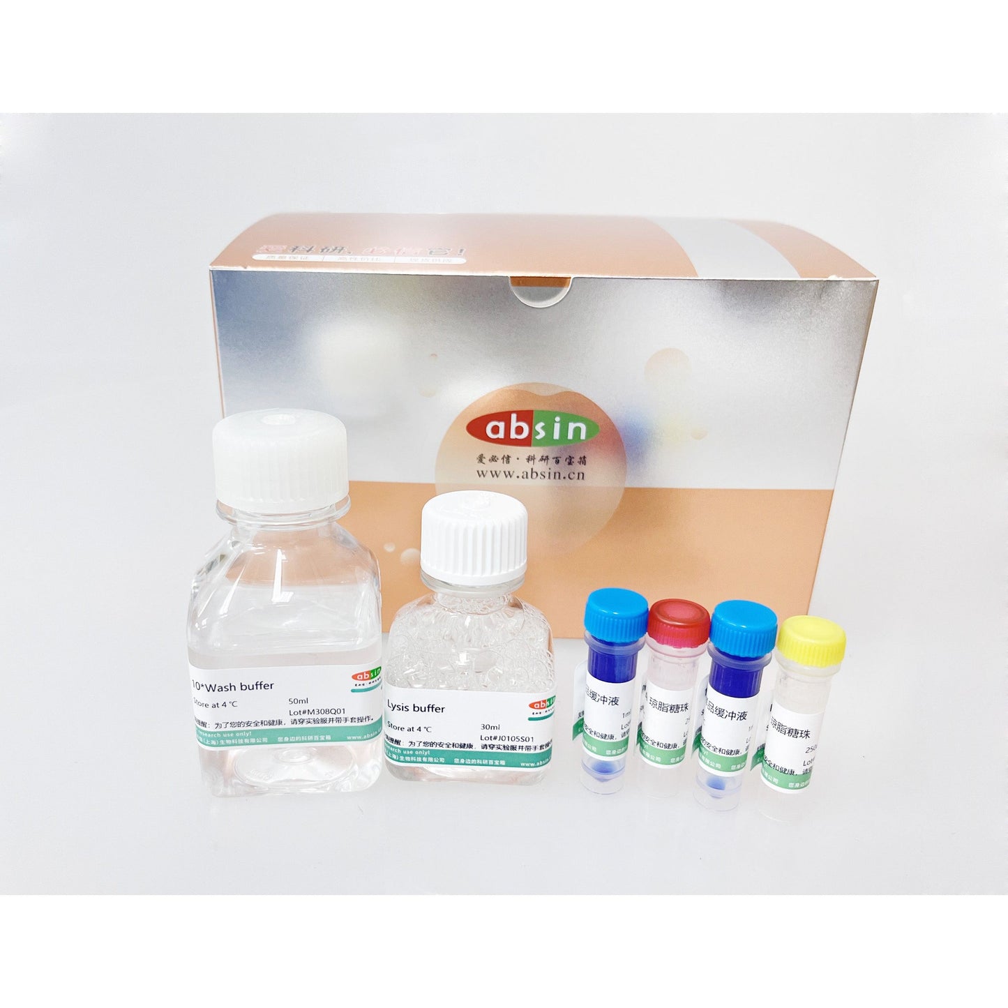
Before experiment: In order to obtain better experimental results, all steps need to be optimized according to the actual situation. For example, more appropriate cell lysis conditions may be selected depending on the cell line or the subsequent use of the trapped antigen. With no cell wall structure of mammalian cells, the stain remover can be directly used for cracking, but the other cells, are in need of some auxiliary mechanical shear force, such as ultrasonic broken. The list below (including lysate, incubation time, volume, and concentration) is recommended as an initial attempt, and the final optimization will need to be adjusted according to the situation. In addition, the cell lysates can be detected by Western blot before immunoprecipitation to confirm the presence of target proteins in the cell lysates. I. Kit components
Note: Add 1 mM PMSF immediately before using Lysis buffer (it is recommended to purchase abs9146 and make your own preparation).
2.Method of Application
A. Preparation of cell lysis content 1. Blot medium. Fresh medium containing regulatory molecules was added to allow treatment of the cells for a predetermined time. 2. Cells were harvested and washed once with ice-precooled 1 X PBS after removing the medium. 3. PBS was removed and 0.5 ml of ice-precooled Lysis buffer was added to each cell plate (10 cm) and incubated on ice for at least 5 min. 4. Scrape down from the board, the cell, the extract to trace centrifuge tube. Place on ice. 5. 3 times on the ice ultrasonic broken, every 5 seconds. (PS: Ultrasound conditions need to be adjusted according to liquid volume, probe diameter, and maximum power. As an example, a 200W power ultrasound instrument was used with a 3 mm probe. We generally recommend three ultrasound pulses at 35% to 40% of the maximum power, with a 10-second pause between ultrasound pulses.) 6. In 4 ° C, the supernatant was transferred to a new tube by microcentrifugation at 14,000 x g for 10 min. The supernatant was the cell lysate. If necessary, lysates could be stored at -80 ° C. Note: After cell lysates are prepared, Western blotting can be performed to confirm the presence of the target protein in the cell lysates. B. Immunoprecipitation a) Preclearance of cell lysates (optional step) 1. To 500 μ l (containing 200-1000 ug of total protein) cell lysates were supplemented with 5μ l Protein A and 5μ l Protein G. 2. In 4° At C, the cells were incubated with rotation for 30 to 60 min. 3. 4° The mixture was centrifuged at 12000g for 1 min at C, and the supernatant was retained until further use. b) antigen-antibody binding 1. Add 500 &mu to a new centrifuge tube; l (containing total protein 200 – 1000 ug) cell lysates (can be pre-washed cell lysates). 2. Monoclonal/polyclonal antibodies (1– 5 μ g). 3. At the same time, the homologous antibody of non-specific immunity was used as a control. 4. In 4° C gently mix for the night. c) immunocomplex precipitation 1. After antibody incubation overnight, 5&mu was added; l Protein A and 5μ l Protein G. 2. In the 4 & deg; C Mix gently for 1-3 hours or overnight. 3. 12000 g centrifugal 1 minute, retain precipitation. 4. The precipitate was washed using 0.5 ml 1*Wash buffer and centrifuged at 12,000 g for 1 min to retain the precipitate. 5. Repeat step 4, a total of three times cleaning. note: clean, go to the qing need to pay attention to avoid suck packing C. The results were analyzed by Western blot 1. The precipitate was resuspended in l 1*SDS sample buffer, vortexed and shaken, and then microcentrifuged for an additional 30 s to bring beads and liquid attached to the tube wall to the bottom of the tube. 2. Heat the sample to 95-100° C and continued for 2-5 min, followed by transient centrifugation at 14,000 g for 1 min to remove the supernatant. 3. The samples (15– 30 μ l) were loaded onto SDS-PAGE gels. 4. Samples were analyzed by Western blotting. 3.Storage This kit is valid for 1 year when stored at 4 ° C.
Note: The matrix of Protein A and Protein G is 20% ethanol, which is more volatile. When used, attention should be paid to sealing. If volatilization occurs, 20% ethanol can be used to resuspend.
|
Picture
Picture
Immunohistochemistry