Product Specification
| Host |
Rabbit |
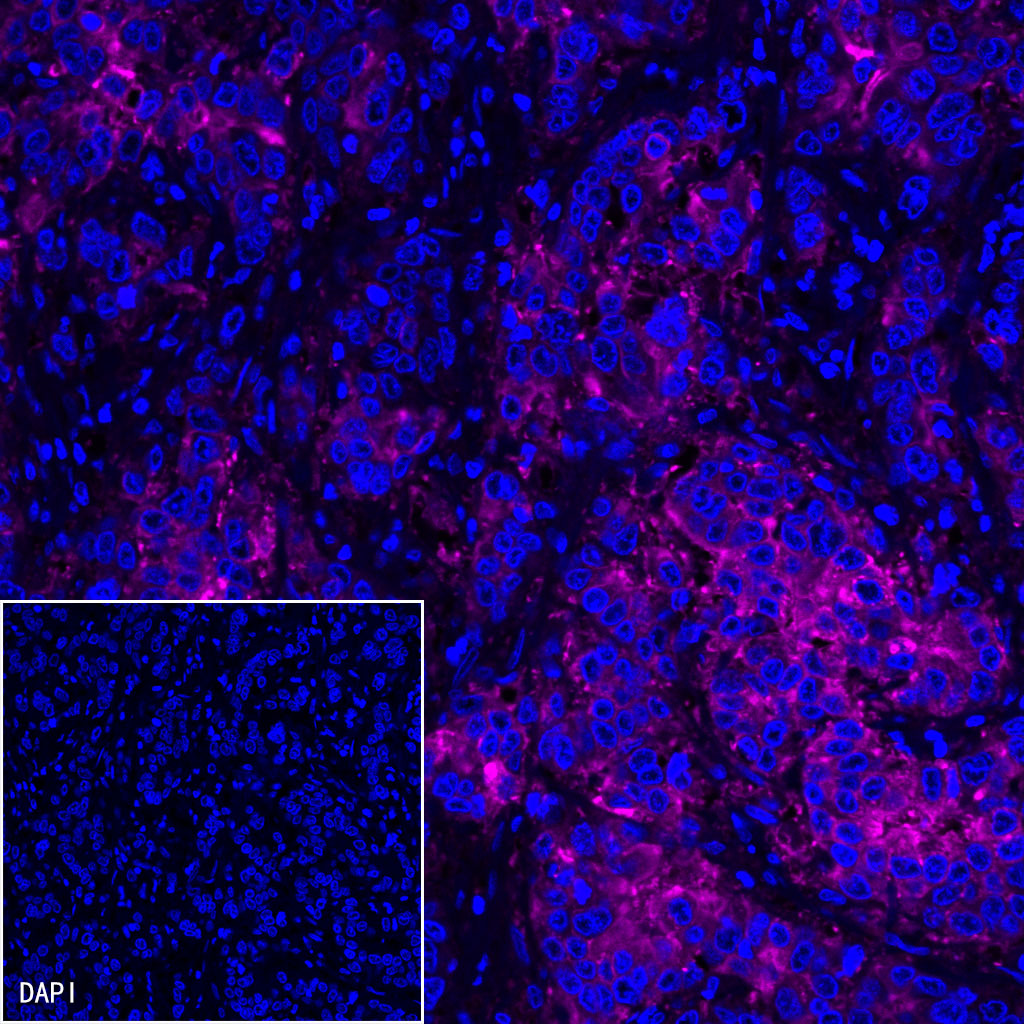
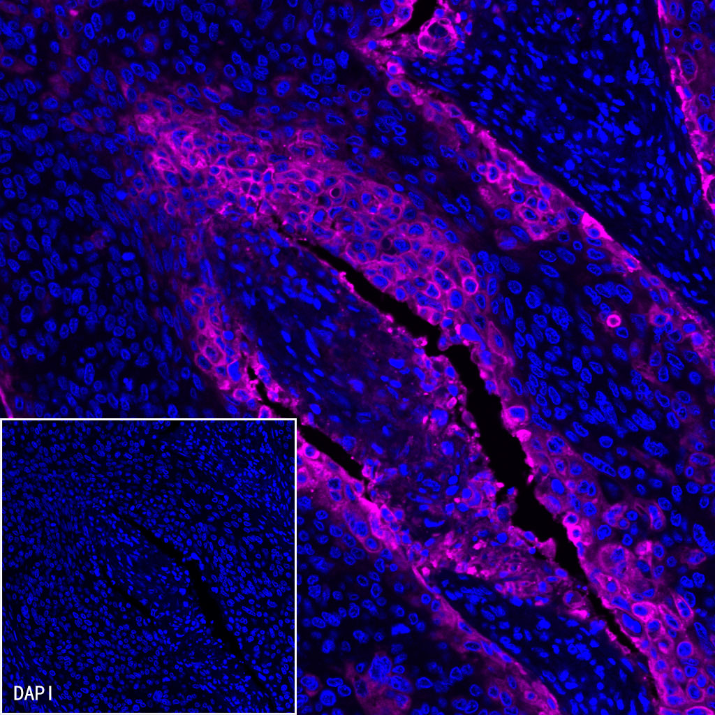
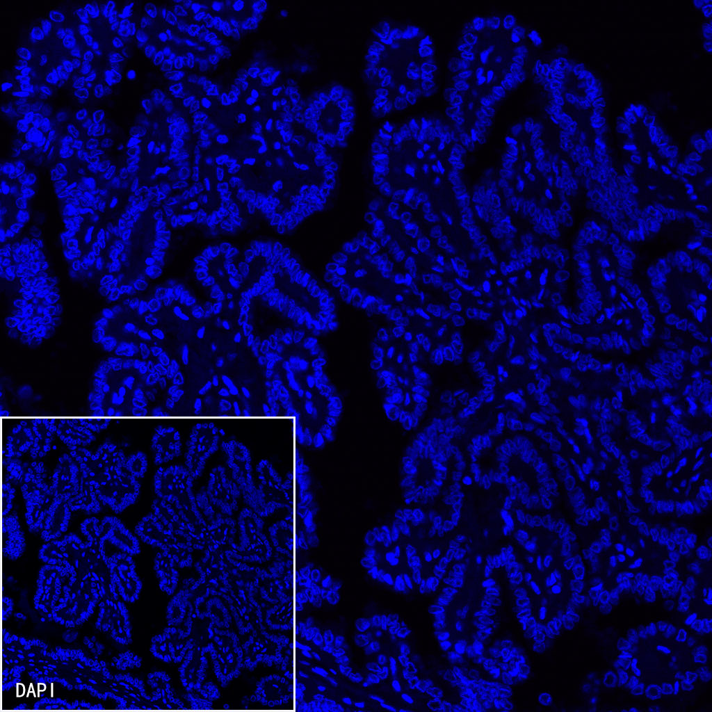
| Antigen |
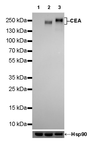
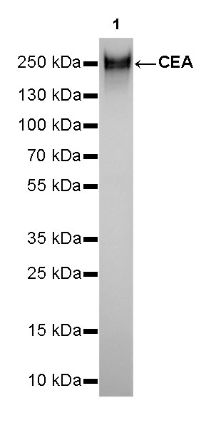
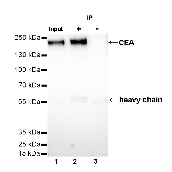
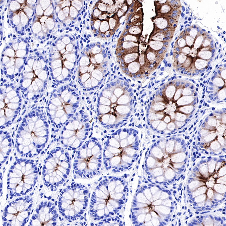
CEA |
| Synonyms |
Carcinoembryonic antigen-related cell adhesion molecule 5, Carcinoembryonic antigen, CD66e |
| Immunogen |
Recombinant Protein |
| Location |
Cell membrane |
| Accession |
P06731 |
| Clone Number |
SDT-098-54 |
| Antibody Type |
Rabbit mAb |
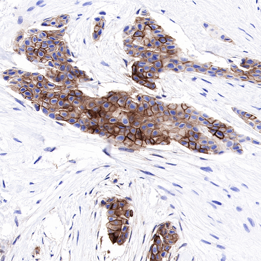
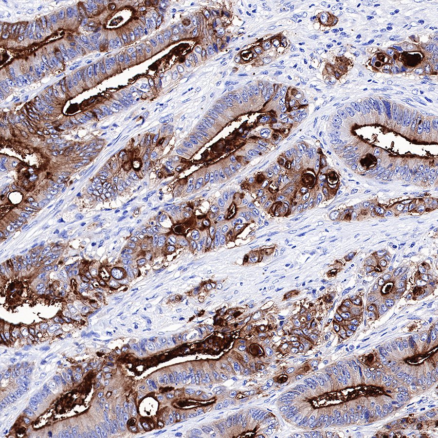
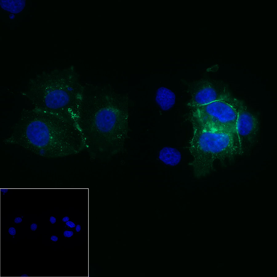
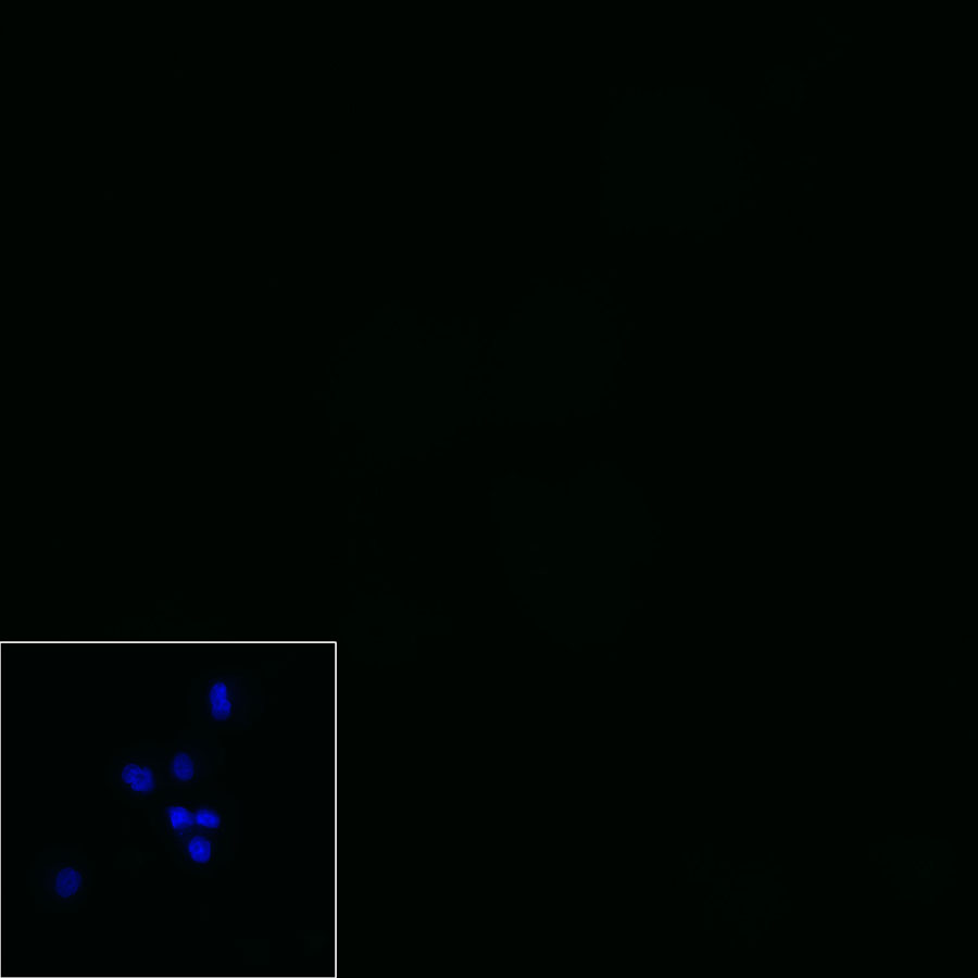
| Application |
WB, IHC-P, ICC, IP, IF |
| Reactivity |
Hu |
| Purification |
Protein A |
| Concentration |
0.25 mg/ml |
| Physical Appearance |
Liquid |
| Storage Buffer |
PBS, 40% Glycerol, 0.05%BSA, 0.03% Proclin 300 |
| Stability & Storage |
12 months from date of receipt / reconstitution, -20 °C as supplied |
Dilution
| application |
dilution |
species |
| ICC |
1:50 |
null |
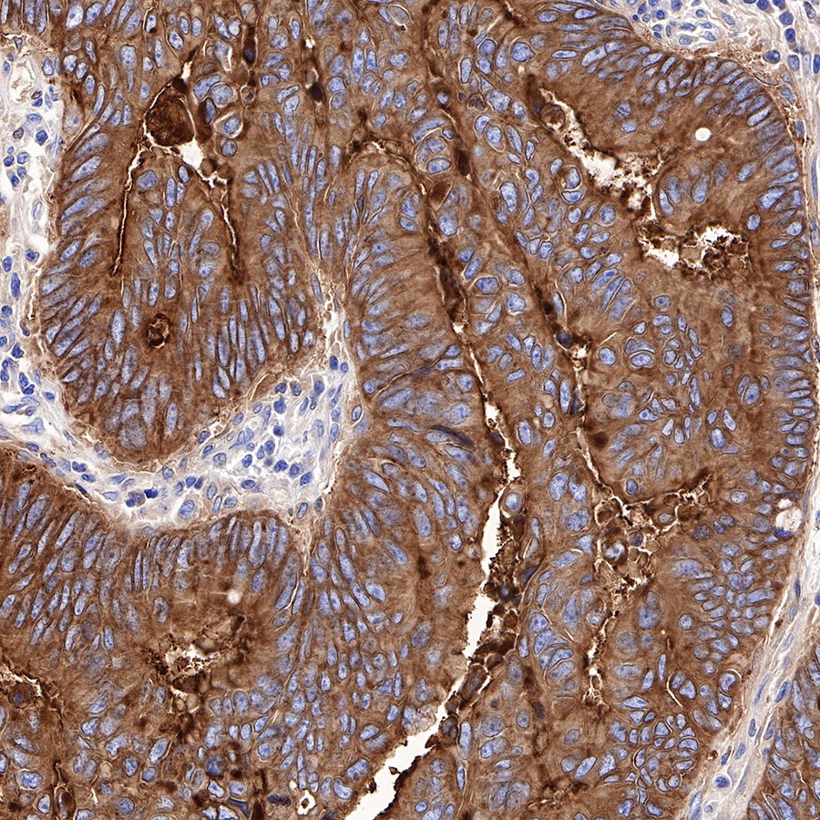
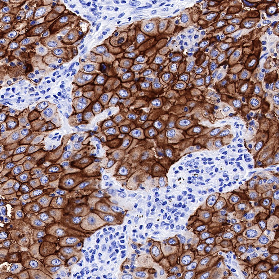
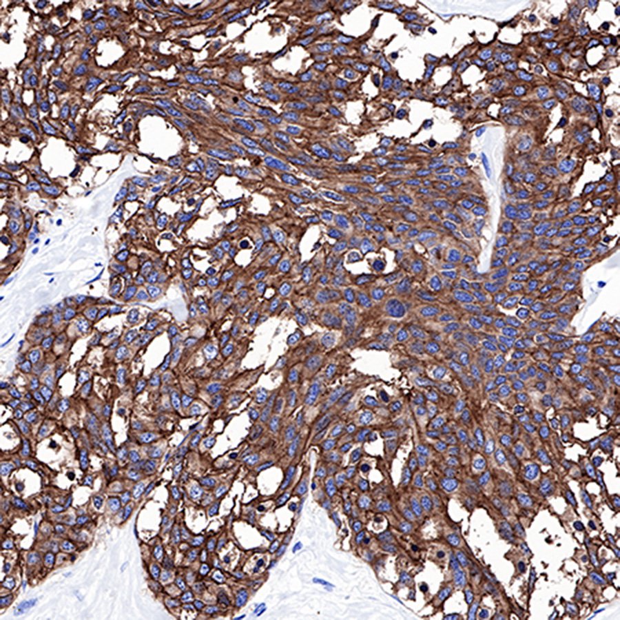
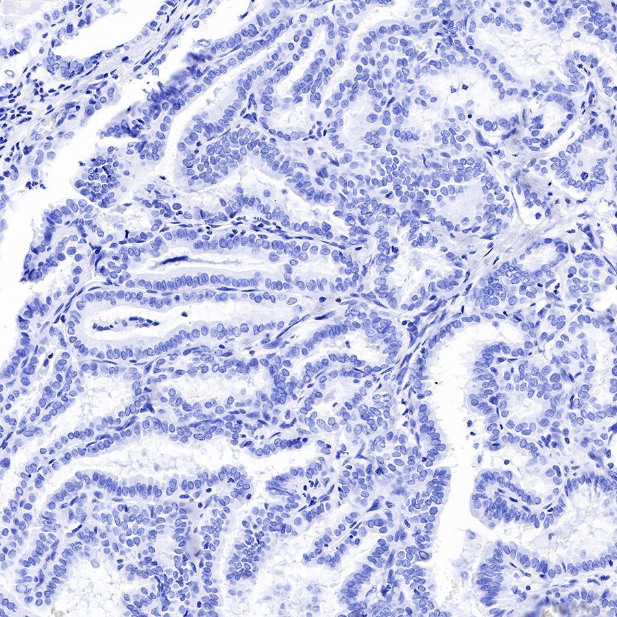
| IHC-P |
1:1000 |
null |
| WB |
1:500-10000 |
null |
| IP |
1:25 |
null |
| IF |
1:500 |
null |
Background
Carcinoembryonic antigen (CEA) describes a set of highly related glycoproteins involved in cell adhesion. CEA is normally produced in gastrointestinal tissue during fetal development, but the production stops before birth. CEA are glycosyl phosphatidyl inositol (GPI) cell-surface-anchored glycoproteins whose specialized sialofucosylated glycoforms serve as functional colon carcinoma L-selectin and E-selectin ligands, which may be critical to the metastatic dissemination of colon carcinoma cells. Immunologically they are characterized as members of the CD66 cluster of differentiation. The proteins include CD66a, CD66b, CD66c, CD66d, CD66e, CD66f.Consequently, CEA is usually present at very low levels in the blood of healthy adults (about 2–4 ng/mL). However, the serum levels are raised in some types of cancer. CEA is an important tumor marker for colorectal and some other carcinomas. The CEA subgroup members are cell membrane associated and show a complex expression pattern in normal and cancerous tissues with notably CEA showing a selective epithelial expression.