Glutathione (GSH) Detection Aids Lung Cancer Treatment Research
|
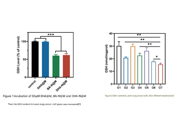
Background Introduction Glutathione (GSH) is an important antioxidant within cells, and its level directly reflects the redox status of tumor cells. Lung cancer cells often rely on high GSH levels to resist the reactive oxygen species (ROS) produced by radiochemotherapy, leading to therapeutic resistance. GSH, as a cofactor of glutathione peroxidase 4 (GPX4), can inhibit ferroptosis, and its depletion can lead to GPX4 inactivation, thereby promoting ferroptosis. Therefore, changes in GSH levels are an important marker of ferroptosis. Dynamic detection of GSH can assess whether the treatment activates oxidative stress and ferroptosis pathways. In addition, detecting GSH can also verify the release efficiency of GSH-responsive drugs, achieving tumor-specific activation. Application of Glutathione Detection in the Study of Lung Cancer Treatment On May 8, 2025, Dr. Chen Yiting and others from the School of Pharmacy, Fudan University, published a research paper titled "Inhalable biomimetic polyunsaturated fatty acid-based nanoreactors for peroxynitrite-augmented ferroptosis potentiate radiotherapy in lung cancer" in the Journal of Nanobiotechnology (IF=10.6). The research team designed a polyunsaturated fatty acid (PUFA)-based nanoreactor, DHA-N@M, which acts on lung tumors through inhalation administration. This nanoreactor, composed of macrophage membranes encapsulating DHA-SNO (a PUFA derivative that releases NO in response to GSH), has both tumor targeting and GSH-responsive characteristics. Experiments showed that DHA-N@M can significantly increase the accumulation of drugs in lung tumors, with better effects than intravenous injection. Its mechanism of action involves inducing tumor cell ferroptosis by consuming GSH, releasing NO, and generating ONOO⁻, and when used in conjunction with radiotherapy, it can synergistically enhance the antitumor effect. In a lung cancer orthotopic model, the combination of DHA-N@M and radiotherapy showed a tumor inhibition rate of 93.91% and good safety. This study provides a new type of inhalable nano drug delivery system for lung cancer treatment, significantly enhancing the effect of radiotherapy by targeting the tumor microenvironment and enhancing ferroptosis, and has significant potential for clinical translation.
Next, let's introduce in detail the usage of the Glutathione (GSH) Detection Kit(abs580006) 一、Components of the Kit
二、Procedure 1、Preparation of Materials and Reagents Materials to be prepared: Microplate reader, distilled water, pipette and tips, mortar and pestle, centrifuge, timer, wet ice, etc.
Standard: Before use, add 0.65 mL of distilled water to the tube, mix well until completely dissolved; then take 0.05 mL of the solution and add it to 0.95 mL of distilled water to mix evenly, to obtain a standard solution with a concentration of 0.5 mmol/L, store at 4°C.
Dye Reagent: Before use, add 4 mL of Diluent to dissolve.
2、Sample Preparation (1) Cell/bacterial samples: Collect cells/bacteria (5×10^6) into a centrifuge tube, centrifuge to remove the supernatant, add 1 mL of Assay Buffer, sonicate (power 20%, sonication 3s, interval 10s, repeat 30 times), then centrifuge at 4°C, 8000g for 10 minutes, transfer the supernatant to a new centrifuge tube, and keep on wet ice for detection.
(2) Tissue samples: Weigh 0.1g of tissue, add 1 mL of Assay Buffer, and homogenize on wet ice, centrifuge at 4°C, 8000g for 10 minutes, transfer the supernatant to a new centrifuge tube, and keep on wet ice for detection.
(3) Serum/plasma samples: Test directly.
3、Loading and Detection Before loading, heat all reagents to 37°C, then add samples according to the following table.
Notes: (1) For the standard, a serial dilution of 2 times (about 6 points) can be performed to create a standard curve.
(2) For unknown samples, we recommend selecting a few samples for a preliminary experiment to determine the sample concentration and dilution factor.
4、Standard Curve The standard curve is for reference only; a new standard curve must be generated for each experiment.
Frequently Asked Questions
Q1: What should I do if there is only one 96-Well Microplate in the kit and it's not enough for the experiment?
A1: The 96-well plate included in the kit is complimentary. For subsequent experiments, you can use regular 96-well plates as substitutes. Q2: The kit's operating manual includes both the standard curve method and the formula method. Which calculation method should I choose?
A2: You can choose either method. The formula is a simplified calculation. It is recommended to use the standard curve method for more accuracy. Note that when using the standard curve method, a new standard curve must be drawn for each experiment. Q3: What should I do if precipitation occurs during the reaction?
A3: Precipitation or flocculation is mostly caused by high protein concentration. It is recommended to aspirate the samples from the plate, centrifuge to obtain the supernatant, and then proceed with the test. Alternatively, you can add the samples to a centrifuge tube, react, centrifuge, and then transfer the supernatant to the plate for testing. Q4: What if the instrument does not have the corresponding detection wavelength?
A4: If the detection wavelength recommended in the kit is not available in the instrument settings, you can find a wavelength close to the recommended value within ±10nm, which should generally be acceptable. References: |
||||||||||||||||||||||||||||||||||||||||||||||||||||


Absin provides antibodies, proteins, ELISA kits, cell culture, detection kits, and other research reagents. If you have any product needs, please contact us.
|
Absin Bioscience Inc. |
 Follow us on Facebook: Absin Bio Follow us on Facebook: Absin Bio |


