Starter's Best - Selling Immune Checkpoint Antibodies
07 Jun 2025

Hangzhou Starter is a high - tech biotechnology company integrating R&D, production, sales, and technical services. It is dedicated to providing global biomedical research institutions, in - vitro diagnostic enterprises, and biopharmaceutical companies with high - quality antibodies, proteins, reagent kits, and customized services. These include research antibodies, CDx and IVD antigens and antibody raw materials, one - step ELISA kits, universal secondary antibody kits for mice and rabbits, and other immunoreagents, offering comprehensive solutions.
With a core team boasting fifteen years of antibody development experience and having successfully developed thousands of precision and research - oriented medical antibodies, the company has established platforms for multi - species single - B - cell antibody development (rabbit and mouse monoclonals), traditional mouse monoclonals, and recombinant protein expression. It also has a robust antibody application validation and testing system covering immunochromatography, ELISA, flow cytometry, IHC, Western blotting, immunofluorescence, and immunoprecipitation. It has passed EU 98/79/EC, ISO9001, and ISO13485 certifications.
What Are Immune Checkpoints?
Immune checkpoints are regulators of the immune system that prevent it from indiscriminately attacking self - antigens. While this is crucial for preventing autoimmune diseases, it can also cause the immune system to be ineffective in eradicating or suppressing cancer.
Tumor cells exploit certain immune checkpoint pathways as a major mechanism of immune resistance, particularly targeting T cells specific to tumor antigens. Since many of these immune checkpoints are initiated by ligand - receptor interactions, they are susceptible to antibody blockade. Blocking these immune checkpoints can restore anti - tumor activity and is one of the most promising methods for activating therapeutic anti - tumor immunity.
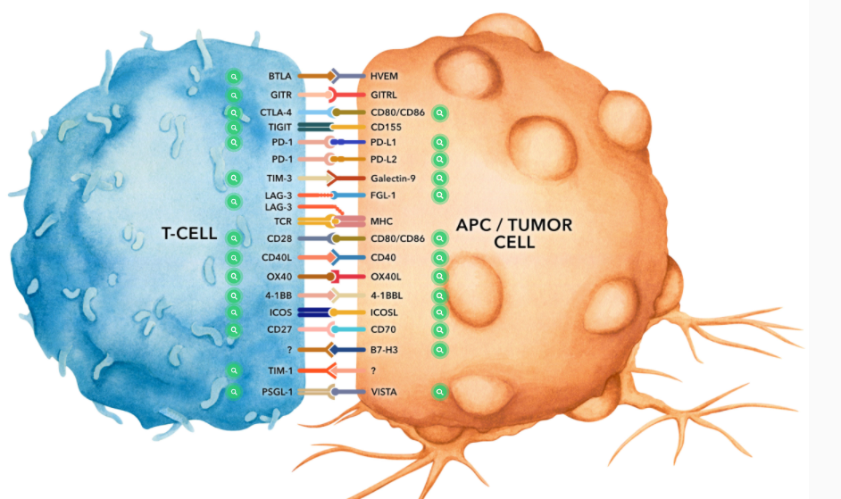
Starter offers a variety of antibodies targeting immune checkpoint proteins in mice.
1. CTLA - 4 (CD152)
CTLA - 4 is expressed on activated T and B lymphocytes. Structurally similar to the T - cell costimulatory protein CD28, both CTLA - 4 and CD28 bind to B7 family members B7 - 1 (CD80) and B7 - 2 (CD86). After ligand binding, CTLA - 4 negatively regulates cell - mediated immune responses. CTLA - 4 plays a role in the induction and/or maintenance of immune tolerance, thymocyte development, and the regulation of protective immunity. Its critical role in immune down - regulation has been confirmed in CTLA - 4 - deficient mice, which die at 3 - 5 weeks of age due to the development of lymphoproliferative disease. CTLA - 4 is one of a set of inhibitory receptors explored as cancer targets through immune checkpoint blockade.
2. 4 - 1BB (CD137)
4 - 1BB is a 39 kDa transmembrane protein expressed on T lymphocytes, NK cells, dendritic cells, granulocytes, and mast cells. After binding to its ligand 4 - 1BBL, 4 - 1BB provides co - stimulatory signals to CD4 and CD8 T cells by activating downstream pathways such as NF - κB, c - Jun, and p38. The importance of the 4 - 1BB pathway has been emphasized in many diseases, including cancer. Agonistic anti - 4 - 1BB antibodies have been reported to induce T - cell - mediated anti - tumor immunity.
3. 4 - 1BBL (CD137L)
4 - 1BBL is a 97 kDa member of the TNF superfamily expressed on dendritic cells, macrophages, and activated B and T lymphocytes. The interaction between 4 - 1BBL and 4 - 1BB (CD137) provides co - stimulatory signals to CD4 and CD8 T cells by activating downstream pathways such as NF - κB, c - Jun, and p38.
4. CD40
CD40 is widely expressed on antigen - presenting cells (APCs), such as dendritic cells, B cells, macrophages, and monocytes, as well as on non - immune endothelial cells, basal epithelial cells, and a variety of tumors. After binding to its ligand CD154, CD40 functions as a co - stimulatory molecule to activate B cells, dendritic cells, monocytes, and other APCs. CD40 plays a role in B - cell activation, differentiation, proliferation, and Ig isotype switching, as well as in dendritic cell maturation. Agonistic CD40 monoclonal antibodies have been shown to activate APCs and promote anti - tumor T - cell responses.
5. CD40L (CD154)
CD154 is primarily expressed on the surface of activated CD4+ T lymphocytes but is also expressed on platelets, mast cells, macrophages, basophils, NK cells, B lymphocytes, CD8+ T lymphocytes, and non - hematopoietic cells, including smooth muscle cells, endothelial cells, and epithelial cells. CD154 signals through CD40 and is believed to play a key role in T - and B - lymphocyte costimulation. Anti - CD154 antibodies have been reported to inhibit germinal center formation and disrupt antigen - specific T - cell responses.
6. CD47 (IAP)
CD47 is universally expressed on hematopoietic cells such as T and B lymphocytes, monocytes, platelets, and red blood cells, as well as on non - hematopoietic cells. CD47 is involved in a variety of cellular processes, including apoptosis, proliferation, adhesion, and migration. Additionally, it plays a key role in immune and angiogenic responses. CD47 is the receptor for thrombospondin - 1 (TSP - 1), a secreted glycoprotein involved in angiogenesis and vascular development. CD47 has been found to be overexpressed in many different tumor cells. Therefore, anti - CD47 monoclonal antibodies have been proposed and studied as a therapeutic approach for human cancers.
7. CD80 (B7 - 1)
CD80 is expressed on activated B cells and is constitutively expressed on monocytes and dendritic cells. This ligand binds to CD28 to provide co - stimulatory signals necessary for T - cell activation, survival, and cytokine production. Additionally, CD80 binds to CTLA - 4, thereby inhibiting T cells.
8. CD86 (B7 - 2)
CD86 is expressed on activated T and B cells, macrophages, and dendritic cells. This ligand binds to CD28 to provide co - stimulatory signals necessary for T - cell activation, survival, and cytokine production. Additionally, CD80 binds to CTLA - 4, thereby inhibiting T cells.
9. CD276 (B7 - H3)
CD276 is weakly expressed on activated lymphocytes, macrophages, dendritic cells, nasal and airway epithelial cells, osteoblasts, and some tumor cell lines. Monocytes, dendritic cells, and activated T cells also secrete a soluble form of CD276. The biological functions of CD276 are still under investigation. However, recent studies suggest that CD276 has a negative regulatory role in T - cell responses.
10. LAG - 3
LAG - 3 is expressed on activated T lymphocytes, NK cells, and T regulatory cells. The primary ligand for LAG - 3 is MHC class II, which it binds with higher affinity than CD4. LAG - 3 is thought to play a role similar to CTLA - 4 and PD - 1, including down - regulating TCR signaling and inhibiting CD4 - dependent T - cell functions. LAG - 3 has also been shown to contribute to the suppressive function of T regulatory cells. Conversely, LAG - 3 has been shown to promote immune responses by activating antigen - presenting cells.
11. OX40 (CD134)
OX40 is expressed on activated CD4 and CD8 T cells but is not found on resting naive T cells or most resting memory T cells. Initially thought to be expressed only on activated conventional T cells, OX40 has now been visualized on activated regulatory T cells, NKT cells, NK cells, and neutrophils. OX40 plays a major role in regulating CD4 and CD8 T - cell clonal expansion. It provides co - stimulatory signals to naive T cells responding to antigens, prolonging proliferation and increasing the production of several cytokines. OX40 knockout mice have demonstrated this, as they generate fewer primary effector CD4 T cells after immunization. Furthermore,
12. OX40L (CD134L)
OX40L is expressed on activated B cells and antigen - presenting cells. OX40L is the ligand for OX40 (CD134). OX40 signaling regulates CD4 and CD8 T - cell clonal expansion. It provides co - stimulatory signals to naive T cells responding to antigens to prolong proliferation and increase the production of several cytokines, including IL - 2.
13. PD - 1 (CD279)
PD - 1 is transiently expressed on CD4 and CD8 thymocytes, as well as on activated T and B lymphocytes and myeloid cells. PD - 1 expression declines after successful antigen elimination. Additionally, Pdcd1 mRNA is expressed in developing B lymphocytes at the pre - B cell stage. The structure of PD - 1 includes an ITIM (immunoreceptor tyrosine - based inhibition motif), indicating that PD - 1 negatively regulates TCR signaling. PD - 1 signals via binding to two ligands of the B7 family, PD - L1 and PD - L2. After ligand binding, PD - 1 signaling inhibits T - cell activation, leading to reduced proliferation, cytokine production, and T - cell death. Moreover, PD - 1 has been shown to play a critical role in peripheral tolerance and the prevention of autoimmune diseases in mice, as PD - 1 knockout animals exhibit dilated cardiomyopathy, splenomegaly, and loss of peripheral tolerance. Induced PD - L1 expression is common in many tumors, including squamous cell carcinomas, colorectal adenocarcinomas, and breast cancers. PD - L1 overexpression results in increased resistance of tumor cells to CD8 T - cell - mediated lysis. In a melanoma mouse model, treatment with antibodies blocking the interaction between PD - L1 and its receptor PD - 1 temporarily halted tumor growth. For these reasons, anti - PD - 1 mediated immunotherapy is currently being explored as a cancer treatment. Treatment with antibodies blocking the interaction between PD - L1 and its receptor PD - 1 can temporarily halt tumor growth. For these reasons, anti - PD - 1 mediated immunotherapy is currently being explored as a cancer treatment. Treatment with antibodies blocking the interaction between PD - L1 and its receptor PD - 1 can temporarily halt tumor growth. For these reasons, anti - PD - 1 mediated immunotherapy is currently being explored as a cancer treatment.
14. PD - L1 (B7 - H1)
PD - L1 is expressed on T lymphocytes, B lymphocytes, NK cells, dendritic cells, and IFNγ - stimulated monocytes, epithelial cells, and endothelial cells. PD - L1 binds to its receptor PD - 1, which is present on CD4 and CD8 thymocytes, as well as on activated T and B lymphocytes and myeloid cells. The binding of PD - L1 to PD - 1 results in the inhibition of TCR - mediated T - cell proliferation and cytokine production. PD - L1 is thought to play an important role in tumor immune evasion. Induced PD - L1 expression is common in many tumors and leads to increased resistance of tumor cells to CD8 T - cell - mediated lysis. In a melanoma mouse model, tumor growth can be temporarily halted by treatment with antibodies blocking the interaction between PD - L1 and PD - 1.
15. PD - L2 (B7 - DC)
PD - L2 is a 25 kDa type I transmembrane protein and a member of the B7 family within the Ig superfamily. PD - L2 is expressed on monocytes, macrophages, and subpopulations of dendritic cells. PD - L2 binds to its receptor PD - 1, which is present on CD4 and CD8 thymocytes, as well as on activated T and B lymphocytes and myeloid cells. The binding of PD - L2 to PD - 1 results in the inhibition of TCR - mediated T - cell proliferation and cytokine production.
16. TIM - 1 (CD365)
TIM - 1 is a type I cell surface glycoprotein and a member of the Ig superfamily. TIM - 1 is preferentially expressed on TH2 cells and has been identified as a stimulatory molecule for T - cell activation. The TIM gene family plays a key role in regulating immune responses to viral infections. TIM - 1 is also associated with allergic reactions, asthma, and transplant tolerance.
17. TIM - 3 (CD366)
TIM - 3 is specifically expressed at high levels on the surface of Th1 lymphocytes, while Th2 lymphocytes express TIM - 1 and TIM - 2. TIM - 3 activation occurs through binding to the cell - associated C - type lectin galectin - 9. Galectin - 9 binding to TIM - 3 induces apoptosis in Th1 cells. Inhibiting TIM - 3 signaling in mice exacerbates experimental autoimmune encephalomyelitis, promoting IFNγ production and Th1 cell proliferation. TIM - 3 has also been shown to be required for the induction of tolerance, as both TIM - 3 knockout animals and mice treated with TIM - 3 - Ig fusion proteins exhibit defects in the induction of antigen - specific tolerance. Furthermore,
18.VISTA
VISTA, an inhibitory molecule of cell activation, also known as PD - 1H and B7 - H5, is a 309 - aa type I transmembrane glycoprotein and a member of the Ig superfamily. VISTA is expressed on naïve and activated T cells, NK cells, macrophages, dendritic cells, and neutrophils. VISTA functions as a negative immune checkpoint protein, inhibiting T - cell cytokine production and proliferation. VISTA is overexpressed on tumor - infiltrating lymphocytes, such as myeloid cells and regulatory T cells. In a melanoma mouse model, blocking VISTA results in delayed tumor growth.
19. ICOS
ICOS is a 47 - 57 kDa homodimeric glycoprotein and a member of the costimulatory molecule CD28 family. ICOS is expressed on activated T cells and costimulates T and B cell responses following ICOSL binding. The ligand is expressed on antigen - presenting cells, including splenic B cells, dendritic cells, and macrophages. ICOS signaling is also thought to be important for maintaining regulatory T cell homeostasis.
20. GITR
GITR is expressed at low levels on resting T lymphocytes and at high levels on regulatory T cells. GITR is upregulated on activated T cells and provides costimulation. GITR ligand (GITRL) is present on B cells, macrophages, dendritic cells, and endothelial cells and is involved in regulating innate and adaptive immune responses. GITR is also believed to play a key role in the maintenance of dominant immune self - tolerance by regulatory T cells. Mouse knockout studies have also indicated that this receptor modulates CD3 - driven T - cell activation and programmed cell death.
21. Galectin - 9
Galectin - 9 binds to β - galactosides and serves as a ligand for TIM - 3 (CD366). This protein is particularly involved in innate and adaptive immune responses, macrophage - induced cytokine secretion, bactericidal functions, dendritic cell maturation promotion, regulatory T cell expansion, and the negative regulation of Th1, Th17, NK, and cytotoxic T cells.
22. CD27
CD27 is a 45 kDa type I transmembrane protein and a member of the TNF superfamily. CD27 is expressed on peripheral T cells, memory B cells, NK cells, and a subset of thymocytes. CD27 is highly induced on T cells following TCR stimulation. CD70 is the ligand for CD27. The interaction with CD70 provides costimulation in T cells and induces signaling events crucial for cell proliferation, the long - term maintenance of antigen - specific T cells, antiviral responses, anti - tumor immunity, and allogeneic reactions. Agonistic antibodies stimulating CD27 are currently being explored as experimental cancer therapies.
23. CD28
CD28 is expressed on thymocytes, most peripheral T cells, and NK cells. CD28 is the receptor for CD80 (B7 - 1) and CD86 (B7 - 2). Signaling through CD28 enhances the expression of IL - 2 and IL - 2 receptors, as well as the cytotoxicity of CD3 - activated T cells.
24. CD70
CD70 is expressed on activated mouse T and B lymphocytes and dendritic cells. CD70 is the ligand for CD27, and their interaction promotes cross - stimulation of T and B cells, as well as costimulation for B cell proliferation and immunoglobulin production. Cells expressing CD70 have been shown to costimulate T cell proliferation and induce cytokine production.
25. ICOSL (CD275)
ICOSL (inducible T - cell costimulator ligand) is also known as CD275, B7RP - 1, and B7 - H2. ICOSL is a 40 kDa immune checkpoint protein belonging to the Ig receptor superfamily. After binding to ICOSL, ICOS signaling costimulates T and B cell responses. The ligand is expressed on antigen - presenting cells, including splenic B cells, dendritic cells, and macrophages. ICOS signaling is also thought to be important for maintaining regulatory T cell homeostasis.
26. BTLA
BTLA is a member of the Ig superfamily and is expressed on B cells, T cells, macrophages, dendritic cells, NK cells, and NKT cells. Like PD - 1 and CTLA - 4, BTLA interacts with B7 - homologous molecule B7 - H4. However, unlike PD - 1 and CTLA - 4, BTLA demonstrates T - cell inhibition by interacting with tumor necrosis family receptors (not just B7 family cell surface receptors). BTLA is the ligand for herpesvirus entry mediator (HVEM). The BTLA - HVEM complex has been shown to negatively regulate T - cell immune responses.
27. FGL - 1
FGL - 1 is a member of the fibrinogen protein family. Under normal physiological conditions, FGL - 1 is primarily secreted from hepatocytes and contributes to mitosis and metabolic functions. FGL - 1 is produced at high levels in various tumors, including lung cancer and melanoma. High expression of FGL - 1 is associated with resistance to anti - PD - 1/PD - L1 therapy and poor prognosis in cancer patients. Recently, FGL - 1 has been identified as the major inhibitory ligand for LAG - 3, a receptor that negatively regulates T - cell proliferation, activation, and effector functions.
28. PSGL - 1
PSGL - 1 is a 230 kDa glycoprotein expressed on bone marrow - derived mast cells and dendritic cells, splenic white blood cells, platelets, peripheral blood neutrophils, and T lymphocytes. PSGL - 1 is the ligand for P - selectin (CD62P) and plays a role in leukocyte rolling, leukocyte migration to inflamed tissues, and responses to vascular injury.
29. TIGIT
TIGIT is a 26 kDa type I transmembrane protein and a member of the poliovirus receptor (PVR) family. TIGIT has been found to be expressed on follicular T helper cells in mice, while in humans, it is expressed by multiple T - cell subsets, including activated T cells, follicular T helper cells, memory T cells, regulatory T cells, and NK cells. TIGIT can interact with certain members of the PVR and PVR - like families, including PVR, PVRL2, PVRL3, CD155, and CD112. TIGIT is believed to have a negative regulatory role in NK and T - cell activation. Dendritic cells binding TIGIT to T cells lead to their differentiation into a tolerogenic phenotype, increased IL - 10 secretion, and reduced IL - 12 production. TIGIT gene knockout mice are more susceptible to autoimmune diseases.
Best - Selling Products
Antibodies
| Starter Catalog Number | Product Name |
|---|---|
| S0B0594 | Invivo anti - mouse PD - 1 Recombinant mAb (D265A) |
| S0B0593 | Invivo anti - mouse PD - L1 Recombinant mAb (D265A) |
| S0B1198 | Invivo Anti - mouse Ly6G monoclonal Antibody |
| S0B0574 | Invivo anti - mouse CTLA - 4 (CD152) mAb |
| S0B0873 | Invivo anti - mouse CD25 Recombinant mAb |
| S0B0690 | Invivo anti - mouse CD4 Recombinant mAb |
| S0B0574 | Invivo anti - mouse CTLA - 4 (CD152) mAb |
Isotype Controls
| Starter Catalog Number | Product Name |
|---|---|
| S0B0787 | Invivo mouse IgG2a isotype control |
| S0B0997 | Invivo rat IgG2b isotype control, anti - keyhole limpet hemocyanin |
| S0B0997 | Invivo rat IgG2b isotype control, anti - keyhole limpet hemocyanin |
| S0B0997 | Invivo rat IgG2b isotype control, anti - keyhole limpet hemocyanin |


