M13 Phage Detection: An Indispensable Component in the Screening of Novel Drugs via Phage Display Antibody Technology.

Recent Advances
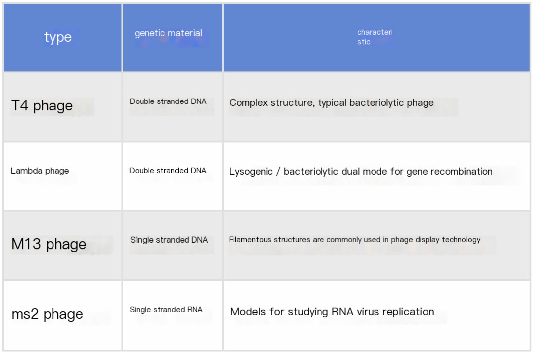
Bacteriophages, commonly referred to as "phages" or "bacteriophage viruses," are a class of viruses that specifically infect bacteria. They consist of a protein capsid enclosing internal genetic material, which may be either DNA or RNA. Bacteriophages are ubiquitously present in nature and possess the ability to attach to the surface of bacterial cells, subsequently injecting their genetic material into the bacterial cytoplasm. This process enables the phage to hijack the bacterial machinery for replication and propagation.
Common Types of Bacteriophages
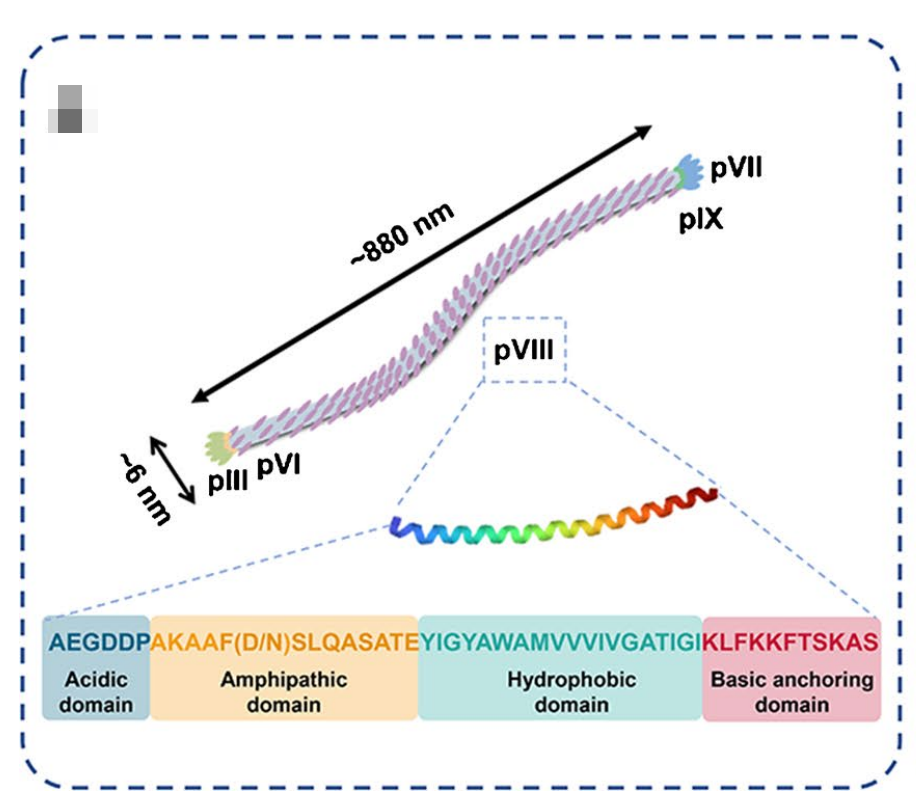
Schematic Diagram of M13 Phage Structure
Introduction to Phage Display Technology
Historical Background
Phage display technology was pioneered by George Smith in 1985. For their contributions to directed molecular evolution, Smith and Gregory Winter were jointly awarded the 2018 Nobel Prize in Chemistry. M13, due to its temperate nature (non-lytic to the host), has become one of the most commonly used display vectors.
Fundamental Principles
M13 phage is a filamentous bacteriophage that infects Escherichia coli and contains a single-stranded DNA genome. The core of the display technology involves genetically engineering the fusion of a foreign protein gene with a phage capsid protein gene (such as pIII or pVIII), enabling the expression of the foreign protein on the phage surface.
-
pIII Protein: Located at the terminus of the phage, it typically displays single copies of large molecular proteins (e.g., antibody fragments).
-
pVIII Protein: Constitutes the major capsid of the phage and is suitable for displaying multiple copies of small peptides (e.g., 10-20 amino acids).
Technical Workflow
-
Gene Cloning: The gene encoding the target protein or peptide is cloned into the M13 phage genome, usually inserted into the gene encoding the phage capsid protein.
-
Phage Library Construction: The engineered M13 phage is transformed into E. coli. After infecting the bacteria, the phage replicates within the bacteria and synthesizes capsid proteins, simultaneously displaying the target protein or peptide on the phage surface.
-
Affinity Screening (Panning): The phage is incubated with immobilized target molecules (e.g., antigens). Unbound phages are washed away, while specifically bound phages are retained.
-
Amplification and Enrichment: Bound phages are recovered and amplified by infecting E. coli. The screening process is repeated for 3-4 rounds to enhance binding affinity.
-
Identification: The foreign genes carried by the selected phages are analyzed through sequencing or functional assays.
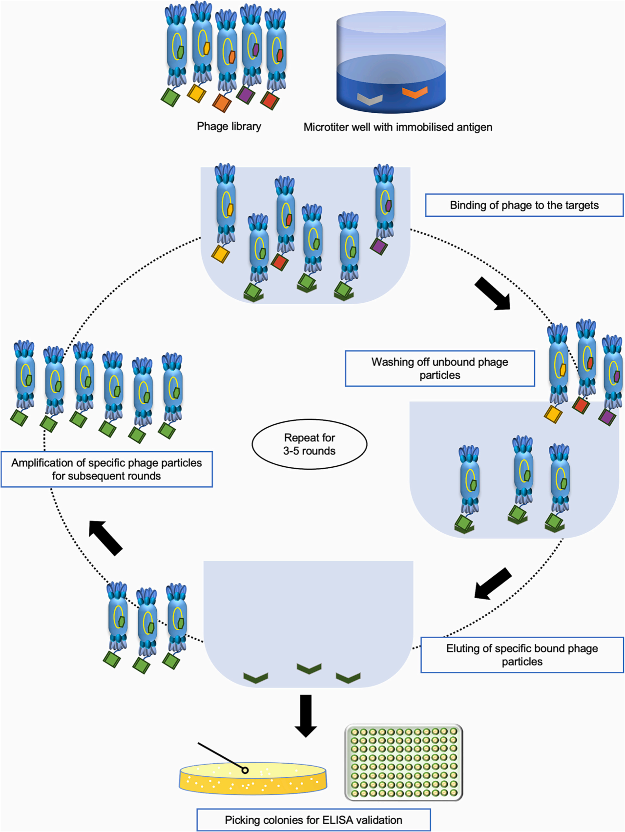
General Process of Biopanning
Core Advantages
-
Direct Genetic Linkage: The phage carries the gene encoding the foreign protein, facilitating subsequent cloning.
-
High-Throughput Screening: Enables rapid screening of millions to billions of molecules.
-
No Protein Purification Required: Displayed proteins are directly used in binding assays.
Applications
-
Antibody Screening: One of the most widespread applications of M13 phage display technology is the screening of monoclonal antibodies. By displaying different antibody fragments (e.g., variable regions of heavy or light chains), antibodies that bind to specific antigens can be rapidly identified. These antibodies are applicable in disease diagnosis, therapy, and other fields.
-
Vaccine Development: Phage display technology can be used to display surface antigens of pathogens. By screening, antigenic peptides that interact with pathogenic substances can be obtained, providing insights for vaccine design.
-
Drug Screening: This technology is also applicable for small-molecule drug screening, particularly in identifying target molecules or protein-protein interactions. For example, small peptides can be displayed to screen for molecules with specific biological activities.
-
Protein-Protein Interaction Studies: Phage display technology can be used to investigate interactions between different proteins. For instance, by displaying a specific fragment of a protein, other proteins that bind to this fragment can be identified.
-
Development of Novel Enzymes: By displaying specific peptides at enzyme active sites, mutants or entirely new enzymes with desired enzymatic activities can be screened.
START has developed M13 antibodies and one-step kits, which are powerful tools for M13 phage detection. We welcome you to explore and purchase these products!
Partial Data Presentation
M13 phage Recombinant Rabbit mAb (Biotin Conjugate) (S-1403-13) (Catalog Number:S0B1686)
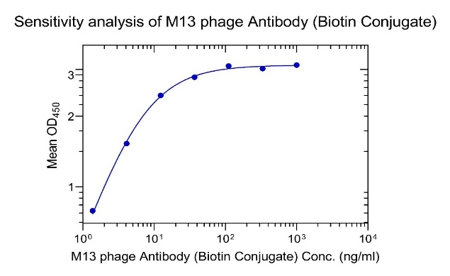
Indirect ELISA binding analysis of M13 phage Recombinant Rabbit mAb (Biotin Conjugate) (S-1403-13)
Coating: M13 phage, 1*10E7 pfu/ml
Primary antibody: M13 phage (Biotin Conjugate) (S-1403-13), from 1μg/ml, 3-fold diluent, 7 points
M13 OneStep ELISA Kit(Catalog Number:S0C3033)
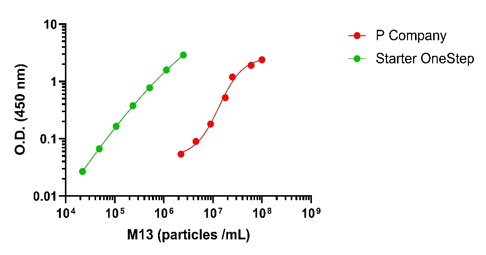
Leading Competitor comparison
Product Information
| Gatalog Num | Product Name | Product Parameters | Price |
| S0C3033 | M13 OneStep ELISA Kit | $535 | |
| S0B1064 | M13 phage Recombinant Rabbit mAb (S-1403-28) | Host : Rabbit | Inquiry |
| Conjugation : Unconjugated | |||
| S0B1063 | M13 phage Recombinant Rabbit mAb (S-1403-13) | Host : Rabbit | Inquiry |
| Conjugation : Unconjugated | |||
| S0B1826 | M13 phage Recombinant Rabbit mAb (HRP Conjugate) (S-1403-13) | Host : Rabbit | $100 |
| Conjugation : HRP | |||
| S0B1686 | M13 phage Recombinant Rabbit mAb (Biotin Conjugate) (S-1403-13) | Host : Rabbit | $100 |
| Conjugation : Biotin |


