Identification and sterilization strategy of organoid contamination (1)
1. Overview
Organoid contamination is a common thing, especially in the gastrointestinal tract, the number of microflora brought by the tissue itself is very large, and once the disinfection is not in place, it will lead to the failure of organoid culture. The common types of organoid pollution include bacteria, fungi, mycoplasma, black gum worms and other pollution sources.
2. Identification of organoid contamination
1. Bacterial contamination

Bacterial contamination - under the naked eye
Contamination characteristics:
● There are obvious black spheres in the matrix visible to the naked eye;
● And it is distributed in multiple black spheres;
● Rapid growth, usually D1 will appear;
● The culture medium will turn red and yellow and turbid overnight.

Bacterial contamination - microscopic primary extraction of organoids D1
Bacterial contamination characteristics:
● The bacterial colony is black and spherical, completely impermeable;
● The periphery is blurry and hazy;
● The outbreak is very fast, and a large number of colonies can appear in 1-2 days.
Bacterial Busters
|
Catalog number |
antibiotic |
Affected object |
Use scales |
Storage conditions |
|
Kanamycin B sulfate |
Bacterial (G+, G-) mycoplasma |
The concentration of the mother liquor is 10mg/mL (dissolved in sterile water), and the |
-20℃ |
|
|
Gentamicin sulfate solution (10 mg/mL) |
Bacterial (G+, G-) mycoplasma |
Dilute 1:200 |
-20℃ |
|
|
Cyan-chain bispecific antibody |
Bacteria (G+, G-) |
Dilute 1:100 |
-20℃ |
|
|
Penicillin-streptomycin-amphotericin B tertiary antibody |
Bacteria (G+, G-), fungi (e.g. yeast, molds) |
Dilute 1:100 |
-20℃ |
|
|
Penicillin-streptomycin-gentamicin triantibody |
Bacteria (G+, G-), mycoplasma |
Dilute 1:100 |
-20℃ |
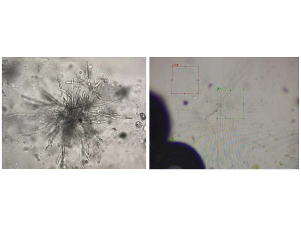
2. Fungal contamination

Left: Fungal contamination characteristics – mold
Right: Fungal contamination feature – Rhizopus vulgaris
Mold contamination characteristics:
● In the early stage, the medium will not turn yellow and turbid, but the mycelium can be observed under the microscope;
● The hyphae formed by the fungus are filamentous, tubular, dendritic, beaded.
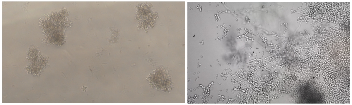
Fungal contamination – yeast

The yeast clusters found in mouse gastric organoid P0D1 are 4X, 10X, and 20X from left to right

Yeast contamination in organoid matrigel
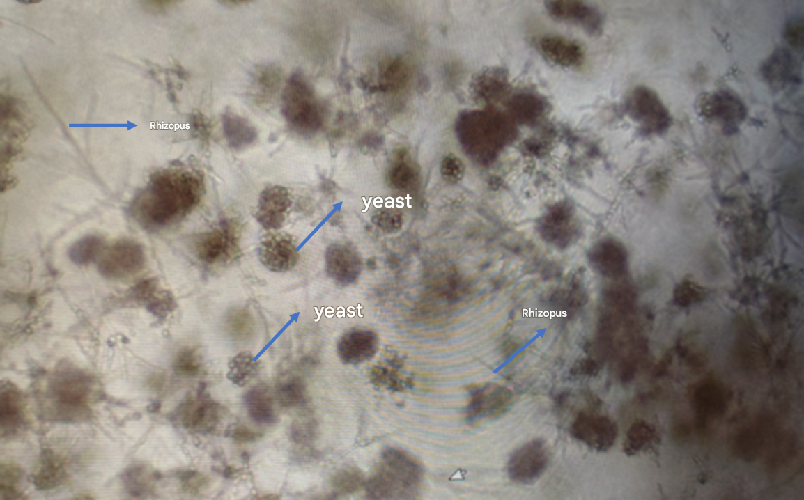
Yeast + Rhizopus contamination in organoid matrigel
Yeast contamination characteristics:
● Yeast is a cotton flocculent-like clump;
● Yeast, etc., can be identified from a large and a small conjoined structure when it sprouts and reproduces;
● After the cells are contaminated, they can still grow, and the viability of the organoids will deteriorate for a long time;
● The pollution rate increases during the rainy season.
Fungal nemesis
|
Catalog number |
antibiotic |
Affected object |
Use scales |
Storage conditions |
|
abs42013298 |
Amphotericin B |
Fungi (e.g. yeasts, molds) |
The concentration of mother liquor was 2.5mg/mL (DMSO dissolved), and the working concentration was diluted 1:1000 |
-20℃ |
|
abs44071148 |
nystatin |
Fungi (e.g. yeasts, molds) |
The concentration of mother liquor was 2mg/mL (DMSO dissolved), and the |
-20℃ |
|
abs9246 |
Penicillin-streptomycin-amphotericin B mixture solution (100× triclonal antibody) |
Bacteria (e.g. G+, G-), |
Dilute 1:100 |
-20℃ |
3. Black gum worms
Characteristics of black gum insect contamination:
● Active twisting;
● Mostly rod-shaped;
● The medium is not turbid.
Black gum worm remover PLUS (500×) abs9841
Benefits:
1) Stable performance: through bacteria, fungi, mycoplasma, endotoxin detection; Through osmotic pressure and pH detection; Pass the product performance test;
2) Efficient and fast: effectively inhibit and kill "black rubber worms" and reduce unnecessary experimental losses;
3) Environmental protection and safety: It has been verified by hundreds of cells to have no toxic effect.
How to Use:
1) Please centrifuge the reagent tube instantaneously (3000rpm, 3~5s) after receiving the goods;
2) When using black gum insect remover, it is recommended to prepare and use it now, and keep the cell density at 50%~60%;
3) The recommended dilution ratio is 1:500. For example, add 10 μL of black gumworm scavenger to 5 mL of medium. Discard the old medium, wash the cells with PBS, and then add fresh medium containing gelatina scavenger once every 2 days for one week.

Black gum insect prophylaxis (1000×) ABS9842
Benefits:
1) Efficient and stable: through product performance testing, it can effectively prevent and control the pollution problem of "black rubber insects" in cell culture;
2) Environmental protection and safety: no toxic effect on cells, will not affect cells in the process of cell culture, this product is a polypeptide, unlike antibiotics will affect the growth state of cells and make cells resistant.
How to Use:
1) When using the prophylaxis of black gum worm (1000X), it is recommended to prepare and use it now;
2) It is recommended to dilute the prophylactic agent (1000X) at a ratio of 1:1000, for example, add 10 μL of prophylactic agent (1000X) to 10 mL of medium.
4. Mycoplasma contamination
Mycoplasma is a common prokaryotic organism in the laboratory that contaminates cells, and the contamination mainly comes from animal raw materials such as tissues and serum, and the operators themselves. Mycoplasma is 0.1-0.3 μm in diameter, and it is difficult to distinguish under the microscope, so it is generally detected by aspirating the supernatant of the culture medium and using qPCR technology to amplify DNA.
1) Detection principle
TaqMan fluorescent probes are oligonucleotide probes that carry fluorophores (reporters) at the 5' end, such as FAM, CY5, VIC, etc., and quencher groups at the 3' end, such as TAMRA, BHQ1, etc. PCR amplification involves the addition of a pair of primers along with a specific fluorescent probe, and when the probe is intact, the fluorescent signal emitted by the fluorophore is absorbed by the quencher group. During PCR amplification, the 5'-3' exonuclease activity of Taq enzyme digests and degrades the probe, separating the fluorophore and quencher group, so that the fluorescence monitoring system (fluorescence quantitative PCR instrument) can receive the fluorescence signal, that is, for each amplified DNA strand, a fluorescent molecule is formed, realizing the complete synchronization of the accumulation of the fluorescence signal with the formation of the PCR product. This kit uses multiplex PCR to detect the target sequence and internal control with two different fluorescent probes. For the specific steps of the experiment, please refer to the product manual (absin.cn) of the Mycoplasma Detection Kit (qPCR method).
2) Removal reagent

Mycoplasma Removal Reagent Plus abs9253
Benefits:
● Specific removal of mycoplasma in the medium;
● It only takes 3-6 days to effectively remove mycoplasma;
● The active ingredient is an antibiotic and will not produce drug resistance;
● Virtually non-toxic to cells, validated on more than 100 kinds of cells, including but not limited to HEK293, Hela, MCF-7, MRC-5, NIH-3T3, CCC-ESF, CHO-S, CHO-K1, CHO-DG44, H295R, HL-60, K562, MDA-MB-231, SP2/0, T47D, BM and BV2, etc.
● Broad-spectrum, can replace bispecific antibodies, can remove common Gram-negative and positive bacteria;
● It can be directly added to serum and culture medium for removing mycoplasma contamination.
3) How to use
A. Preparation of storage solution:
Weigh an appropriate amount of mycoplasma removal reagent and prepare a 10mg/mL stock solution. Store at -20 °C after proper aliquoting. If not used for a long time, it can also be stored at -80°C;
B. Prevention of mycoplasma contamination or inhibition of mycoplasma:
Add an appropriate amount of mycoplasma scavenging reagent stock to the cell culture medium to a final concentration of 0.01-0.5 μg/mL, with a typical recommended concentration of 0.2 μg/mL or 0.5 μg/mL. In the case of the addition of the aforementioned dose of mycoplasma scavenging reagent, it can effectively prevent mycoplasma contamination or inhibit mycoplasma proliferation;
C. Removal of contaminated mycoplasma:
For cells where mycoplasma contamination is found or suspected, add an appropriate amount of mycoplasma removal reagent stock to the cell culture medium to a final concentration of 0.5-250 μg/mL. It is recommended to try the 4 concentrations of 25, 50, 100, and 250 μg/mL in order. After 1 week of incubation in a cell culture medium supplemented with the aforementioned dose of mycoplasma removal reagent, mycoplasma contamination was detected. If mycoplasma contamination is still present, the working concentration of the mycoplasma removal reagent can be increased as appropriate.
5. Non-polluting situation
Because there will be a lot of impurities, tissue debris, fibroblasts, etc. in the primary culture of organoids, it is easy to cause the illusion of cell "pollution", which is not actually pollution, as follows:
1) Fibroblasts
Fibroblasts have elongated and fibrous bodies that can easily be mistaken for mycelium and mistaken for fungal contamination. In fact, if you look closely, fibroblasts are spindle-shaped, with thick cells in the middle and slender ends, while the mycelium is uniform in thickness.
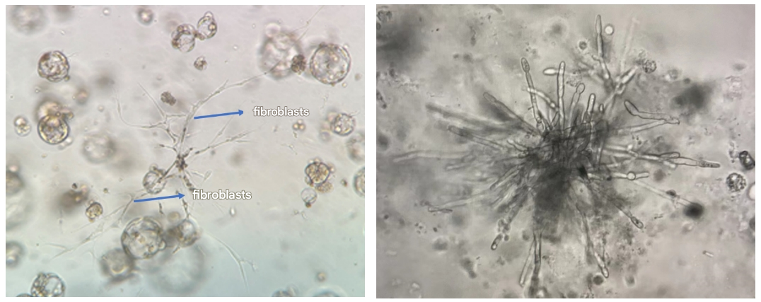
Left: Fibroblasts growing adherently on the stromal gum layer
Right: Fungal contamination characteristics – mold
2) Tissue fragments
In the process of extracting primary cells, a lot of cell debris and impurities are generated during the dissociation process of tissue, so the primary culture background will be dirty, and this situation is easy to be mistaken for contamination. Next, Xiao Ai will take you to understand the characteristics of tissue fragmentation:

● Primary tissue fragments tend to be irregular in shape, which is different from the oval colony morphology of the bacterial cluster;
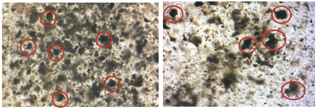
● Primary tissue impurities often vary in size and morphology; The morphology of the bacterial clusters is uniform and all oval.
That's all for today's explanation, if you have experimental questions, welcome to join the group to communicate!
This issue is recommended by Xiaoai
|
Catalog number |
name of article |
specification |
Lead time |
|
abs9841 |
Black Gumworm Remover PLUS(500×) |
400uL×5 |
Spot |
|
abs9842 |
Black gum insect prophylaxis(1000×) |
500uL×5 |
Spot |
|
abs9925 |
Mycoplasma Detection Kit (qPCR) |
50T |
1-2 weeks |
|
abs9253 |
Mycoplasma Clearance Reagent Plus |
20mg/100mg |
1-2 weeks |
|
abs42020125 |
Kanamycin B sulfate |
250mg |
Spot |
|
abs42018967 |
Gentamicin sulfate solution (10 mg/mL) |
10mL |
Spot |
|
abs9244 |
Cyan-chain bispecific antibody |
100mL |
Spot |
|
abs9246 |
Penicillin-streptomycin-amphotericin B tertiary antibody |
100mL |
Spot |
|
abs9245 |
Penicillin-streptomycin-gentamicin triantibody |
100mL |
Spot |
|
abs42013298 |
Amphotericin B |
1g/5g |
Spot |
|
nystatin |
1g/5g |
Spot |
Absin provides antibodies, proteins, ELISA kits, cell culture, detection kits, and other research reagents. If you have any product needs, please contact us.
|
Absin Bioscience Inc. Email: worldwide@absin.cn |
 Follow us on Facebook: Absin Bio Follow us on Facebook: Absin Bio |


