Detailed IHC Secondary Antibody Kit Instructions

Concept
Immunohistochemistry (IHC) is a technique that utilizes antigen-antibody reactions to visualize antigens within tissue cells through the use of chromogenic substrates for labeled antibodies, enabling the localization and qualitative analysis of proteins.
Advantages of Absin IHC Secondary Antibody Kits
1. Based on polymer technology + primary antibody enhancer, high sensitivity, primary antibody can be further diluted 2-4 times!
2. Non-biotin detection system, lower background!
3. Quick and convenient, shorter incubation time!
4. abs957 has over 90 citations, highly recognized and reliable!
Components
|
Component Code |
Component Name |
Specification |
Quantity |
|
A |
Hydrogen Peroxide |
5mL |
1 |
|
B |
Super Blocking Solution |
5mL |
1 |
|
C |
Primary Antibody Enhancer |
5mL |
1 |
|
D |
Enzyme-Linked Secondary Antibody Polymer |
5mL |
1 |
|
E |
DAB A Solution |
5mL |
1 |
|
F |
DAB B Solution |
160uL |
1 |
Prepare primary antibodies (from mouse or rabbit) and PBS additionally.
The components of this kit are sufficient for 50 slides and are suitable for samples with primary antibodies from mouse and rabbit (note: not the sample source), with DAB chromogen.
Preparation Before Experiment
1. Remove the fixed sample and rinse it with running water 3 times, each for 5 minutes;
2. Dehydration, clearing, and paraffin immersion
75% ethanol (1h) —— 85% ethanol (1h) —— 95% ethanol (1h) —— 100% ethanol (50min) —— 100% ethanol (50min) —— 100% ethanol (50min) —— xylene (40min) —— xylene (40min) —— xylene (40min) —— paraffin at 63℃ (50min) —— paraffin at 63℃ (50min) —— paraffin at 63℃ (50min).
3. Embedding
After dehydration, place the sample into the preheated embedding machine and embed it according to the required orientation;
4. Label the glass slides with numbers;
5. Sectioning: Prepare sections according to the requirements, with a typical thickness of 3-5um;
6. Spreading: Gently place the sections on a 46°C water bath using a brush to spread them;
7. Retrieving and baking: After the tissue on the section is fully spread, retrieve it and place it in a 60°C oven for baking (the baking time depends on the tissue type; generally, 30 minutes for most tissues, and up to 1 hour for bone tissues), followed by dewaxing.
Operational Steps
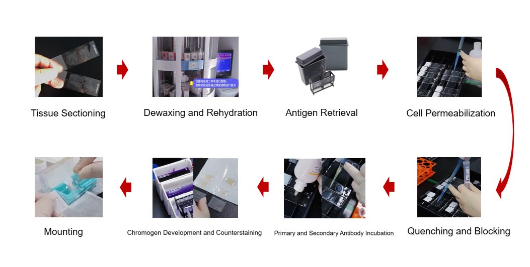
Figure IHC Experimental Workflow
Note: All steps are performed at room temperature. Reagents can be added directly from the dropper bottle or using a pipette, with one drop equal to 30-40uL.
1. Dewaxing and rehydration of tissue sections xylene (5min) —— xylene (5min) —— xylene (5min) —— 100% ethanol (5min) —— 100% ethanol (5min) —— 95% ethanol (3min) —— 85% ethanol (3min) —— 75% ethanol (3min) —— deionized water (5min)
2. Wash with PBS 2-3 times, each for 5 minutes;
3. Antigen retrieval: Pre-treat the tissue sections according to the specific requirements of the primary antibody; (Antigen retrieval: Place the dewaxed sections into antigen retrieval solution (choose between sodium citrate or EDTA according to the primary antibody instructions), perform high-temperature antigen retrieval in a pressure cooker. Heat the pressure cooker to full pressure, then continue heating for 5 minutes. Turn off the power and let the staining box cool at room temperature for 30 minutes after 10 minutes, or use microwave retrieval. Wipe the tissue around the antigen-retrieved sections dry and draw a hydrophobic barrier with an IHC pen)
4. Wash with PBS 2-3 times, each for 5 minutes;
5. Endogenous peroxidase blocking: Add 100uL of Solution A and incubate for 10 minutes to block endogenous peroxidase to reduce non-specific background staining;
6. Wash with PBS 2-3 times, each for 5 minutes;
7. Serum blocking: Add 100uL of Solution B and incubate for 5 minutes to reduce non-specific staining. Note: Do not exceed 10 minutes for this step; if the primary antibody is diluted in a buffer containing 5% to 10% normal goat serum, this step can be omitted.
8. Wash with PBS 2-3 times, each for 5 minutes;
9. Primary antibody incubation: Add primary antibody and incubate at room temperature or 37℃ for 20 minutes, with the amount sufficient to cover the sample area, which can be adjusted flexibly;
10. Wash with PBS 2-3 times, each for 5 minutes;
11. Primary antibody enhancer incubation: Add 100uL of Solution C and incubate for 10 minutes;
12. Wash with PBS 2-3 times, each for 5 minutes;
13. Secondary antibody incubation: Add 100uL of Solution D and incubate at room temperature or 37℃ for 10 minutes. Note: This solution is light-sensitive, avoid light.
14. Wash with PBS 2-3 times, each for 5 minutes;
15. DAB reagent preparation: Prepare fresh substrate solution by mixing 30uL of Solution F with 1mL of Solution E thoroughly (this substrate solution can be stored for 2 weeks).
16. DAB staining: Add 100uL of fresh substrate solution and incubate for 5 minutes. Note: The DAB staining time should be controlled under the microscope to select the optimal staining time and take precautions.
17. Stop staining: Rinse with deionized water;
18. Counterstaining: Hematoxylin counterstaining for about 3 minutes, rinse with tap water, differentiate in 1% hydrochloric acid alcohol for 1 second, rinse with tap water;
19. Dehydration and clearing: 80%, 95%, 100%, 100% ethanol, each for 5 minutes, xylene for 5 minutes, twice;
20. Mounting with neutral gum: When covering the slide, tilt it at 45° from one end to avoid bubbles.
Recommended IHC Products
|
Catalog Number |
Product Name |
Specification |
|
Ready-to-Use High-Performance IHC Secondary Antibody Kit |
5mL |
|
|
Ready-to-Use IHC Secondary Antibody Kit (Mouse/Rabbit Universal) |
100mL |
|
|
Ready-to-Use IHC Secondary Antibody Kit (Anti-Rabbit) |
100mL |
|
|
Ready-to-Use IHC Secondary Antibody Kit (Anti-Mouse) |
100mL |
|
|
IHC Double Staining Kit (Mouse/Rabbit Universal) |
50T |
AntBio provides antibodies, proteins, ELISA kits, cell culture, detection kits, and other research reagents. If you have any product needs, please contact us.


