Application of mIHC in Exploring the Phenotypic and Functional Characteristics of Distinct T Cell Subsets

Literature Overview
This featured study focuses on the critical role of T cells in cancer immunotherapy, with a specific emphasis on colorectal cancer (CRC). The research integrates single-cell RNA sequencing, flow cytometry, and multiplex immunohistochemistry (mIHC) to dissect the heterogeneity, developmental trajectories, and functional states of T cell subsets within the tumor microenvironment, providing novel insights for CRC immunotherapy strategies.
Research Background
T cells represent the cornerstone of anti-tumor immune responses, yet fundamental biological processes, including their developmental dynamics, migratory patterns, and phenotypic transitions within solid tumors, remain incompletely understood. T cell receptors (TCRs) serve as unique lineage identifiers, enabling precise tracking of clonal T cell populations in tumor tissues. Elucidating the spatiotemporal distribution and functional crosstalk of T cell subsets is essential for optimizing cancer immunotherapies, particularly immune checkpoint blockade.
Research Strategy and Experimental Design
The research team analyzed transcriptomic profiles of 11,138 single T cells isolated from 12 colorectal cancer patients. They established the STARTRAC (Single T-cell Analysis by RNA-sequencing and TCR Tracking) index, a quantitative tool to characterize dynamic interrelationships among 20 functionally and clonally distinct T cell subsets.
Complementary to high-throughput sequencing and flow cytometric analysis, multiplex immunofluorescence immunohistochemistry (mIHC) was employed for in situ spatial localization and phenotypic validation of T cell subsets, preserving the architectural context of the tumor microenvironment.
Key Research Outcomes
Multicolor IHC staining targeting CD3, CD4, CD8, and FOXP3 was performed to validate the presence and spatial distribution of major T cell subsets in colorectal cancer tissues.
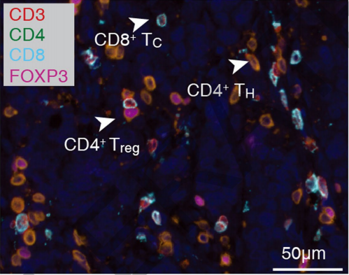 Multicolor IHC staining for CD3, CD4, CD8, FOXP3 in CRC tissues
Multiplex immunofluorescence imaging demonstrated the co-expression of Ki-67, CD8, PD-1, and TIM-3 in CD8+ exhausted T (TEX) cells, defining the proliferative and dysfunctional phenotype of tumor-infiltrating CD8+ T cells.
 Co-expression of Ki-67, CD8, PD-1, TIM-3 in CD8+ TEX cells
Additional mIHC analysis revealed the co-localization of CD3, FOXP3, and RORγ in specific T cell populations, identifying a phenotypically unique regulatory T cell subset within the tumor niche.
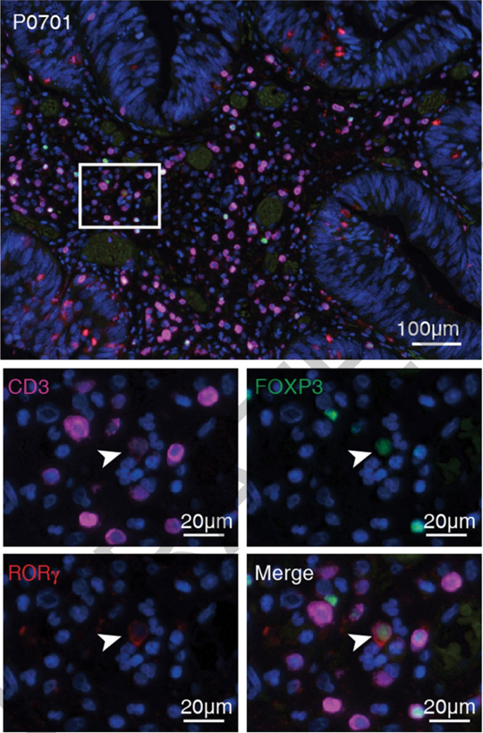 Co-expression of CD3, FOXP3, RORγ in CRC T cells
STARTRAC index analysis uncovered critical functional, migratory, and developmental connections between T cell subsets in CRC. The data defined TCR-dependent developmental trajectories of tumor-infiltrating CD8+ TEM cells differentiating into TEMRA and TEX subsets, proposing novel therapeutic strategies to redirect TEX cell fate toward TEMRA. Enrichment of CXCL13+BHLHE40+IFNG+ TH1-like cells in MSI patients explains the superior efficacy of checkpoint blockade in this subgroup and highlights a promising cellular therapeutic target. The comprehensive gene expression dataset, including the novel marker IGFLR1, is publicly accessible via the interactive portal: http://crc.cancer-pku.cn.
ANT BIO PTE. LTD. Products Empowering This Research
The high-quality immunoassay reagents and antibodies from ANT BIO PTE. LTD. were instrumental in the success of this study's mIHC and immunophenotyping experiments. These reagents ensured reliable detection of co-expressed proteins in formalin-fixed paraffin-embedded (FFPE) tissue sections, validating single-cell sequencing findings at the spatial protein level and supporting the robust conclusions of the research.
Related High-Quality Reagents from ANT BIO PTE. LTD.
| Catalog No. | Product Name | Application | Sub-Brand |
|---|---|---|---|
| abs50012 | 4-Color Multiplex Fluorescent IHC Kit (Mouse-Rabbit Universal Secondary Antibody) | mIHC | Absin |
| abs50013 | 5-Color Multiplex Fluorescent IHC Kit (Mouse-Rabbit Universal Secondary Antibody) | mIHC | Absin |
| abs50014 | 6-Color Multiplex Fluorescent IHC Kit (Mouse-Rabbit Universal Secondary Antibody) | mIHC | Absin |
| abs50015 | 7-Color Multiplex Fluorescent IHC Kit (Mouse-Rabbit Universal Secondary Antibody) | mIHC | Absin |
| abs50028 | 4-Color Multiplex Fluorescent IHC Kit (Anti-Rabbit Secondary Antibody) | mIHC | Absin |
| abs50029 | 5-Color Multiplex Fluorescent IHC Kit (Anti-Rabbit Secondary Antibody) | mIHC | Absin |
| abs50030 | 6-Color Multiplex Fluorescent IHC Kit (Anti-Rabbit Secondary Antibody) | mIHC | Absin |
| abs50031 | 7-Color Multiplex Fluorescent IHC Kit (Anti-Rabbit Secondary Antibody) | mIHC | Absin |
ANT BIO PTE. LTD. – Empowering Scientific Breakthroughs
At ANTBIO, we are committed to advancing life science research through high-quality, reliable reagents and comprehensive solutions. Our specialized sub-brands (Absin, Starter, UA) cover a full spectrum of research needs, from general reagents and kits to antibodies and recombinant proteins. With a focus on innovation, quality, and customer-centricity, we strive to be your trusted partner in unlocking scientific mysteries and driving medical progress. Explore our product portfolio today and elevate your research to new heights.
Disclaimer
This article was partially created with the assistance of artificial intelligence. If any content involves copyright or intellectual property issues, please inform us, and we promise to verify and remove it immediately.


