The actual WB stripe is not as expected? It's smooth

1. The protein of interest exists in other forms, most commonly
● Post-translational modification: such as glycosylation, the common amino acids that accept glycosylation are serine (Ser), threonine (Thr), asparagine (Asn) and hydroxylysine (Hydroxylysine), such as CD2, according to uniprot, its predicted molecular weight should be about 39kda, but the apparent molecular weight is generally around 45kDa, which is due to the glycosylation modification at multiple sites of CD2. If you want to explore whether the molecular weight difference is caused by glycosylation, you can add the glycosidase PNGase F to remove the glycosyl and proceed to WB, and in addition to glycosylation, there are also various post-translational modifications such as phosphorylation, ubiquitination, methylation, and acetylation. Among them, glycosylation, acetylation, and methylation usually make the apparent molecular weight larger, and phosphorylation usually remains unchanged or larger.
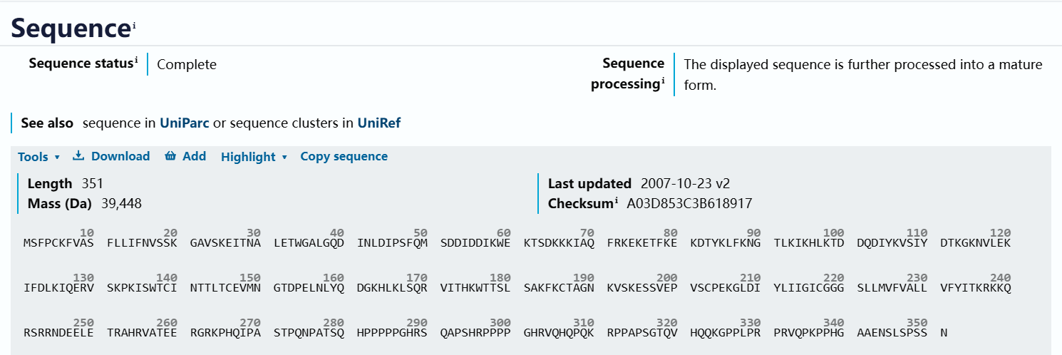
The theoretical molecular weight of CD2 is about 39 kDa
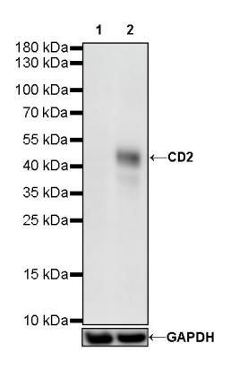
The apparent molecular weight is about 45kDa
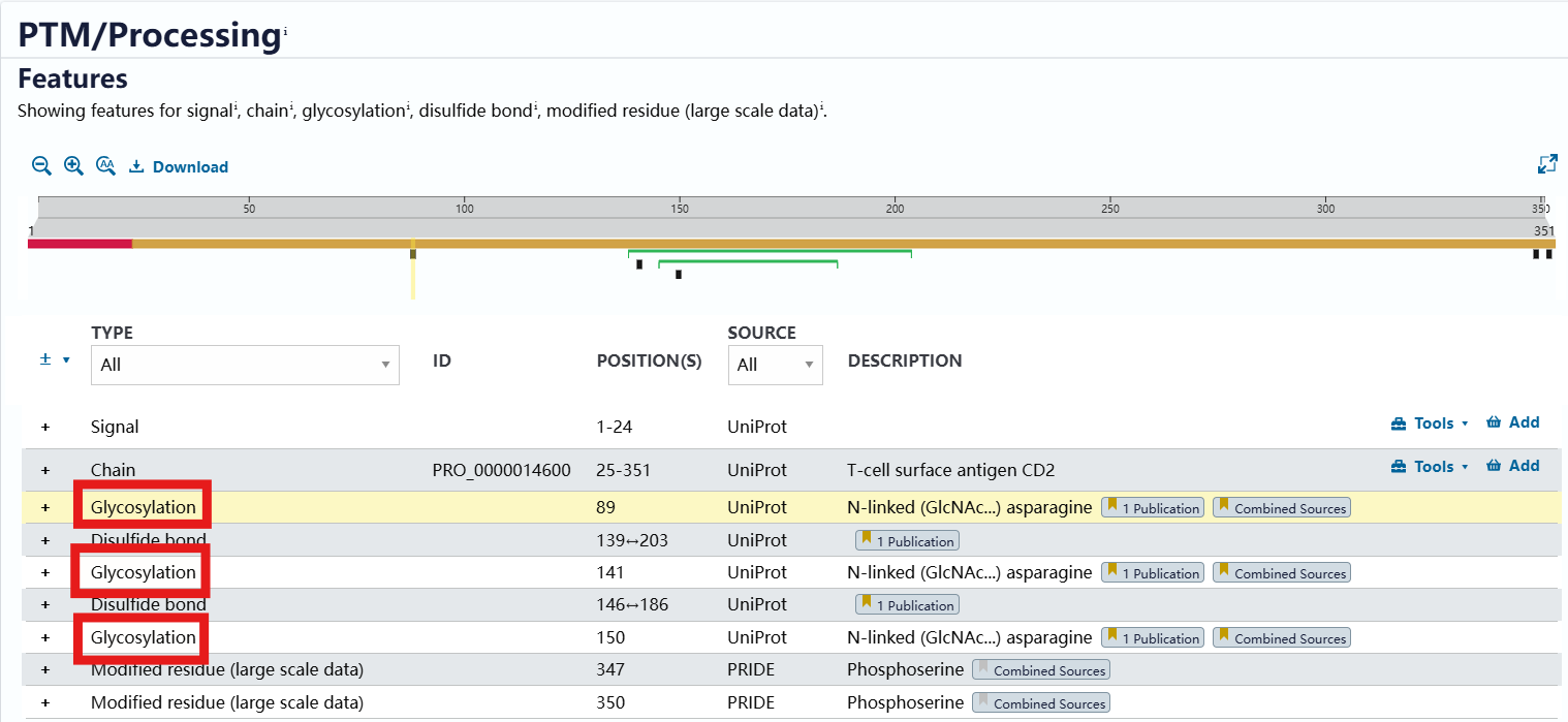
Glycosylation of CD2 is shown in Uniprot
● Shearing: Caspase-7 exists in many forms:
1) Procaspase-7: a precursor form of Caspase-7 with a molecular weight of approximately 37 kDa;
2) Intermediate caspase-7: the intermediate form of caspase-7 with a molecular weight of about 28 kDa;
3) Caspase-7 p20: one of the fragments of activated Caspase-7 with a molecular weight of approximately 20 kDa.
Procaspase-7 is an inactivated precursor form of caspase-7 with a large molecular weight. When cells are stimulated by apoptotic signals, procaspase is activated by cleavage by itself or by cleavage by other caspas such as initiator caspases, forming smaller active subunits that combine together to form viable heterodimers. In this case, both induction and non-induction groups can be set up for validation. The apparent molecular weight after shearing will be smaller.
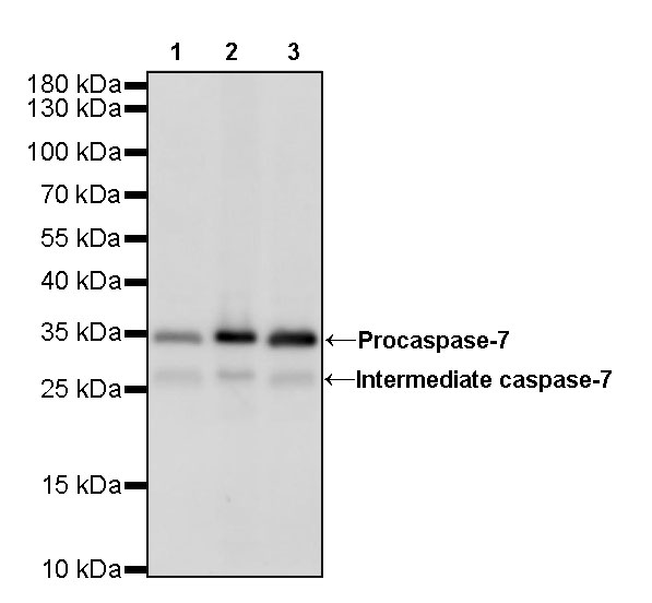
WB results of Caspase-7 rabbit monoclonal antibody
Lane 1: HeLa whole cell lysate 20 ug
Lane 2: Jurkat whole cell lysate 20 ug
Lane 3: HEK-293 whole cell lysate 20 ug
Secondary antibody: goat anti-rabbit IgG, (H+L), 1/10,000 dilution conjugated HRP
Predicted molecular weight: 34 kDa
Observed molecular weight: 28,35 kDa
Exposure time: 180s
● Polymers and covalent binders: If the apparent molecular weight and predicted molecular weight show a fold increase, it is necessary to consider whether it is a multimer, and the addition of reducing agents such as DTT can verify whether it is a non-covalent binder.
2. Abnormal protein migration
● For proteins with a molecular weight of less than 20 kDa
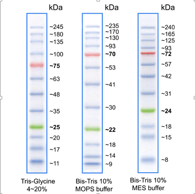
When the traditional Tris-Glycine system is used in SDS-PAGE (sodium dodecyl sulfate-polyacrylamide gel electrophoresis), the separation of small molecular weight proteins is not good, mainly due to the following reasons:
1) Effect of ionic pre-peaks: In the Tris-Glycine system, Tris-HCl buffer pH 6.8 is usually used for stacking gels, while Tris-Glycine buffer at pH 8.3 is used for electrophoresis buffers and resolving gels. When electrophoresis begins, glycine dissociates less in the stacking gel and moves slowly, forming a moving ionic propeak. With the action of the electric field, glycine gradually dissociates and accelerates its movement, in the process a large number of free dodecyl sulfate ions (DS-) are produced, which will bind to small molecular weight proteins, causing them to aggregate or denature, thus affecting the resolution;
2) Migration of low-molecular-weight proteins: Due to the above-mentioned reasons, low-molecular-weight proteins may be hindered in the process of entering the separating gel through the stacking gel, resulting in blurred bands, tailing, or reduced resolution. This phenomenon is especially evident when separating proteins or peptides smaller than 20 kDa;
3) Effect of pH value of buffer system:The high pH of the Tris-Glycine system may lead to certain modifications or degradation of small molecular weight proteins during electrophoresis, further affecting their separation.
Therefore, it is recommended to use the Tricine-SDS-PAGE system for the separation of small molecular weight proteins or peptides below 20 kDa. Tricine, as one of the components of the electrophoresis buffer, has a low pKa value (8.15), which makes it less likely to produce large amounts of free DS-ions during electrophoresis like glycine, which reduces the impact on small molecular weight proteins and is able to provide higher resolution. In addition, Tricine-SDS-PAGE typically uses a lower concentration of acrylamide gel to accommodate the separation needs of small molecular weight proteins.
● The special arrangement of protein charge or special structure, such as amino acid side chains, may migrate too fast or too slowly during electrophoresis.
3. The problem of protein marker
Although the prestained marker is more convenient to use than the non-prestained marker, there is also a molecular weight shift due to the coupling of the dye, and the migration rate of the same marker will be different in different electrophoresis systems.
abs923 prestained protein marker (10-245 kDa)
4. Sample preparation problems
● The denaturation of membrane protein at 95 degrees of heating will cause aggregation, resulting in an increase in the apparent molecular weight of the detection band;
● Incomplete denaturation or reduction of the sample will affect the exposure of the epitope, and then affect its migration rate, so it is necessary to ensure that the sample loading buffer is sufficient and within the shelf life when preparing the sample, and the right lysate should be selected to see the specific components;
● The sample must be fresh, some proteins are easy to degrade, and attention should be paid when preparing.
5. To eliminate the above problems, the most important thing is to fully understand whether your sample expresses the target protein and what the expression abundance is, and check the manufacturer's verification chart to understand the specificity of the antibody.
WB good things recommended
|
category |
Catalog number |
Product name |
specification |
|
Sample processing |
abs9229 |
RIPA lysate (strong) |
100mL |
|
abs9116 |
Lysates for western blotting and immunoprecipitation (WB/IP). |
100mL |
|
|
abs9161 |
Broad Spectrum Protease Inhibitor Cocktail (EDTA-free, 100×DMSO stock) |
1mL |
|
|
abs9162 |
Broad spectrum phosphatase inhibitor cocktail (100× stock) |
1mL |
|
|
Protein quantification |
BCA Protein Assay Kit |
500T |
|
|
Loading |
abs953 |
Loading buffer (2×) |
10mL |
|
abs9829 |
Loading buffer (5×) |
10mL |
|
|
Marker |
Prestained protein marker (10-180 kDa) |
250uL |
|
|
Prestained protein marker (10-245 kDa) |
500uL |
||
|
abs90004 |
Prestained luminescent Western Marker |
500uL |
|
|
electrophoresis |
Electrophoresis Solution(10×) |
1L |
|
|
abs9359 |
Tris-Glycine-SDS Running Buffer (10×) |
500mL |
|
|
Precast gels |
Plastic gel, glass gel and other precast adhesives |
||
|
Gel kits |
abs9367 |
Color PAGE Rapid Gel Kit (10%) |
1kit |
|
abs9368 |
Color PAGE Rapid Gel Kit (12%) |
1kit |
|
|
abs9366 |
Color PAGE Rapid Gel Kit (8%) |
1kit |
|
|
Transfer |
abs932 |
PVDF film (0.45 μm) |
1 volume |
|
abs931 |
PVDF film (0.22 μm) |
1 volume |
|
|
abs960 |
NC membrane (0.45 μm) |
1 volume |
|
|
NC membrane (0.22 μm) |
1 volume |
||
|
abs950 |
Electroporation (10×) |
100mL |
|
|
Closed |
abs9175 |
Skim milk powder |
500g |
|
abs9915 |
Western Blot Rapid Blocking Solution |
500mL |
|
|
Primary antibody incubation |
Primary and secondary antibody dilutions for WB |
100mL |
|
|
WB primary antibody (more than 30,000 products, multi-index coverage) |
|||
|
Internal control antibodies |
abs132001 |
Rabbit anti-β-Actin Polyclonal Antibody |
100ug |
|
abs830030 |
Mouse anti-GAPDH Monoclonal Antibody |
1mL |
|
|
abs830032 |
Mouse anti-β-Tubulin Monoclonal Antibody |
1mL |
|
|
Secondary antibodies |
abs20040 |
Goat anti-Rabbit IgG-HRP Antibody |
500uL |
|
abs20001 |
Goat anti-Mouse IgG-HRP Antibody |
1mL |
|
|
Color development |
abs9213 |
DAB chromogenic kit (violet blue, WB) |
1kit |
|
abs920 |
ECL Chemiluminescence Detection Kit |
25mL×2 |
|
|
abs9434 |
Ultrasensitive ECL chemiluminescence detection kit |
25mL×2 |
|
|
abs9435 |
Extremely sensitive ECL chemiluminescence detection kit |
25mL×2 |
|
Absin provides antibodies, proteins, ELISA kits, cell culture, detection kits, and other research reagents. If you have any product needs, please contact us.
|
Absin Bioscience Inc. Email: worldwide@absin.cn |
 Follow us on Facebook: Absin Bio Follow us on Facebook: Absin Bio |


