TDP-43 and ALS: Mechanistic Insights & ANT BIO PTE. LTD. Reagent Empowerment

1. Research Background
TDP-43 is a 414-amino-acid multifunctional protein belonging to the heterogeneous nuclear ribonucleoprotein (hnRNP) family. Characterized by two conserved RNA recognition motifs (RRMs), it mediates sequence-specific single-stranded RNA binding and governs core RNA metabolic processes, including transcription, translation, alternative splicing, and mRNA stability regulation. Discovered by Virginia Lee in 2006, TDP-43 has been identified as a pivotal pathological hallmark of neurodegenerative diseases, particularly ALS and frontotemporal lobar degeneration (FTLD).
Under physiological conditions, TDP-43 predominantly localizes to the nucleus to regulate gene expression; under stress, it translocates to the cytoplasm to form reversible stress granules via liquid-liquid phase separation, maintaining cellular homeostasis. In disease states, aberrant cytoplasmic aggregation of TDP-43 becomes a defining pathological feature of ALS/FTLD, driving neuronal dysfunction and degeneration.
2. Research Strategy
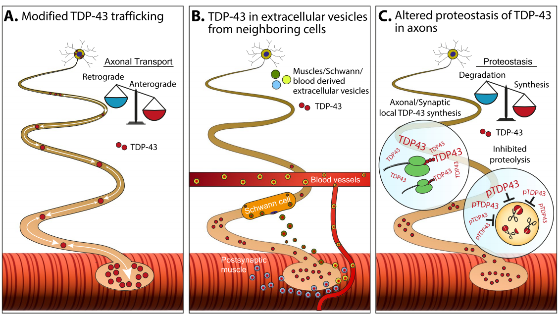
The study adopted a multi-dimensional research approach to decode TDP-43 pathogenesis: (1) Investigated the subcellular localization and functional transformation of TDP-43 in normal and stressed cells; (2) Analyzed three potential mechanisms of pathological TDP-43 accumulation in axons, including axonal transport dysregulation, extracellular vesicle-mediated propagation, and protein homeostasis imbalance; (3) Verified TDP-43 aggregation and post-translational modifications in clinical ALS-FTLD patient tissues; (4) Explored the impact of TDP-43 abnormalities on astrocyte function and neuronal homeostasis; (5) Validated TDP-43 expression and localization via multiple experimental techniques.
3. Key Research Findings
3.1 Axonal and Neuromuscular Pathological Mechanisms
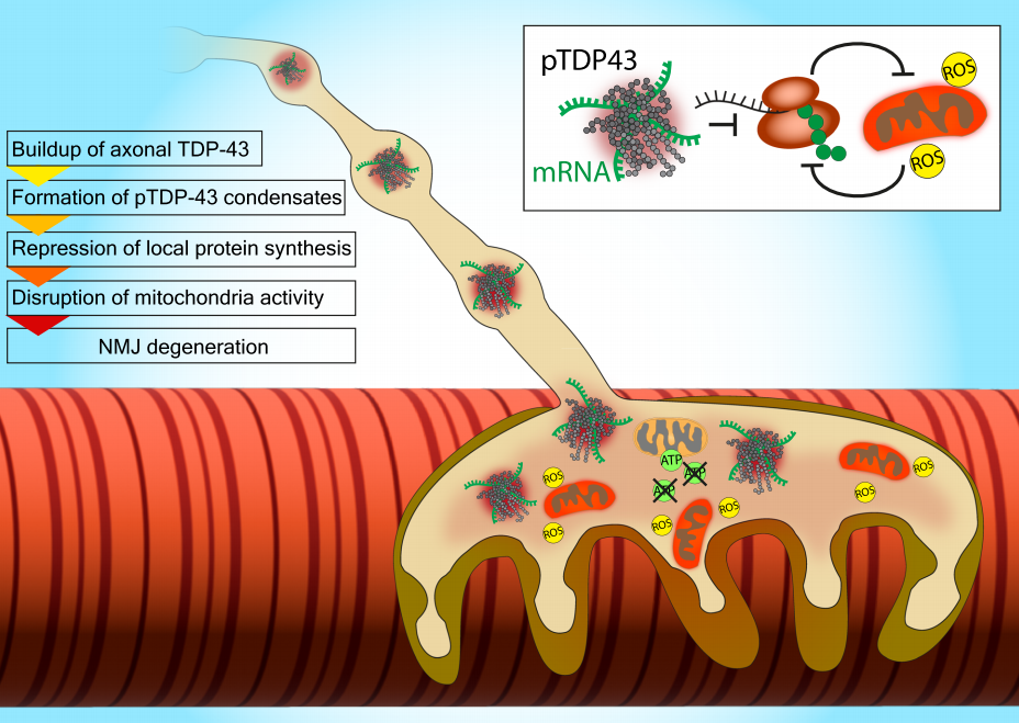
Pathological TDP-43 accumulates in the distal axons and presynaptic compartments of motor neurons, forming phosphorylated TDP-43 condensates that sequester mRNAs essential for mitochondrial and ribosomal protein synthesis. This disruption triggers a vicious cycle of mitochondrial dysfunction, reactive oxygen species (ROS) overproduction, and impaired local protein synthesis, ultimately inducing neuromuscular junction (NMJ) degeneration and axonal loss.
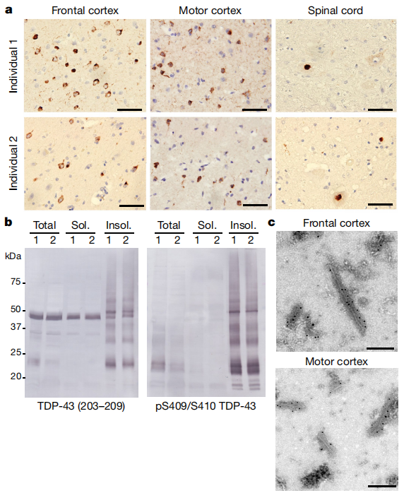
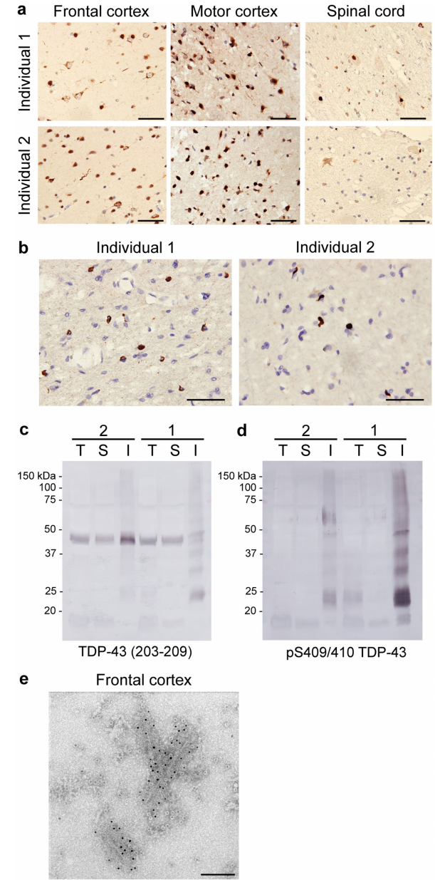
3.2 Clinical Pathological Validation
Analysis of ALS-FTLD patient brain and spinal cord tissues revealed abundant TDP-43 neuronal cytoplasmic inclusions (NCIs) and glial inclusions, consistent with stage 4 ALS TDP-43 pathology. Immunological and electron microscopy analyses confirmed that TDP-43 aggregates consist of full-length protein and C-terminal fragments (CTFs) phosphorylated at S409/S410, forming helical filaments with a width of 10-15 nm.
3.3 Glial Cell Dysfunction
Mutated or abnormally expressed TDP-43 impairs astrocyte function by upregulating interferon-induced chemokine genes. Excessive chemokines activate neuronal receptors, triggering presynaptic dysfunction and neuronal hyperexcitability, leading to cognitive impairment in neurodegenerative conditions.
3.4 Core Conclusion
TDP-43 acts as a "double-edged sword": a guardian of RNA metabolism in normal cells, but a betrayer driving neurodegeneration when pathologically aggregated. Its abnormalities are nearly ubiquitous in ALS, representing a critical therapeutic target for neurodegenerative diseases.
4. ANT BIO PTE. LTD. Product Empowerment
The high-quality experimental data in this study relies on the TDP-43 Recombinant Rabbit mAb (Catalog No.: S0B0731) independently developed by ANT BIO PTE. LTD., which provides robust technical support for multi-scenario TDP-43 detection:
- Immunocytochemistry (ICC): Specific green fluorescence staining in HeLa cells, clearly visualizing TDP-43 subcellular localization;
- Immunohistochemistry (IHC): High-specificity positive staining in human paraffin-embedded colon and testis tissues;
- Western Blot (WB): Specific detection of endogenous TDP-43 in HeLa and SH-SY5Y cell lysates (observed molecular weight ~40 kDa);
- Flow Cytometry: Accurate quantitative analysis of TDP-43 expression in fixed and permeabilized HeLa cells.
This antibody exhibits high specificity, sensitivity, and stability, enabling reliable detection of TDP-43 in cellular and tissue samples, and strongly supporting the mechanistic research and pathological validation of TDP-43 in ALS.


6. ANT BIO PTE. LTD. Related Product Portfolio
| Catalog Number | Product Name | Host | Conjugation | Application |
|---|---|---|---|---|
| S0B0731 | TDP-43 Recombinant Rabbit mAb (S-989-41) | Rabbit | Unconjugated | ICC, IHC, WB, Flow Cytometry |
ANT BIO PTE. LTD. provides a full range of supporting secondary antibodies, buffer reagents, and detection kits to meet the full chain needs of neurodegenerative disease research.
ANT BIO PTE. LTD. – Empowering Scientific Breakthroughs
At ANTBIO, we are committed to advancing life science research through high-quality, reliable reagents and comprehensive solutions. Our specialized sub-brands (Absin, Starter, UA) cover a full spectrum of research needs, from general reagents and kits to antibodies and recombinant proteins. With a focus on innovation, quality, and customer-centricity, we strive to be your trusted partner in unlocking scientific mysteries and driving medical progress. Explore our product portfolio today and elevate your research to new heights.
Disclaimer
This article was partially created with the assistance of artificial intelligence. If any content involves copyright or intellectual property issues, please inform us, and we promise to verify and remove it immediately.


