Innovative Approaches to Building and Validating Alzheimer's Disease Models

Introduction to AD
Alzheimer's disease (AD) is a primary neurodegenerative disorder with unknown etiology and complex pathogenesis. It is one of the most common types of dementia. Clinically, it is characterized by progressive memory decline, cognitive impairment, behavioral abnormalities, and personality changes. Its typical pathological changes include neuronal loss, amyloid β-protein (Aβ) deposition forming senile plaques (SP), and hyperphosphorylation of tau protein forming neurofibrillary tangles (NFT)[1]. With the increasing aging population, AD has become a hot topic of concern both domestically and internationally.

Figure 1 a) Healthy brain; b) Physiological structure of the brain and neurons in AD brain[2]
Methods for AD Modeling
The current approaches for preparing AD animal models mainly focus on reflecting the pathological, physiological, and clinical characteristics of AD. Modeling to simulate the pathological features of AD is an important means of studying this disease. The methods for establishing AD animal models include aging models, transgenic models, and exogenous harmful substance injection models (Table 1). For example, researchers have established AD models by directly injecting Aβ into the brain to induce Aβ deposition and senile plaques. The main types of Aβ used for injection are Aβ1-42, Aβ1-40, and Aβ25-35. The injection sites are primarily the unilateral/bilateral hippocampal CA1 region or the lateral ventricle, with injection durations ranging from 3 days to several weeks[3-4]. Among them, Aβ1-42 is the primary form used in AD neuropathological research. It forms nuclei (seeds) that initiate fiber formation, leading to the classic amyloidosis process.
Table 1 Comparison of Advantages and Disadvantages of Common AD Animal Models
|
Induction Principle |
Model |
Advantages |
Disadvantages |
|
Aging |
Natural aging |
Neuronal atrophy, cholinergic dysfunction, cognitive impairment |
Difficult to obtain large numbers of aged animals, minimal neurofibrillary tangles and amyloid deposition, long modeling time |
|
Accelerated senescence (SAM) |
Aβ deposition, abnormal tricarboxylic acid cycle in glucose metabolism, immune dysfunction |
High cost, short lifespan |
|
|
D-Galactose model |
Cost-effective, neuronal loss, reduced protein synthesis, learning and memory impairment |
Cannot produce AD-specific senile plaques and neurofibrillary tangles, many uncertain factors |
|
|
Transgenic |
APP transgenic |
Extracellular Aβ deposition, neuroinflammatory plaques, synaptic loss |
No tau protein phosphorylation or NFT, no AD-specific neuronal loss in hippocampus and cortex |
|
APP/PS-1 transgenic |
Aβ deposition in cortex and hippocampus, neuronal changes, neurobehavioral dysfunction |
Unstable exogenous gene expression, time-consuming modeling, poor fertility, high cost |
|
|
Tau transgenic |
Formation of NFT in brainstem and spinal cord, memory impairment |
Lack of Aβ deposition |
|
|
Exogenous harmful substance injection |
Aβ injection |
Aβ deposition, immune-inflammatory response, astrocyte proliferation, tau protein hyperphosphorylation, significant decrease in ChAT activity |
Does not fit the chronic onset of AD, Aβ aggregates locally at injection site, non-target damage to brain tissue |
|
Aluminum intoxication |
Aβ aggregation, neuronal degeneration, spatial learning impairment, and memory damage |
Long modeling cycle, no decrease in central cholinergic activity, NFT different from AD patients |
|
|
Streptozocin (STZ) |
High tau protein phosphorylation, Aβ deposition, cholinergic deficit, oxidative stress |
High mortality in animals, no neurofibrillary tangles or senile plaques |
|
|
Scopolamine |
Simple and cost-effective method, cholinergic nervous system dysfunction, cognitive impairment |
Lack of typical AD pathological features |
Below, we share the method of inducing AD models using Aβ1-42[3].
1. Preparation of Aβ1-42
(1) Remove the lyophilized powder of artificially synthesized Aβ1-42 stored at -80°C and equilibrate at room temperature for 30 min;
(2) In a fume hood, suspend Aβ1-42 in 100% HFIP (1.596 g/mL) at a concentration of 1 mM, mix thoroughly, and aliquot into equal portions;
(3) Evaporate HFIP from the aliquoted portions using a vacuum centrifugal evaporator and store at -20°C;
(4) Before use, resuspend the -20°C stored Aβ1-42 in DMSO at a concentration of 5 mM;
(5) Dilute to the experimental concentration with saline or artificial cerebrospinal fluid as needed.
2. Unilateral lateral ventricle cannulation in mice
(1) Anesthetize mice with isoflurane and fix them on a stereotactic apparatus;
(2) Disinfect the scalp with 75% ethanol, shave the hair, and disinfect again;
(3) Incise the scalp to expose the skull, remove the connective tissue on the skull surface with hydrogen peroxide. After the skull is dry, disinfect again;
(4) Using the stereotactic apparatus, point the positioning needle to the bregma and use it as the starting point. Move the positioning needle to determine the cannula position (0.46 mm anterior to bregma, 1 mm lateral to the midline on both sides, 2.3 mm deep). Drill a hole in the skull and randomly place the cannula in the left or right lateral ventricle (cannula diameter 0.48 mm, inner core diameter 0.3 mm);
(5) Seal the gap between the cannula and the skull with bone glue. After the glue dries, fix the cannula to the skull with dental cement;
(6) Transfer the mouse to a heating pad, and after it wakes up, return it to the breeding box for individual housing;
(7) For drug injection, remove the inner core of the cannula, connect the mouse brain cannula to a microinjection needle with a suitable length of PE tube, and inject at a speed of 0.5 μL/min using a microinjection pump.
3. Behavioral tasks
Before the task, inject saline or Aβ1-42 into the mice that have undergone cannula implantation using a microinjection pump at a speed of 0.5 μL/min, with a volume of 2 μL. Keep the needle stationary for 5 min after injection to prevent backflow of the injected solution, then replace the inner core and tighten the nut. Spontaneous alternation Y-maze test and novel object recognition task: 10 min after drug injection, begin testing for spontaneous alternation Y-maze test and novel object recognition task.
AD Validation Methods
An ideal model should possess at least the following three characteristics:
① Presence of major neuropathological features of AD: SP and NFT.
② Occurrence of important pathological changes in AD: neuronal death, synaptic loss, and astrocyte proliferation.
③ Presence of cognitive impairment.
In the basic research and drug discovery of AD, mouse models are important resources for elucidating biological mechanisms, validating molecular targets, and screening potential compounds. Both transgenic and non-transgenic mouse models can be used to obtain different types of AD-like pathology in vivo. Despite extensive genetic and biochemical research on AD pathogenic pathways, as a disease primarily characterized by cognitive impairment, the ultimate readout of any intervention should be the detection of learning and memory[4].
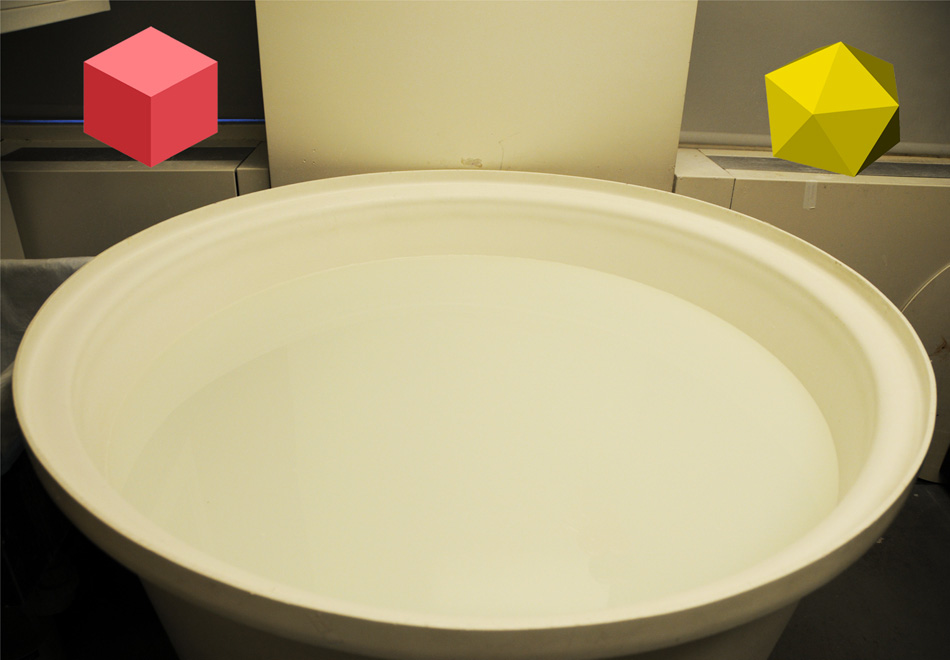
Common behavioral tests used for analyzing AD mouse models include contextual fear conditioning, radial arm water maze, and Morris water maze (Figure 2, 3, and 4). Below, we provide a detailed introduction to the equipment and operational steps of the contextual fear conditioning test based on reference[4].
|
Figure 2 Fear memory equipment |
Figure 3 Radial arm water maze equipment |
Figure 4 Morris water maze equipment |
Mice are tested individually in a conditioning chamber (Figure 2) located within a sound-attenuating box with a transparent Plexiglas window for video recording of the experiment. The conditioning chamber has a 36-bar grid floor that is removable and cleaned with 75% ethanol followed by water after each subject. To provide background white noise (72 dB), a single computer fan is installed on one side of the sound-attenuating chamber.
Before behavioral experiments, mice are handled once daily for 3 consecutive days. Only one animal is placed in the testing chamber at a time. The animal is placed in the conditioning chamber for 2 min, followed by a discrete tone (CS) at 2800 Hz and 85 dB for 30 s. During the last 2 s of the tone, the mouse receives a foot shock (US, 0.50-0.80 mA, lasting 2 s) through the floor bars. The intensity of the foot shock can be adjusted to induce stronger memory. After the CS/US pairing, the mouse remains in the conditioning chamber for an additional 30 s before being returned to its home cage. Freezing behavior can be scored using dedicated software. It is useful to double-check freezing time by manual scoring based on experience. Twenty-four hours after training, the mouse is placed back into the conditioning chamber. Freezing time is recorded for a continuous 5 min. Thirty-six hours after training, the mouse is transferred to a different context by inserting a triangular cage with a smooth floor and vanilla odor. After 2 min (pre-CS test), the mouse is exposed to a tone with the same characteristics as used during training for 3 min (CS test), and freezing time is measured. Typically, 15-20 animals are used in each condition.
An important control during the testing of fear memory is the assessment of sensory thresholds. Alterations in sensory perception of the shock could lead to misattribution of the observed phenotypic memory. A series of single foot shocks are delivered to animals placed on the same grid used for fear conditioning. Initially, a 0.1 mA shock lasting 1 s is administered to assess the animal's withdrawal, jumping, and vocalization behaviors. Every 30 s, the shock intensity is increased by 0.1-0.7 mA, followed by a return to 0 mA every 30 s in increments of 0.1 mA. The threshold for vocalization, withdrawal, and jumping is quantified for each animal based on the average shock intensity at which the animal exhibits behavioral responses to the foot shock.
Treatment of AD
It is reported that there are currently about 24 million cases of Alzheimer's disease worldwide, and the total number of dementia patients is estimated to increase fourfold by 2050. Despite being a public health issue, only two classes of drugs have been approved for the treatment of AD to date, including cholinesterase inhibitors(natural derivatives, synthetic, and hybrid analogs)and N-methyl-D-aspartate (NMDA) antagonists[5].
Several physiological processes in AD can destroy acetylcholine-producing cells, reducing cholinergic transmission throughout the brain. Acetylcholinesterase inhibitors (AChEIs) are classified as reversible, irreversible, and pseudo-reversible. They function by blocking the breakdown of acetylcholine (ACh) by acetylcholinesterase (AChE) and butyrylcholinesterase (BChE), thereby increasing ACh levels in the synaptic cleft. On the other hand, overactivation of NMDA receptors (NMDARs) leads to increased intracellular calcium ion levels, promoting cell death and synaptic dysfunction. NMDAR antagonists prevent the overactivation of NMDAR glutamate receptors, thereby inhibiting calcium ion influx and restoring normal activity. Although these two classes of drugs are effective, they only alleviate the symptoms of Alzheimer's disease but cannot cure or prevent the disease.

Figure 5 Chemical structures of small-molecule drugs approved for the treatment of AD
Establishing ideal animal models that simulate human disease states is conducive to in-depth research on the etiology, pathogenesis, and prevention of AD. Currently, due to the inability to establish a perfect AD animal model, using two or more AD animal models in drug screening is more persuasive than using a single model, and it can further confirm the efficacy of the drug.
References
[1] Puzzo D, Gulisano W, Palmeri A, et al. Rodent models for Alzheimer's disease drug discovery[J]. Expert opinion on drug discovery, 2015, 10(7):703-711.
[2] Breijyeh Z, Karaman R. Comprehensive Review on Alzheimer's Disease: Causes and Treatment [J]. Molecules, 2020, 25, (24).
[3] Lv J. Mechanisms of Aβ1-42 or Aβ1-15 in improving memory in APP/PS1/Tau transgenic Alzheimer's disease mice[D]. East China Normal University, 2023.
[4] A D P, B L L, A A P, et al. Behavioral assays with mouse models of Alzheimer's disease: Practical considerations and guidelines-ScienceDirect[J]. Biochemical Pharmacology, 2014, 88(4):450-467.
[5] Breijyeh Z, Karaman R. Comprehensive Review on Alzheimer's Disease: Causes and Treatment[J]. Molecules, 2020, 25:5789
| Catalog No. | Product Name |
| abs113188 | Rabbit anti-APP (C-term D672) Polyclonal Antibody |
| abs119698 | Mouse anti-β Amyloid 1-16 Polyclonal Antibody |
| abs122984 | Rabbit anti-Cathepsin B Polyclonal Antibody |
AntBio provides antibodies, proteins, ELISA kits, cell culture, detection kits, and other research reagents. If you have any product needs, please contact us.


