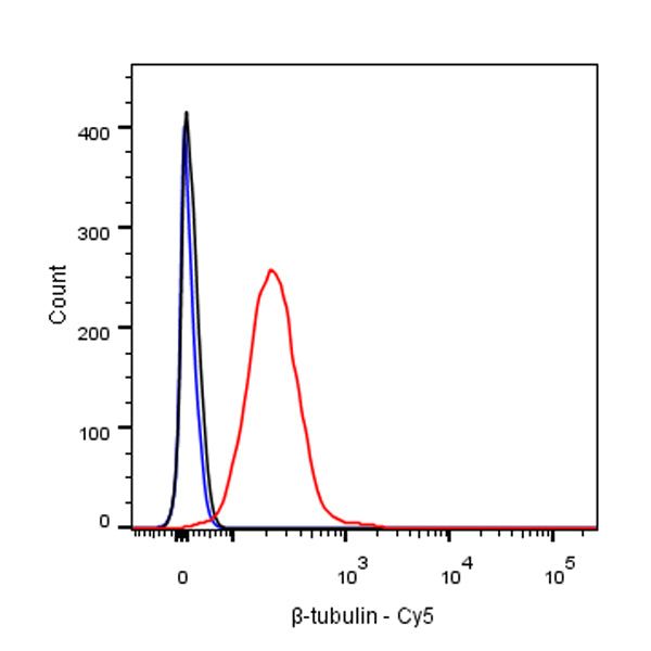
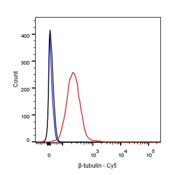
Flow cytometric analysis of 4% PFA fixed 90% methanol permeabilized HepG2 (Human hepatocellular carcinoma epithelial cell) cells labeldcfxling β-tubulin (Cy5 Conjugate) antibody at 1/20000 dilution (0.01 μg)/ (Red) compared with a Rabbit monoclonal IgG (Black) isotype control and an unlabelled control (cells without incubation with primary antibody and secondary antibody) (Blue).
β-tubulin Recombinant Rabbit mAb (Cy5 Conjugate) (S-312-113)
β-tubulin Recombinant Rabbit mAb (Cy5 Conjugate) (S-312-113)
Price:
Regular price
$80 USD
Regular price
Sale price
$80 USD
Unit price
per
For shipping services or bulk orders, you may request a quotation.
Secure checkout with

View full details
Product Details
Product Details
Product Specification
| Host | Rabbit |
| Antigen | β-tubulin |
| Synonyms | TUBB, TUBB5, Tubulin beta-5 chain |
| Immunogen | Synthetic Peptide |
| Location | Cytoplasm, Cytoskeleton |
| Accession | P07437 |
| Clone Number | S-312-113 |
| Antibody Type | Recombinant mAb |
| Isotype | IgG |
| Application | ICC, ICFCM |
| Reactivity | Hu, Ms, Rt |
| Predicted Reactivity | Dr, Lob, Pz, Ar, Av, Pl, Fu, Ys, SeUr, Bv, Pg, Fs |
| Purification | Protein A |
| Concentration | 2 mg/ml |
| Conjugation | Cy5 |
| Physical Appearance | Liquid |
| Storage Buffer | PBS, 0.1% BSA, 0.01% Proclin 300 |
| Stability & Storage | 12 months from date of receipt / reconstitution, 2 to 8 °C as supplied. |
Dilution
| application | dilution | species |
| ICC | 1:500 | |
| ICFCM | 1:20000 |
Background
α- and β-tubulin polymerize into dynamic microtubules. In eukaryotes, microtubules are one of the major components of the cytoskeleton, and function in many processes, including structural support, intracellular transport, and DNA segregation. To form microtubules, the dimers of α- and β-tubulin bind to GTP and assemble onto the (+) ends of microtubules while in the GTP-bound state. The β-tubulin subunit is exposed on the plus end of the microtubule, while the α-tubulin subunit is exposed on the minus end. After the dimer is incorporated into the microtubule, the molecule of GTP bound to the β-tubulin subunit eventually hydrolyzes into GDP through inter-dimer contacts along the microtubule protofilament.
Picture
Picture
FC

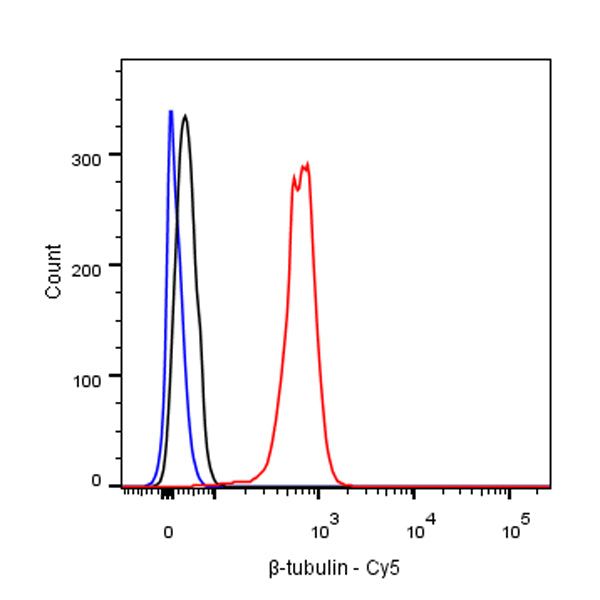
Flow cytometric analysis of 4% PFA fixed 90% methanol permeabilized NIH/3T3 (Mouse embryonic fibroblast) cells labelling β-tubulin (Cy5 Conjugate) antibody at 1/20000 dilution (0.01 μg)/ (Red) compared with a Rabbit monoclonal IgG (Black) isotype control and an unlabelled control (cells without incubation with primary antibody and secondary antibody) (Blue).
Immunocytochemistry
ICC shows positive staining in HepG2 cells. Anti-β-tubulin (Cy5 Conjugate) antibody was used at 1/500 dilution (magenta) and incubated overnight at 4°C. The cells were fixed with 100% ice-cold methanol and permeabilized with 0.1% PBS-Triton X-100. Nuclei were counterstained with DAPI (Blue).
ICC shows positive staining in NIH/3T3 cells. Anti-β-tubulin (Cy5 Conjugate) antibody was used at 1/500 dilution (magenta) and incubated overnight at 4°C. The cells were fixed with 100% ice-cold methanol and permeabilized with 0.1% PBS-Triton X-100. Nuclei were counterstained with DAPI (Blue).