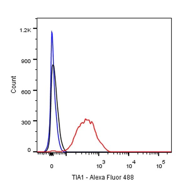
Flow cytometric analysis of Jurkat cells labelling TIA1 antibody at 1/500 dilution/ (red) compared with a Rabbit monoclonal IgG (Black) isotype control and an unlabelled control (cells without incubation with primary antibody and secondary antibody) (Blue). Goat Anti-Rabbit IgG Alexa Fluor® 488 at 1/1000 dilution was used as the secondary antibody.
TIA1 Recombinant Rabbit mAb (S-R026)
TIA1 Recombinant Rabbit mAb (S-R026)
Price:
Regular price
$100 USD
Regular price
Sale price
$100 USD
Unit price
per
For shipping services or bulk orders, you may request a quotation.
Secure checkout with

View full details
Product Details
Product Details
Product Specification
| Host | Rabbit |
| Antigen | TIA1 |
| Synonyms | p40-TIA-1 |
| Immunogen | N/A |
| Location | Cytoplasm, Nucleus |
| Accession | P31483 |
| Clone Number | SDT-R026 |
| Antibody Type | Rabbit mAb |
| Application | WB, ICC, FC, IP |
| Reactivity | Hu |
| Purification | Protein A |
| Concentration | 0.5 mg/ml |
| Physical Appearance | Liquid |
| Storage Buffer | PBS, 40% Glycerol, 0.05%BSA, 0.03% Proclin 300 |
| Stability & Storage | 12 months from date of receipt / reconstitution, -20 °C as supplied |
Dilution
| application | dilution | species |
| ICC | 1:500 | null |
| WB | 1:1000 | null |
| IP | 1:25 | null |
| FC | 1:500 | null |
Background
TIA1 or Tia1 cytotoxic granule-associated RNA binding protein is a 3'UTR mRNA binding protein that can bind the 5'TOP sequence of 5'TOP mRNAs. It is associated with programmed cell death (apoptosis) and regulates alternative splicing of the gene encoding the Fas receptor, an apoptosis-promoting protein. Under stress conditions, TIA1 localizes to cellular RNA-protein conglomerations called stress granules. It is encoded by the TIA1 gene.Mutations in the TIA1 gene have been associated with amyotrophic lateral sclerosis, frontotemporal dementia, and Welander distal myopathy. It also plays a crucial role in the development of toxic oligomeric tau in Alzheimer's disease.
Picture
Picture
Validation Data

Western Blot
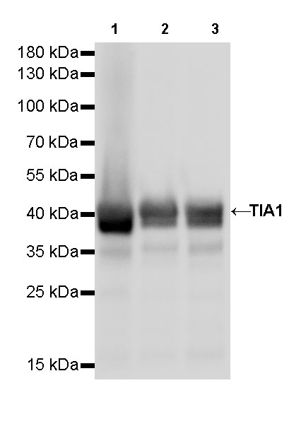
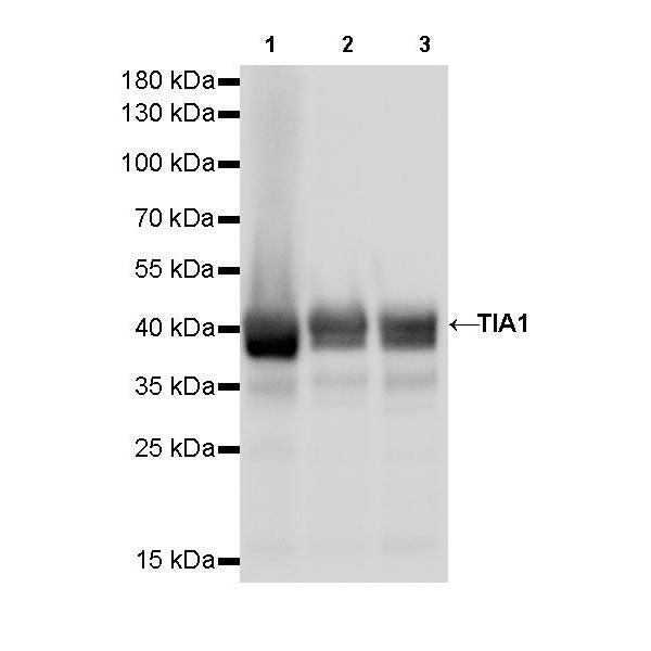
WB result of TIA1 Rabbit mAb
Primary antibody: TIA1 Rabbit mAb at 1/1000 dilution
Lane 1: Jurkat whole cell lysate 20 µg
Lane 2: MOLT-4 whole cell lysate 20 µg
Lane 3: Hela whole cell lysate 20 µg
Secondary antibody: Goat Anti-Rabbit IgG, (H+L), HRP conjugated at 1/10000 dilution
Predicted MW: 42~43 kDa
Observed MW: 42~43 kDa
Exposure time: 2.5s
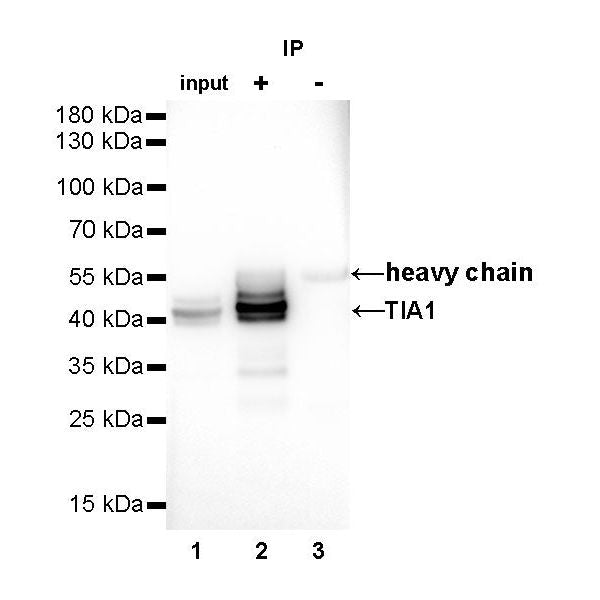
IP
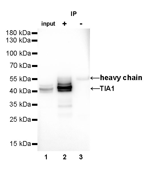
TIA1 Rabbit mAb at 1/25 dilution (2µg) immunoprecipitating TIA1 in 0.4mg Molt-4 whole cell lysate.
Western blot was performed on the immunoprecipitate using TIA1 Rabbit mAb at 1/1000 dilution.
Secondary antibody (HRP) for IP was used at 1/400 dilution.
Lane 1 : Molt-4 whole cell lysate 10µg (input)
Lane 2 : TIA1 Rabbit mAb IP in Molt-4 whole cell lysate
Lane 3 : Rabbit monoclonal IgG IP in Molt-4 whole cell lysate
Predicted MW: 42~43 kDa
Observed MW: 42~43 kDa
Exposure time: 60s
Immunocytochemistry
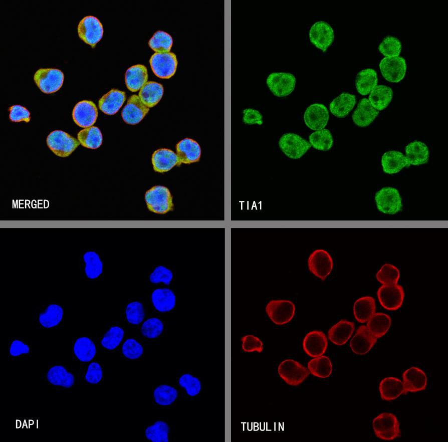
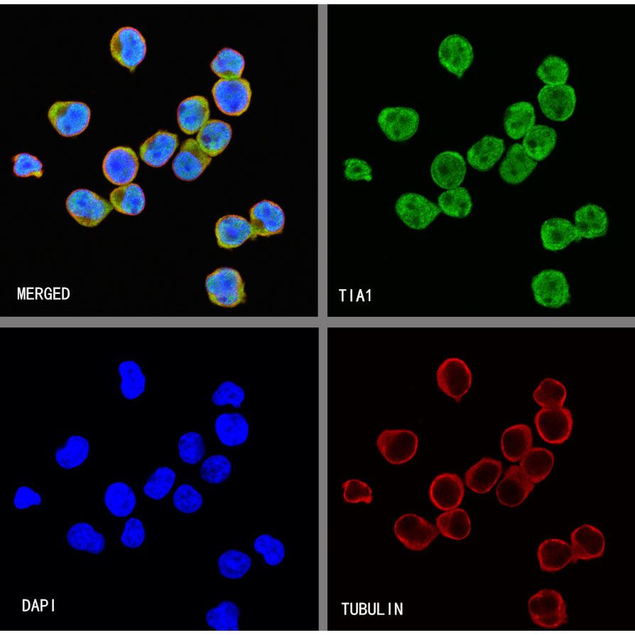
ICC shows positive staining in Jurkat cells. Anti-TIA1 antibody was used at 1/500 dilution (Green) and incubated overnight at 4°C. Goat polyclonal Antibody to Rabbit IgG - H&L (Alexa Fluor® 488) was used as secondary antibody at 1/1000 dilution. The cells were fixed with 4% PFA and permeabilized with 0.1% PBS-Triton X-100. Nuclei were counterstained with DAPI (Blue).Counterstain with tubulin (Red).