Product Specification
| Host |
Rabbit |
| Antigen |
GCDFP-15 |
| Synonyms |
Prolactin-inducible protein,Secretory actin-binding protein (SABP),gp17,GPIP4,PIP |
| Immunogen |
Synthetic Peptide |
| Location |
Secreted |
| Accession |
P12273 |
| Clone Number |
SDT-010-579 |
| Antibody Type |
Rabbit mAb |
| Isotype |
IgG |
| Application |
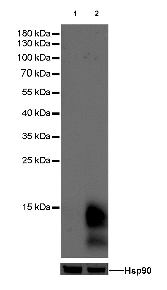
WB, IHC-P |
| Reactivity |
Hu |
| Purification |
Protein A |
| Research Area |
Immunology |
| Concentration |
0.5mg/ml |
| Molecular Weight |
16kDa |
| Physical Appearance |
Liquid |
| Storage Buffer |
PBS, 40% Glycerol, 0.05%BSA, 0.03% Proclin 300 |
| Stability & Storage |
12 months from date of receipt / reconstitution, -20 °C as supplied |
Dilution
| application |
dilution |
species |
| WB |
1:1000 |
|
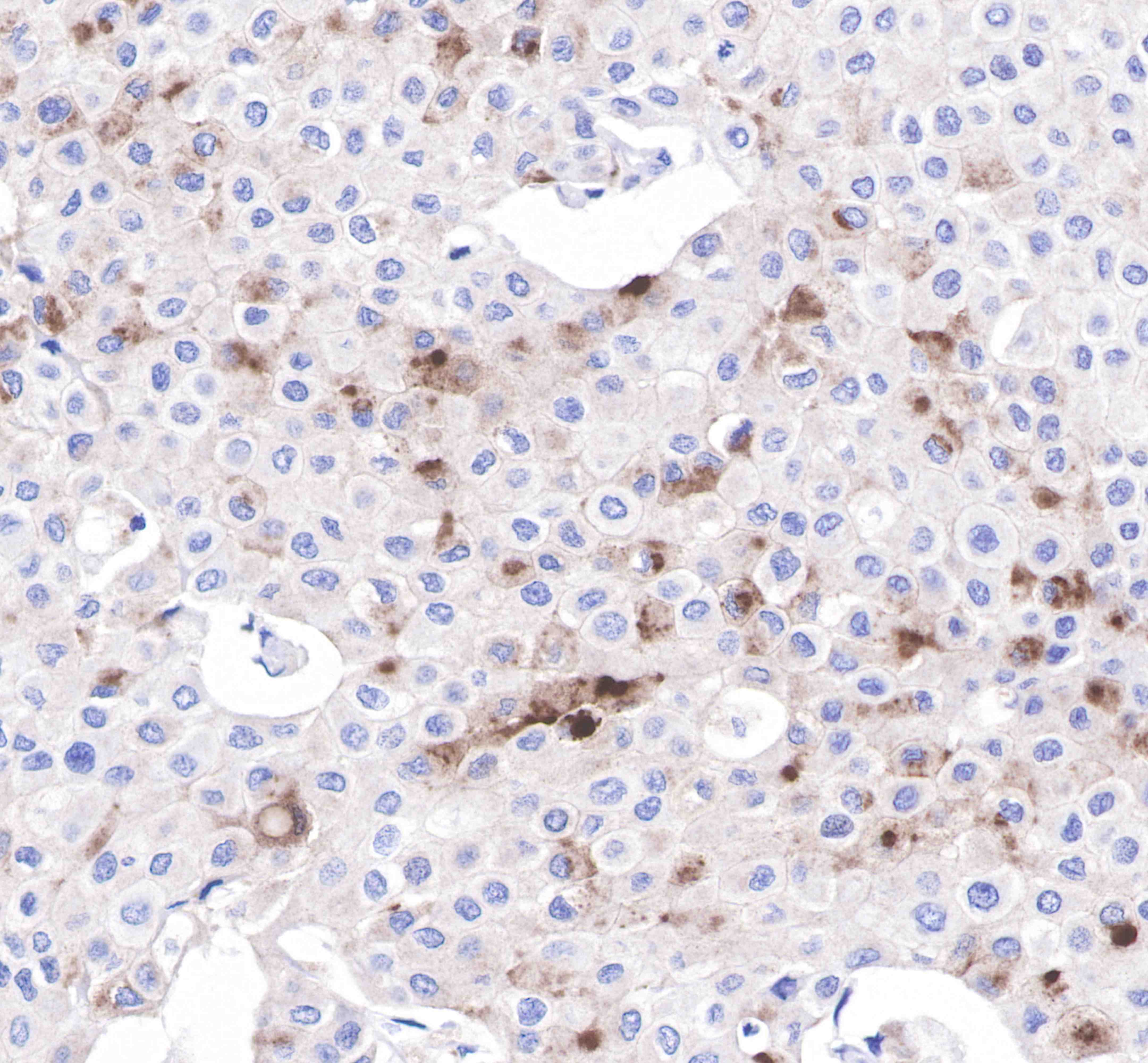
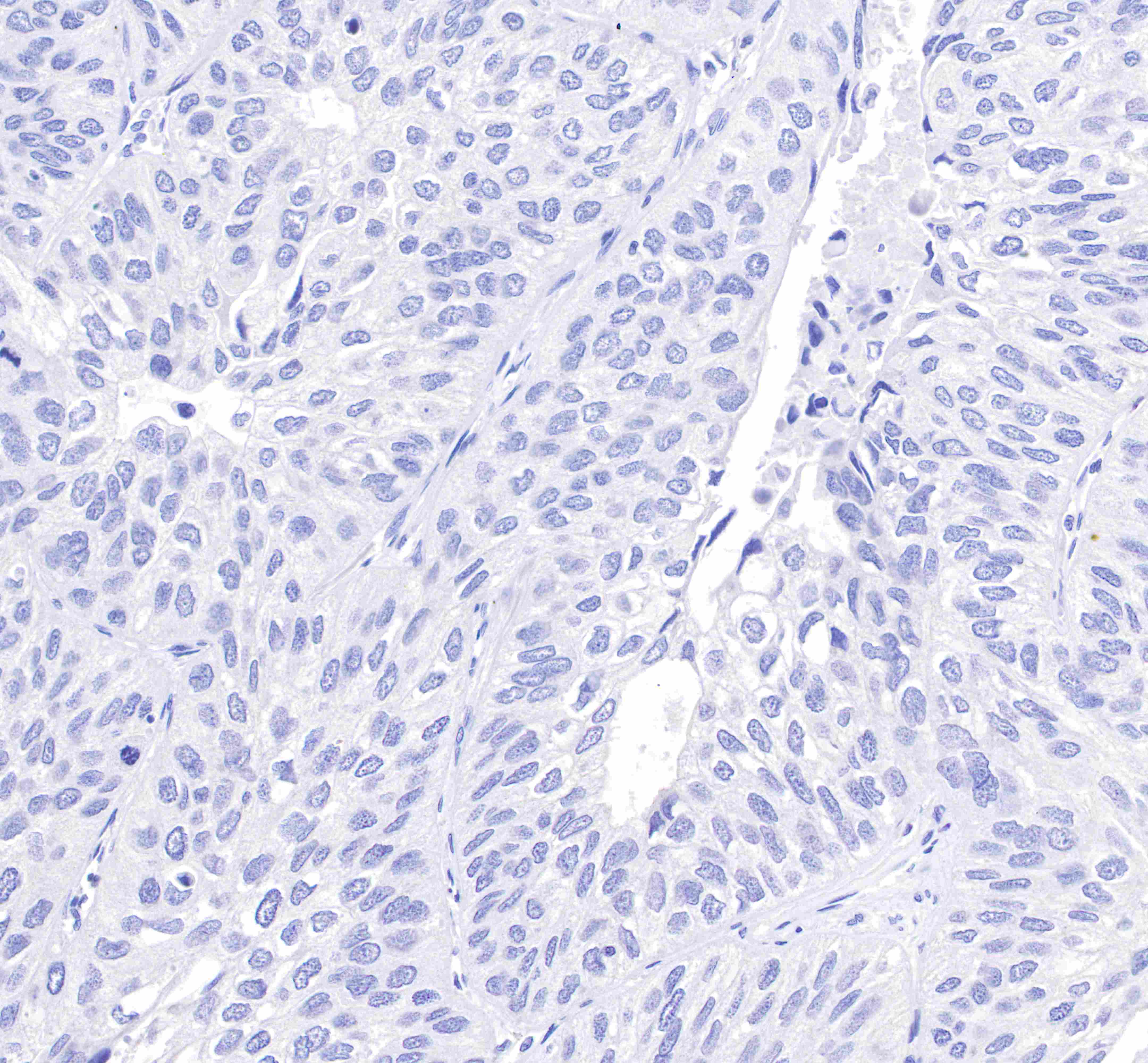
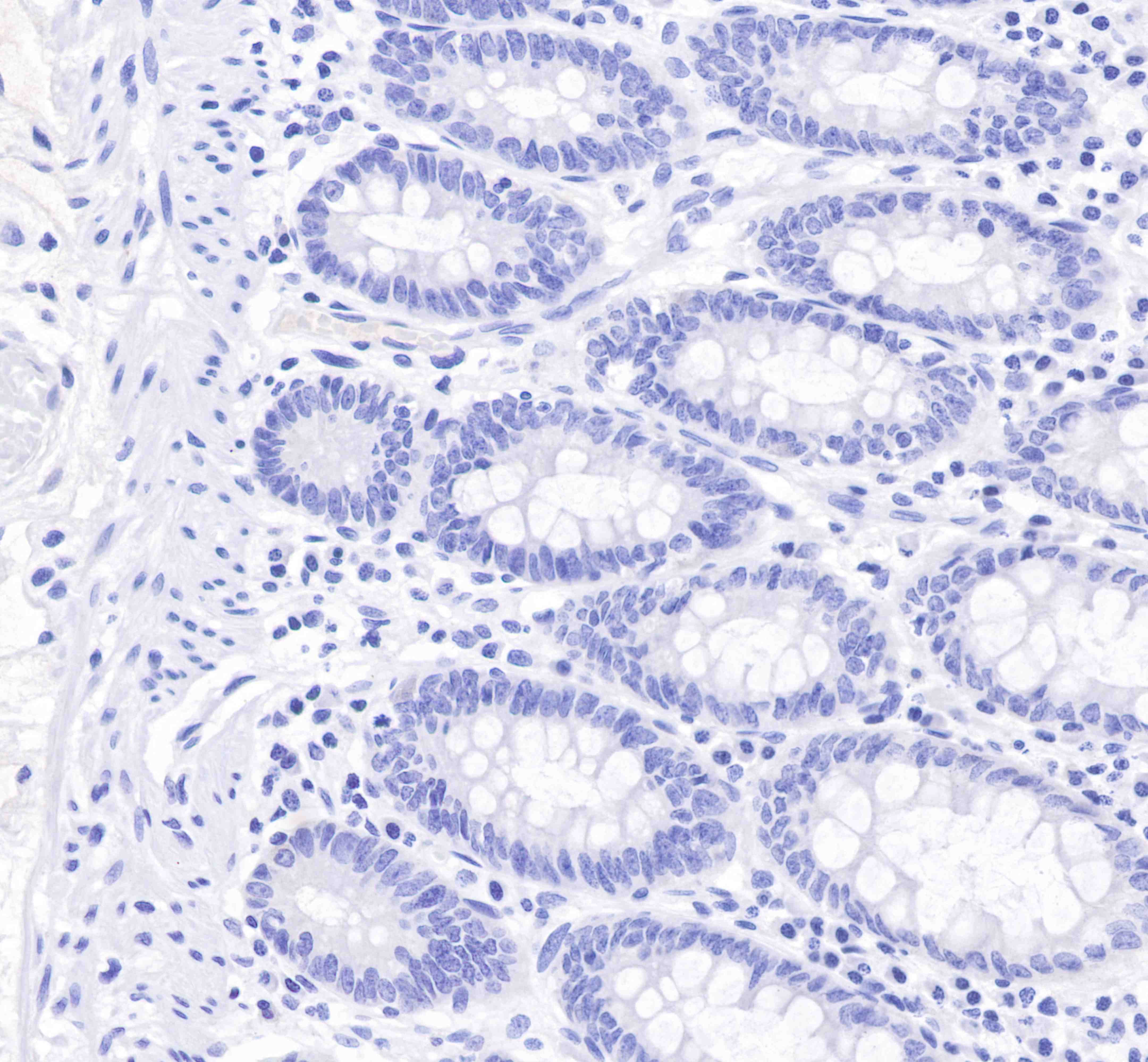
| IHC-P |
1:100 |
|
Background
Gross cystic disease fluid protein 15 (GCDFP-15) also known as Prolactin-inducible protein, extra-parotid glycoprotein (EP-GP), gp17 seminal actin-binding protein (SABP) or BRST2 is a protein that in humans is encoded by the PIP gene. It is upregulated by prolactin and androgens and downregulated by estrogen. The protein has a physiological function in regulation of water transport mainly in apocrine glands in the axilla, vulva, eyelid and ear canal, serous cells of the submandibular salivary gland, serous cells of the submucosal glands of the bronchi, and accessory lacrimal glands as well as cutaneous eccrine glands. It is also found in amniotic fluid and seminal fluid. PIP has the ability to bind immunoglobulin G (IgG), IgG-Fc, CD4-T cell receptor suggesting a wide range of immunological functions. PIP also binds to AZGP1. PIP exerts aspartyl proteinase activity able to cleave fibronectin. Mitogenic effect of PIP was observed on both normal and malignant breast epithelial cells.