Product Specification
| Host |
Rabbit |
| Antigen |
CD3 EPSILON |
| Synonyms |
T-cell surface antigen T3/Leu-4 epsilon chain, CD3e, T3E, CD3E |
| Immunogen |
N/A |
| Location |
Cell membrane |
| Accession |
P07766 |
| Clone Number |
SDT-R137 |
| Antibody Type |
Recombinant mAb |
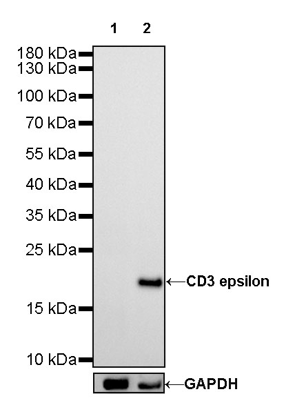
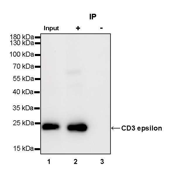
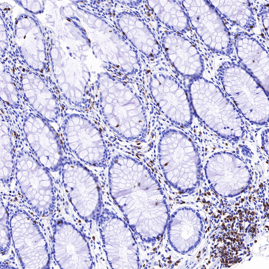
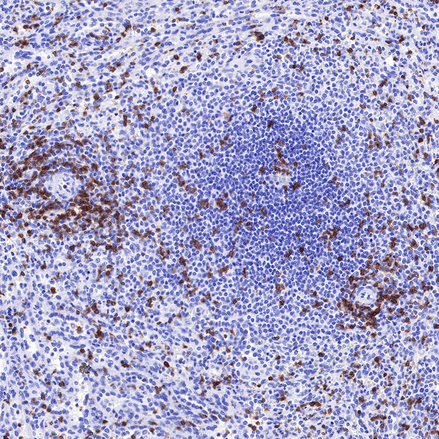
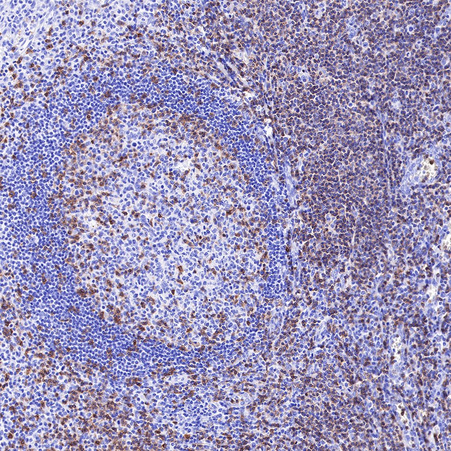
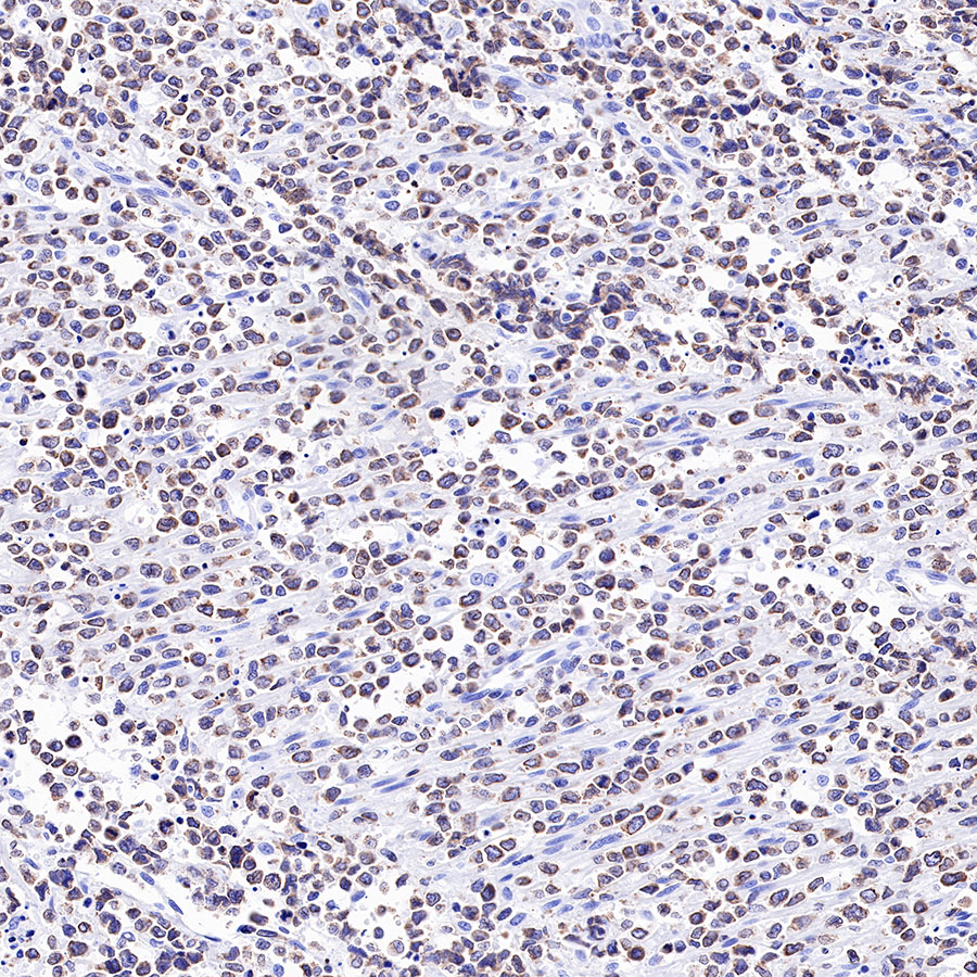
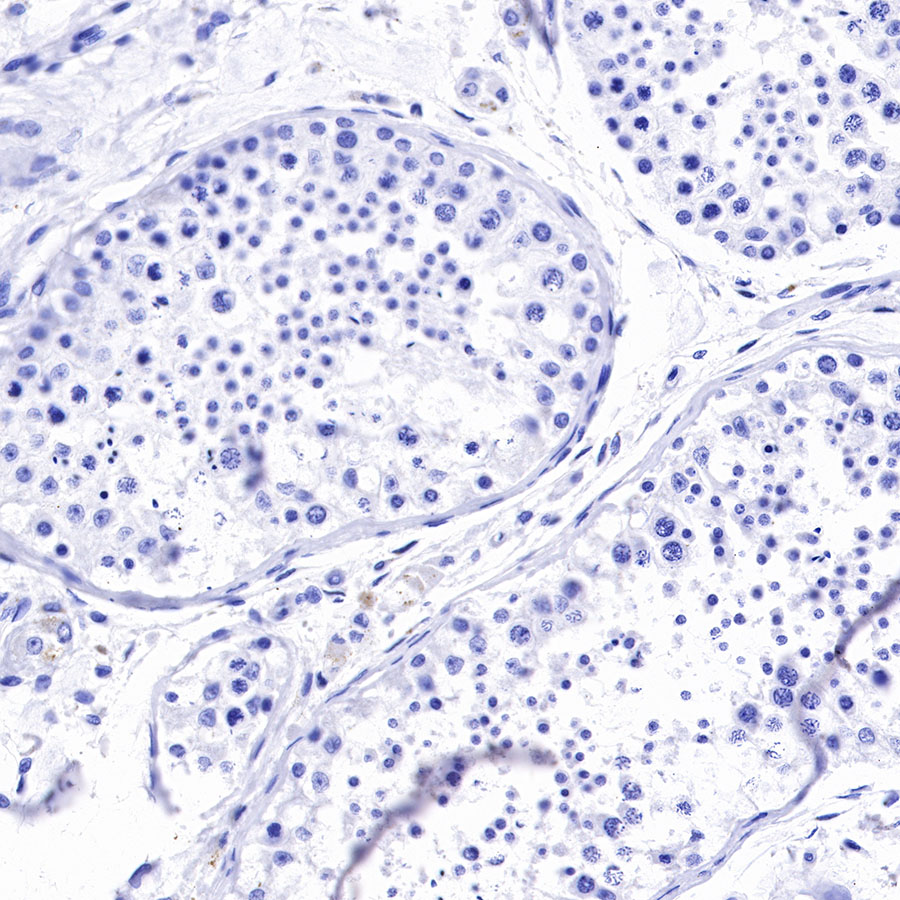
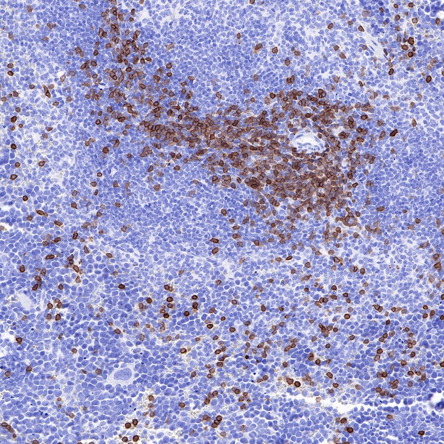
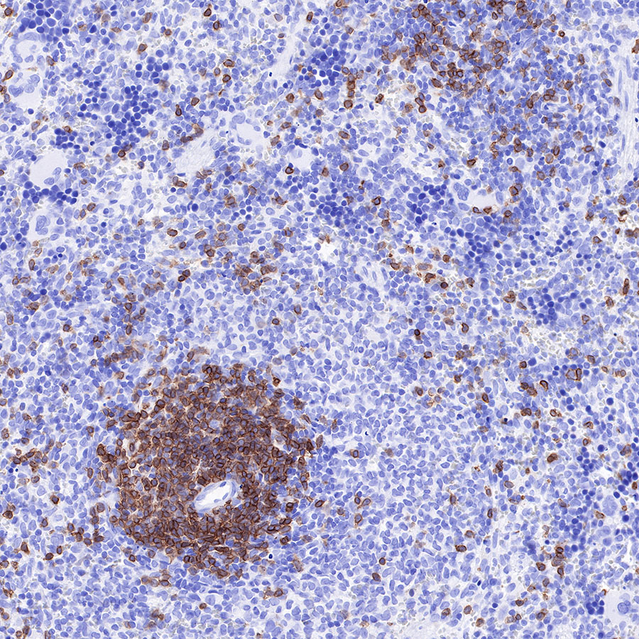
| Application |
WB, IHC-P, IP |
| Reactivity |
Hu, Ms, Rt |
| Purification |
Protein A |
| Concentration |
0.25 mg/ml |
| Tag |
N/A |
| Physical Appearance |
Liquid |
| Storage Buffer |
PBS, 40% Glycerol, 0.05% BSA, 0.03% Proclin 300 |
| Stability & Storage |
12 months from date of receipt / reconstitution, -20 °C as supplied |
Dilution
| application |
dilution |
species |
| WB |
1:500 |
|
| IHC-P |
1:1000-1:2000 |
|
| IP |
1:25 |
|
Background
CD3 (cluster of differentiation 3) is a protein complex and T cell co-receptor that is involved in activating both the cytotoxic T cell (CD8+ naive T cells) and T helper cells (CD4+ naive T cells). It is composed of four distinct chains. In mammals, the complex contains a CD3γ chain, a CD3δ chain, and two CD3ε chains. These chains associate with the T-cell receptor (TCR) and the CD3-zeta (ζ-chain) to generate an activation signal in T lymphocytes. The TCR, CD3-zeta, and the other CD3 molecules together constitute the TCR complex. The CD3–T cell receptor (TCR) complex plays a central role in the T-cell-mediated immunoresponse as it is involved in the recognition of antigens and subsequent signal transduction and activation of immunocompetent T lymphocytes. Because CD3 is required for T cell activation, drugs (often monoclonal antibodies) that target it are being investigated as immunosuppressant therapies (e.g., otelixizumab, teplizumab) for type 1 diabetes and other autoimmune diseases.