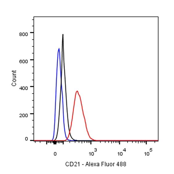
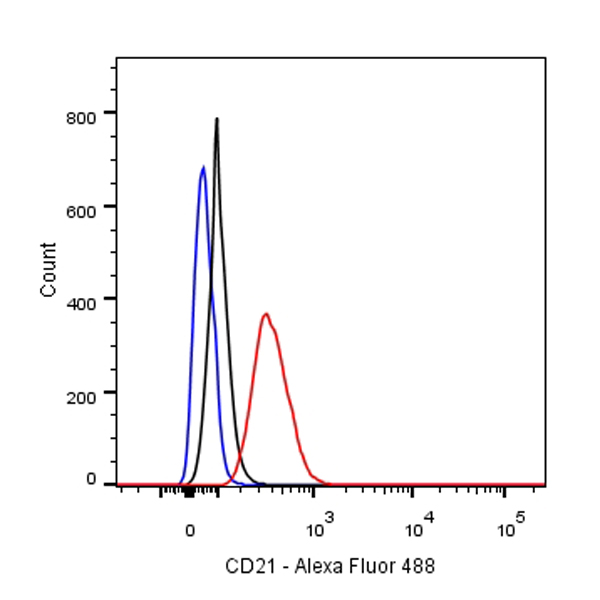
Flow cytometric analysis of Raji cells labelling CD21 antibody at 1/80 dilution (0.1ug)/ (red) compared with a Rabbit monoclonal IgG (Black) isotype control and an unlabelled control (cells without incubation with primary antibody and secondary antibody) (Blue). Goat Anti-Rabbit IgG Alexa Fluor® 488 at 1/1000 dilution was used as the secondary antibody.
S-RMab® CD21 Recombinant Rabbit mAb(SDT-007-47)
S-RMab® CD21 Recombinant Rabbit mAb(SDT-007-47)
Price:
Regular price
$100 USD
Regular price
Sale price
$100 USD
Unit price
per
For shipping services or bulk orders, you may request a quotation.
Secure checkout with

View full details
Product Details
Product Details
Product Specification
| Host | Rabbit |
| Antigen | CD21 |
| Synonyms | CR2,CD3R, Complement C3d receptor, Epstein-Barr virus receptor (EBV receptor) |
| Immunogen | Synthetic Peptide |
| Location | Membrane |
| Accession | P20023 |
| Clone Number | SDT-007-47 |
| Antibody Type | Rabbit mAb |
| Isotype | IgG |
| Application | WB, IHC-P, ICC, FCM |
| Reactivity | Hu, Ms, Rt |
| Purification | Protein A |
| Concentration | 0.08mg/ml |
| Conjugation | Unconjugated |
| Physical Appearance | Liquid |
| Storage Buffer | PBS, 40% Glycerol, 0.05%BSA, 0.03% Proclin 300 |
| Stability & Storage | 12 months from date of receipt / reconstitution, -20 °C as supplied |
Dilution
| application | dilution | species |
| IHC-P | 1:1000 | |
| FCM | 1:80 | |
| ICC | 1:50 | |
| WB | 1:200 |
Background
CD21 is involved in the complement system. It binds to iC3b (inactive derivative of C3b), C3dg, or C3d.B cells express CD21 receptors on their surfaces, allowing the complement system to play a role in B-cell activation and maturation. CD21 interacts with CD19 and on mature B cells forms a complex with CD81 (TAPA-1). The CD21-CD19-CD81 complex is often called the B cell co-receptor complex, because CD21 binds to opsonized antigens through attached C3d (or iC3b or C3dg) when the B-cell receptor binds antigen. This results in the B cell having greatly enhanced response to the antigen. Epstein-Barr virus (EBV) can bind CD21, enabling EBV to enter and infect B cells.
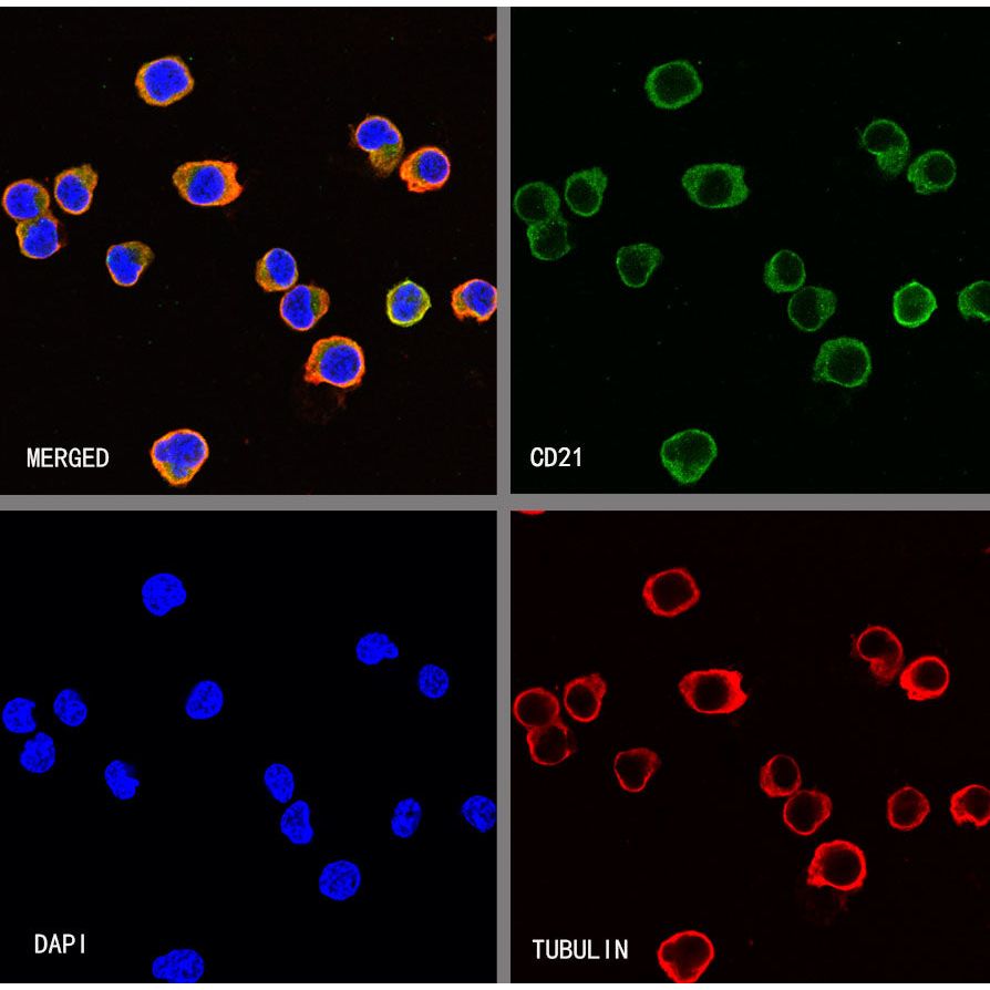
Picture
Picture
Validation Data
Western Blot
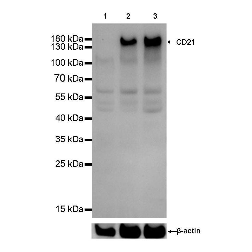
WB result of CD21 Rabbit mAb
Primary antibody: CD21 Rabbit mAb at 1/200 dilution
Lane 1: K562 whole cell lysate 20 µg
Lane 2: Daudi whole cell lysate 20 µg
Lane 3: Raji whole cell lysate 20 µg
Negative control: K562 whole cell lysate
Secondary antibody: Goat Anti-Rabbit IgG, (H+L), HRP conjugated at 1/10000 dilution
Predicted MW: 113 kDa
Observed MW: 160 kDa
Exposure time: 20s
Immunohistochemistry
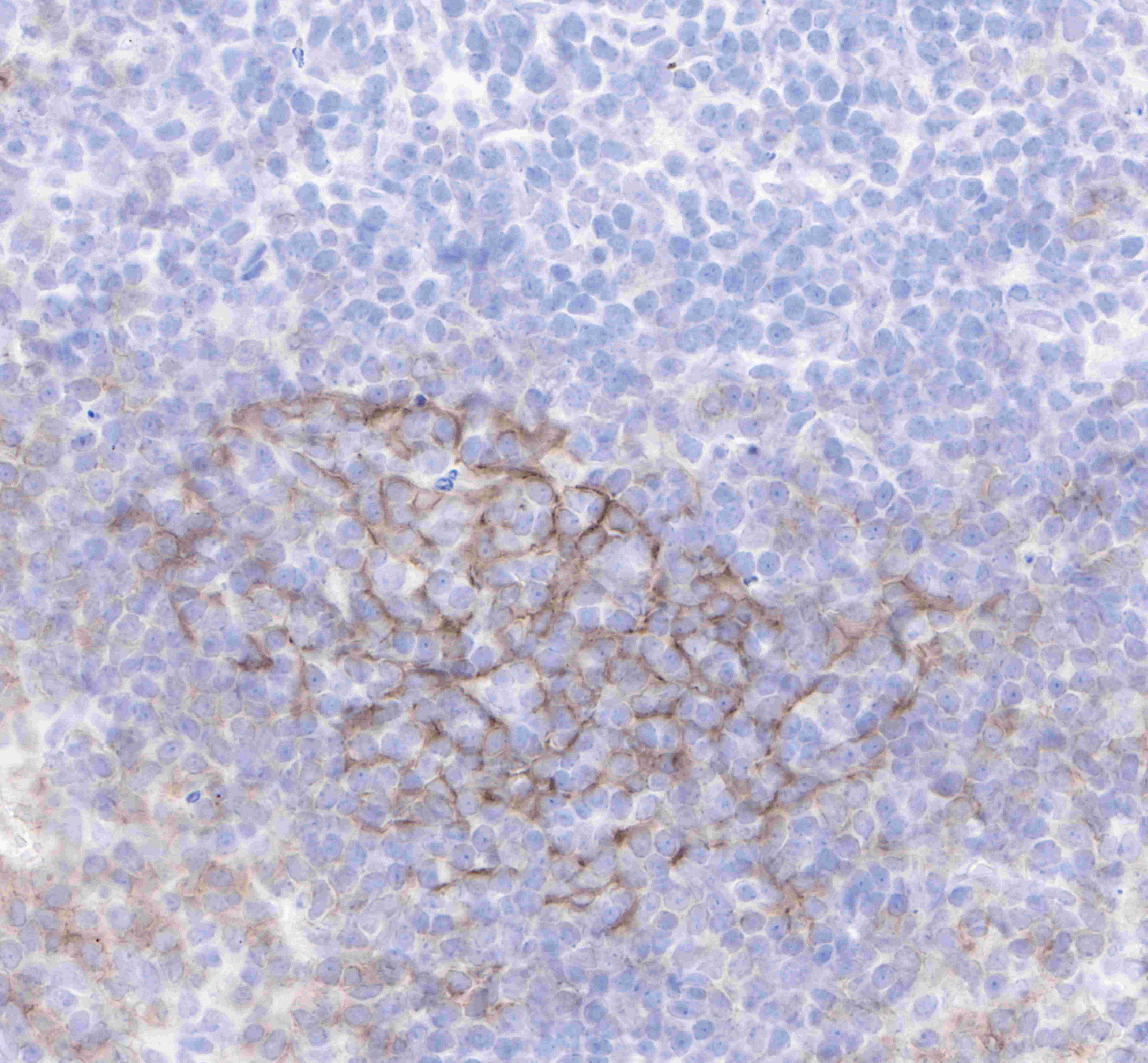
IHC shows positive staining in paraffin-embedded human tonsil. Anti-CD21 antibody was used at 1/1000 dilution, followed by a Goat Anti-Rabbit IgG H&L (HRP) ready to use. Counterstained with hematoxylin.
Heat mediated antigen retrieval with Tris/EDTA buffer pH9.0 was performed before commencing with IHC staining protocol.
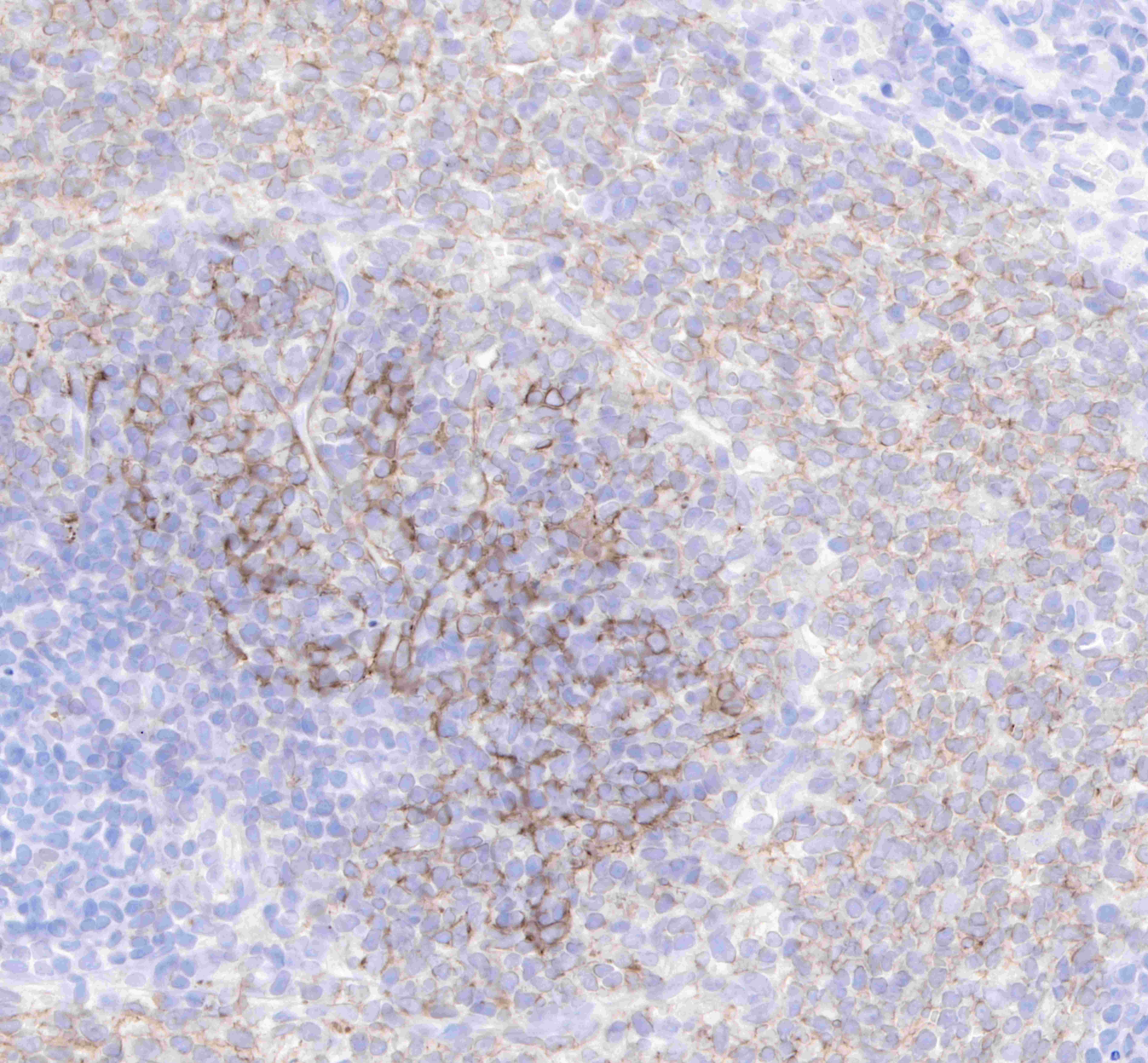
IHC shows positive staining in paraffin-embedded human spleen. Anti-CD21 antibody was used at 1/1000 dilution, followed by a Goat Anti-Rabbit IgG H&L (HRP) ready to use. Counterstained with hematoxylin. Heat mediated antigen retrieval with Tris/EDTA buffer pH9.0 was performed before commencing with IHC staining protocol.
Negative tissue: IHC shows negative staining in paraffin-embedded human kidney. Anti-CD21 antibody was used at 1/1000 dilution, followed by a Goat Anti-Rabbit IgG H&L (HRP) ready to use. Counterstained with hematoxylin. Heat mediated antigen retrieval with Tris/EDTA buffer pH9.0 was performed before commencing with IHC staining protocol.
IHC shows positive staining in paraffin-embedded mouse spleen. Anti-CD21 antibody was used at 1/1000 dilution, followed by a Goat Anti-Rabbit IgG H&L (HRP) ready to use. Counterstained with hematoxylin. Heat mediated antigen retrieval with Tris/EDTA buffer pH9.0 was performed before commencing with IHC staining protocol.
IHC shows positive staining in paraffin-embedded rat spleen. Anti-CD21 antibody was used at 1/1000 dilution, followed by a Goat Anti-Rabbit IgG H&L (HRP) ready to use. Counterstained with hematoxylin. Heat mediated antigen retrieval with Tris/EDTA buffer pH9.0 was performed before commencing with IHC staining protocol.