Product Details
Product Details
Product Specification
| Synonym | Reactive Oxygen Species Assay Kit (DHE) |
| Detection Type | Fluorescence method |
| Description | This kit uses fluorescent probe DHE to detect reactive oxygen species. DHE can freely enter the cell through the membrane of living cells. It shows blue fluorescence in the cytoplasm, and is oxidized by intracellular ROS to form oxyethidium, which is incorporated into the chromosomal DNA, making the nucleus appear bright red fluorescence. At the excitation wavelength of 535nm and emission wavelength of 610nm, the fluorescence intensity was detected by fluorescence microscope, laser confocal microscope, fluorescence spectrophotometer, fluorescence microplate reader and flow cytometry, and the intracellular reactive oxygen species level was measured. It has the advantages of low background, high sensitivity, wide linear range, and easy to use. It is a fast and simple classical method for the detection of ROS in tissues or cultured living cells. |
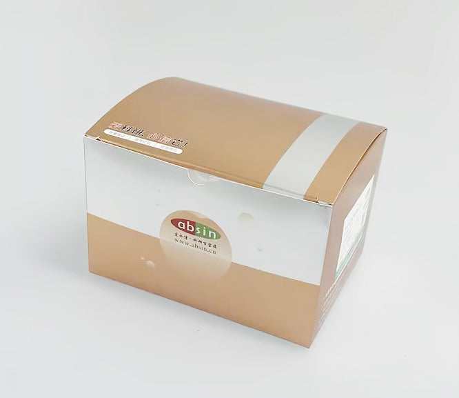
| Composition | 0.2ml, 5mM DHE |
| Background | The optimal excitation wavelength is 480-535nm.The optimal emission wavelength is 590-610nm. |
| General Notes | 1. Before opening the cover, please briefly centrifuge, and collect the liquid on the inner wall of the cover at the bottom of the tube to avoid the loss of the staining solution when opening the cover. 2, there should be a control without DHE incubation, that is, without probe, only cells resuspended in 0.01M PBS. 3, After labeling with fluorescent probes, it is important to wash any remaining probes that have not entered the cell to avoid background elevation. Pay attention to the operation standard when washing many times to avoid washing the cells off. 4. For cells with short drug treatment (incubation) time (within 2 hours, it is also recommended that within 4 hours), you can use fluorescent dye to mark the cells first, and then stimulate the cells with drugs. For cells treated with drugs for a long time (more than 4 hours or more than 6 hours), it is recommended to treat (stimulate) the cells with drugs first and then label them with the probe. 5, DHE is easy to be oxidized in light and air, so attention should be paid to avoid light during storage and operation. 6, for different cells and tissues, the appropriate incubation time and concentration should be selected to observe the changes in ROS. 7, for your safety and health, please wear a laboratory coat and wear disposable gloves to operate. |
| Instructions | Equipment required: Flow cytometer, fluorescence microplate reader, laser confocal microscope, fluorescence spectrophotometer, etc. Steps: I. Cell samples: 1. Collection of cells for reactive oxygen species determination (1) Cell collection: suspension cells: 2000 RPM, centrifugal 5 min, collect precipitation, with 0.01 M PBS serum-free culture or washing twice, 1000 RPM, centrifugal 5 min, abandon supernatant, take cells precipitation; Adherent cells: remove the culture medium by suction, blow with 0.01M PBS or serum-free culture medium repeatedly, make the cell layer all into PBS or culture medium, collect the cell suspension, wash with 0.01M PBS or serum-free culture medium twice, centrifuge at 1000rpm for 5min, discard the supernatant, take cell precipitation; (2) Adding DHE probe: resuspend the cell precipitate with the diluted DHE fluorescent probe. In general, the cell density is required to be 1*106-2*107/ml, and the recommended initial working concentration of probe is 10µ Can be in 1 M (DHE work concentration & micro; M~100 µ In the range of M ~ 100 & micro, pre-experiment is needed to determine the optimal concentration). (3) 37º C The cells were incubated for 10 min to 90 min. Typically, 10-30 minutes will do. Note: The length of incubation time depends on cell type, stimulation condition, DHE concentration, etc. It can be reversed and mixed every 5min so that the probe is in full contact with the sample. (4) was centrifuged at 1000g for 5min, cell precipitates were collected by removing the supernatant, washing twice with PBS buffer, and cells were resuspended. (5) was used for fluorescence detection, and the results were expressed as fluorescence value. 2, without collecting the cells, directly add the probe into the culture medium for determination (1) Add the DHE probe: remove the cell culture medium supernatant, add the DHE probe diluted in serum-free medium (recommended final concentration of probe is 10µ M), the volume of the probe added should be suitable to cover the cells, usually no less than 500 µ per well in 6-well plate; l. (2) 37º C The cells were incubated for 10 min to 90 min. Typically, 10-30 minutes will do. Note: The length of incubation time depends on cell type, stimulation condition, DHE concentration, etc. It can be reversed and mixed every 5min so that the probe is in full contact with the sample. (3) Discard the upper culture medium and wash twice with 0.01M PBS or serum-free culture medium to remove the probe that has not entered the cells. (4) was used for fluorescence detection, and the results were expressed as fluorescence value. II. Animal tissue samples 1. Cell suspension preparation: Single cell suspension preparation instrument or traditional tissue processing methods such as enzymatic hydrolysis and grinding method can be used to prepare single cell suspension; 2, adding DHE probe, adding DHE probe: remove the cell culture medium supernatant, add the DHE probe diluted in serum-free culture medium (recommended final concentration of probe is 10µ M). 3, 37º C The cells were incubated for 10 min to 90 min. Typically, 10-30 minutes will do. Note: The length of incubation time depends on cell type, stimulation condition, DHE concentration, etc. It can be reversed and mixed every 5min so that the probe is in full contact with the sample. 4, 1000g, centrifugation for 5min, remove the supernatant to collect cell precipitation, wash twice with PBS buffer, resuspend the cells 5, fluorescence detection, the results are expressed as fluorescence value. |
| Storage Temp. | -20 ° C in the dark, valid for 12 months. |
| Applications | Adherent cells, suspended cells, fresh animal tissues |
Picture
Picture
Immunohistochemistry


