WB result of PKC eta Recombinant Rabbit mAb
Primary antibody: PKC eta Recombinant Rabbit mAb at 1/1000 dilution
Lane 1: MCF7 whole cell lysate 20 µg
Lane 2: PC-3 whole cell lysate 20 µg
Secondary antibody: Goat Anti-rabbit IgG, (H+L), HRP conjugated at 1/10000 dilution
Predicted MW: 78 kDa
Observed MW: 45, 78 kDa
This blot was developed with high sensitivity substrate
PKC eta Recombinant Rabbit mAb (S-1053-35)
PKC eta Recombinant Rabbit mAb (S-1053-35)
Price:
Regular price
$45 USD
Regular price
Sale price
$45 USD
Unit price
per
For shipping services or bulk orders, you may request a quotation.
Secure checkout with

View full details
Product Details
Product Details
Product Specification
| Host | Rabbit |
| Antigen | PKC eta |
| Synonyms | Protein kinase C eta type, PKC-L, nPKC-eta, Prkch, Pkch |
| Immunogen | Synthetic Peptide |
| Location | Cytoplasm |
| Accession | P23298 |
| Clone Number | S-1053-35 |
| Antibody Type | Recombinant mAb |
| Isotype | IgG |
| Application | WB, IP |
| Reactivity | Hu, Ms, Rt |
| Purification | Protein A |
| Concentration | 0.5 mg/ml |
| Conjugation | Unconjugated |
| Physical Appearance | Liquid |
| Storage Buffer | PBS, 40% Glycerol, 0.05% BSA, 0.03% Proclin 300 |
| Stability & Storage | 12 months from date of receipt / reconstitution, -20 °C as supplied |
Dilution
| application | dilution | species |
| WB | 1:1000 | |
| IP | 1:50 |
Background
PKC eta, also known as Protein Kinase C eta, is a member of the Protein Kinase C (PKC) family of enzymes. This serine/threonine-specific protein kinase plays crucial roles in various cellular processes, particularly in signal transduction, cell differentiation, and gene transcription regulation. PKC eta regulates keratinocyte differentiation by activating the MAPK13 (p38delta) activated protein kinase cascade. PKC eta mediates transcriptional activation of the transglutaminase 1 (TGM1) gene. Mutations in the gene have been linked to susceptibility to cerebral infarction.
Picture
Picture
Western Blot
WB result of PKC eta Recombinant Rabbit mAb
Primary antibody: PKC eta Recombinant Rabbit mAb at 1/1000 dilution
Lane 1: mouse lung lysate 20 µg
Secondary antibody: Goat Anti-rabbit IgG, (H+L), HRP conjugated at 1/10000 dilution
Predicted MW: 78 kDa
Observed MW: 45, 78 kDa
This blot was developed with high sensitivity substrate
WB result of PKC eta Recombinant Rabbit mAb
Primary antibody: PKC eta Recombinant Rabbit mAb at 1/1000 dilution
Lane 1: rat lung lysate 20 µg
Lane 2: rat skin lysate 20 µg
Secondary antibody: Goat Anti-rabbit IgG, (H+L), HRP conjugated at 1/10000 dilution
Predicted MW: 78 kDa
Observed MW: 45, 78 kDa
This blot was developed with high sensitivity substrate
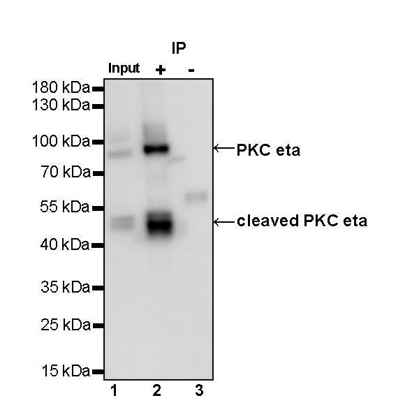
IP
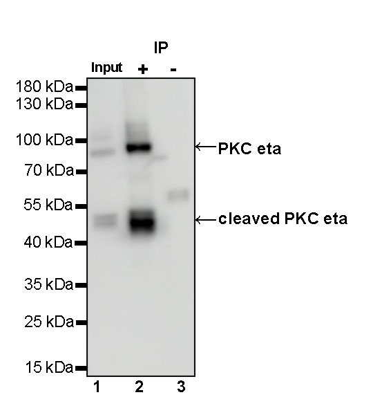
PKC eta Rabbit mAb at 1/50 dilution (1 µg) immunoprecipitating PKC eta in 0.4 mg mouse lung lysate.
Western blot was performed on the immunoprecipitate using PKC eta Rabbit mAb at 1/1000 dilution.
Secondary antibody (HRP) for IP was used at 1/1000 dilution.
Lane 1: mouse lung lysate 20 µg (Input)
Lane 2: PKC eta Rabbit mAb IP in mouse lung lysate
Lane 3: Rabbit monoclonal IgG IP in mouse lung lysate
Predicted MW: 78 kDa
Observed MW: 45, 78 kDa