WB result of Phospho-S6 Ribosomal Protein (Ser235/236) Rabbit mAb Primary antibody: Phospho-S6 Ribosomal Protein (Ser235/236) Rabbit mAb at 1/1000 dilution Lane 1: HeLa whole cell lysate 5 µg Lane 2: HeLa serum starvation 16 hour then treated with calyculin (100 nM, 30 min) whole cell lysate 5 µg Secondary antibody: Goat Anti-Rabbit IgG, (H+L), HRP conjugated at 1/10000 dilution Predicted MW: 29 kDa Observed MW: 32 kDa
Phospho-S6 Ribosomal Protein (Ser235/236) Recombinant Rabbit mAb (S-R203)
Phospho-S6 Ribosomal Protein (Ser235/236) Recombinant Rabbit mAb (S-R203)
Price:
Regular price
$100 USD
Regular price
Sale price
$100 USD
Unit price
per
For shipping services or bulk orders, you may request a quotation.
Secure checkout with

View full details
Product Details
Product Details
Product Specification
| Host | Rabbit |
| Antigen | Phospho-S6 Ribosomal Protein (Ser235/236) |
| Synonyms | Phospho-Small ribosomal subunit protein eS6 (Ser235/236), Phospho-40S ribosomal protein S6 (Ser235/236), Phospho-Phosphoprotein NP33 (Ser235/236) |
| Location | Cytoplasm, Nucleus, Nucleolus |
| Accession | P62753 |
| Clone Number | S-R203 |
| Antibody Type | Rabbit mAb |
| Application | WB, ICC, IP |
| Reactivity | Hu, Ms, Rt |
| Purification | Protein A |
| Concentration | 0.5 mg/ml |
| Conjugation | Unconjugated |
| Physical Appearance | Liquid |
| Storage Buffer | PBS, 40% Glycerol, 0.05% BSA, 0.03% Proclin 300 |
| Stability & Storage | 12 months from date of receipt / reconstitution, -20 °C as supplied |
Dilution
| application | dilution | species |
| WB | 1:1000 | |
| ICC | 1:500 | |
| IP | 1:50 |
Background
Ribosomal protein S6 (rpS6 or eS6) is a component of the 40S ribosomal subunit and is therefore involved in translation. Mouse model studies have shown that phosphorylation of eS6 is involved in the regulation of cell size, cell proliferation, and glucose homeostasis [PMID: 16166381, PMID: 16679021, PMID: 19479038]. p70S6K is a serine/threonine kinase that phosphorylates the S6 protein of the 40S ribosomal subunit (phosphorylated S6 ribosomal protein (phospho-S6rp)) at several sites, including serines 235 and 236, leading to initiation of protein synthesis [PMID: 16860938].
Picture
Picture
Western Blot
WB result of Phospho-S6 Ribosomal Protein (Ser235/236) Rabbit mAb Primary antibody: Phospho-S6 Ribosomal Protein (Ser235/236) Rabbit mAb at 1/1000 dilution Lane 1: NIH/3T3 whole cell lysate 5 µg Lane 2: NIH/3T3 treated with CA (100 nM, 30 min) whole cell lysate 5 µg Secondary antibody: Goat Anti-Rabbit IgG, (H+L), HRP conjugated at 1/10000 dilution Predicted MW: 29 kDa Observed MW: 32 kDa
WB result of Phospho-S6 Ribosomal Protein (Ser235/236) Rabbit mAb Primary antibody: Phospho-S6 Ribosomal Protein (Ser235/236) Rabbit mAb at 1/1000 dilution Lane 1: C6 whole cell lysate 5 µg Lane 2: C6 treated with CA (100 nM, 30 min) whole cell lysate 5 µg Secondary antibody: Goat Anti-Rabbit IgG, (H+L), HRP conjugated at 1/10000 dilution Predicted MW: 29 kDa Observed MW: 32 kDa
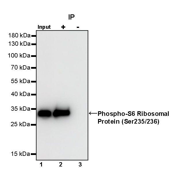
IP
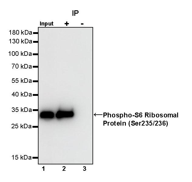
Phospho-S6 Ribosomal Protein (Ser235/236) Rabbit mAb at 1/50 dilution (1 µg) immunoprecipitating Phospho-S6 Ribosomal Protein (Ser235/236) in 0.4 mg HeLa serum starvation 16 hour then treated with calyculin (100 nM, 30 min) whole cell lysate.
Western blot was performed on the immunoprecipitate using Phospho-S6 Ribosomal Protein (Ser235/236) Rabbit mAb at 1/1000 dilution.
Secondary antibody (HRP) for IP was used at 1/1000 dilution.
Lane 1: HeLa serum starvation 16 hour then treated with calyculin (100 nM, 30 min) whole cell lysate 20 µg (Input)
Lane 2: Phospho-S6 Ribosomal Protein (Ser235/236) Rabbit mAb IP in HeLa serum starvation 16 hour then treated with calyculin (100 nM, 30 min) whole cell lysate
Lane 3: Rabbit monoclonal IgG IP in HeLa serum starvation 16 hour then treated with calyculin (100 nM, 30 min) whole cell lysate
Predicted MW: 29 kDa
Observed MW: 32 kDa
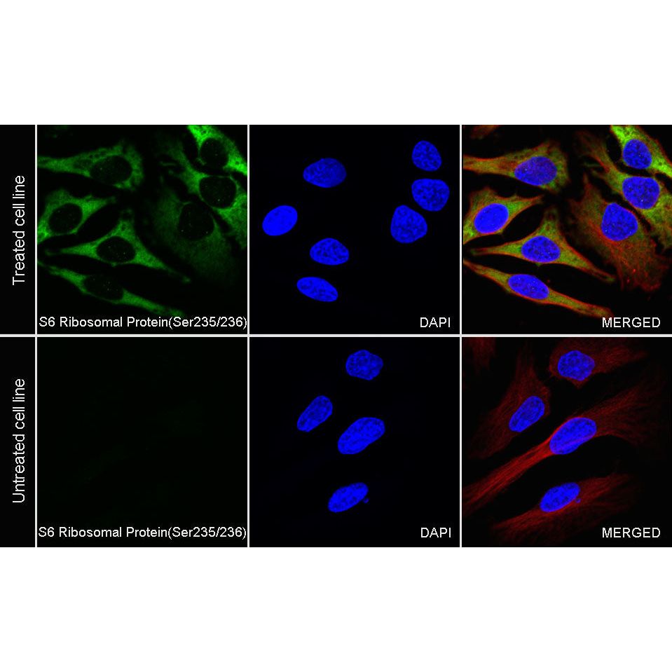
Immunocytochemistry
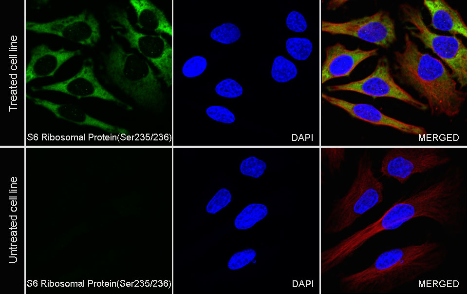
ICC analysis of HeLa cells treated with insulin (150nM,
6mins) (top panel) and HeLa cells treated with Rapamycin (10nM,2h) (below panel). Anti- Phospho-S6 Ribosomal Protein (Ser235/236) antibody was used at 1/500 dilution (Green) and incubated overnight at 4°C. Goat polyclonal Antibody to Rabbit IgG - H&L (Alexa Fluor® 488) was used as secondary antibody at 1/1000 dilution. The cells were fixed with 100% ice-cold methanol and permeabilized with 0.1% PBS-Triton X-100. Nuclei were counterstained with DAPI (Blue). Counterstain with tubulin (Red).