Skip to product information
1 of 1

PD-1 Recombinant Rabbit mAb (SDT-035-25)

PD-1 Recombinant Rabbit mAb (SDT-035-25)

Catalog Number: S0B2037 Application: IHC-P Reactivity: Human Conjugation: Unconjugated Brand: Starter
Price:
Regular price $100 USD
Regular price Sale price $100 USD
Size:
For shipping services or bulk orders, you may request a quotation.
Secure checkout with
View full details

Product Details

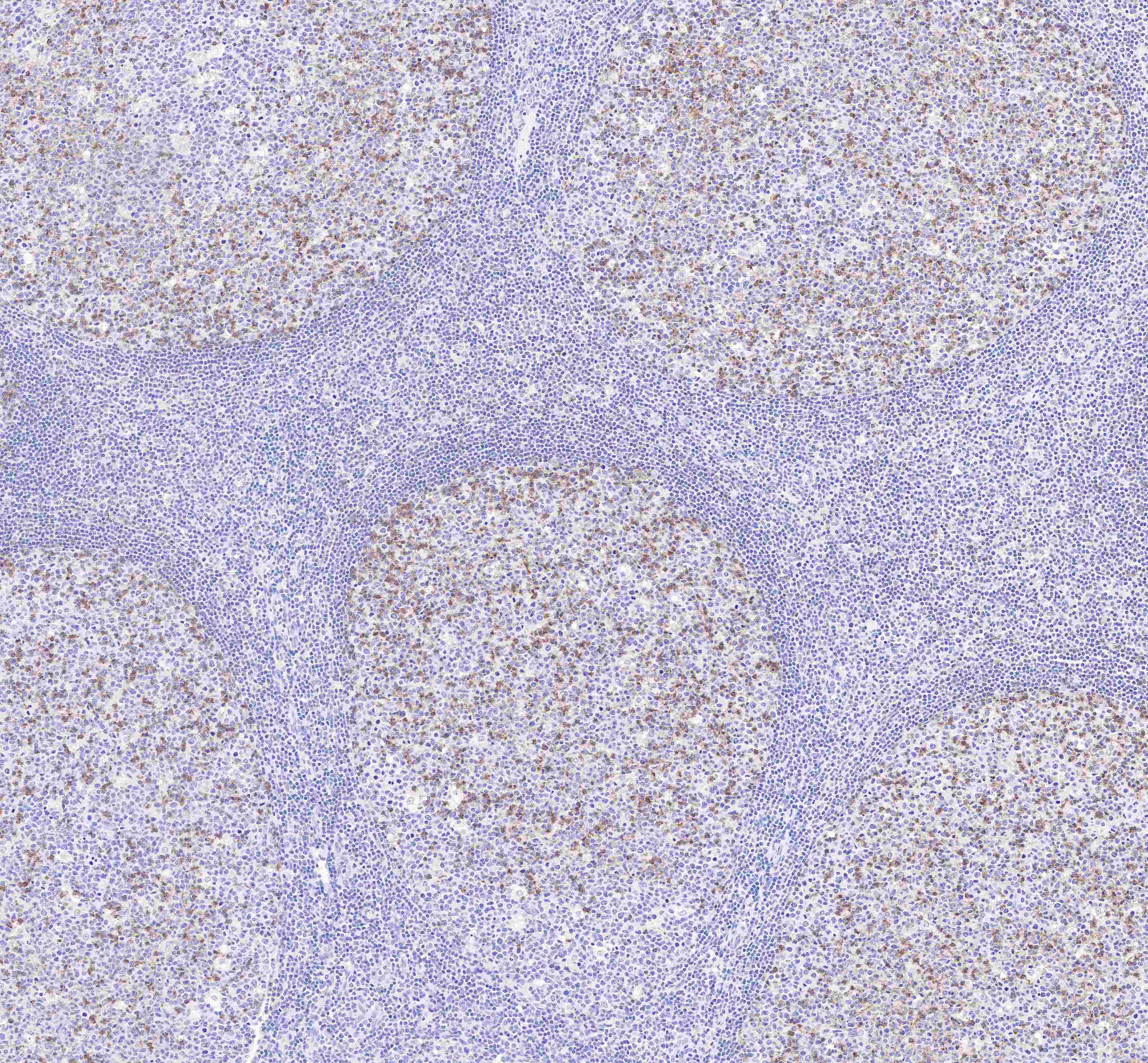
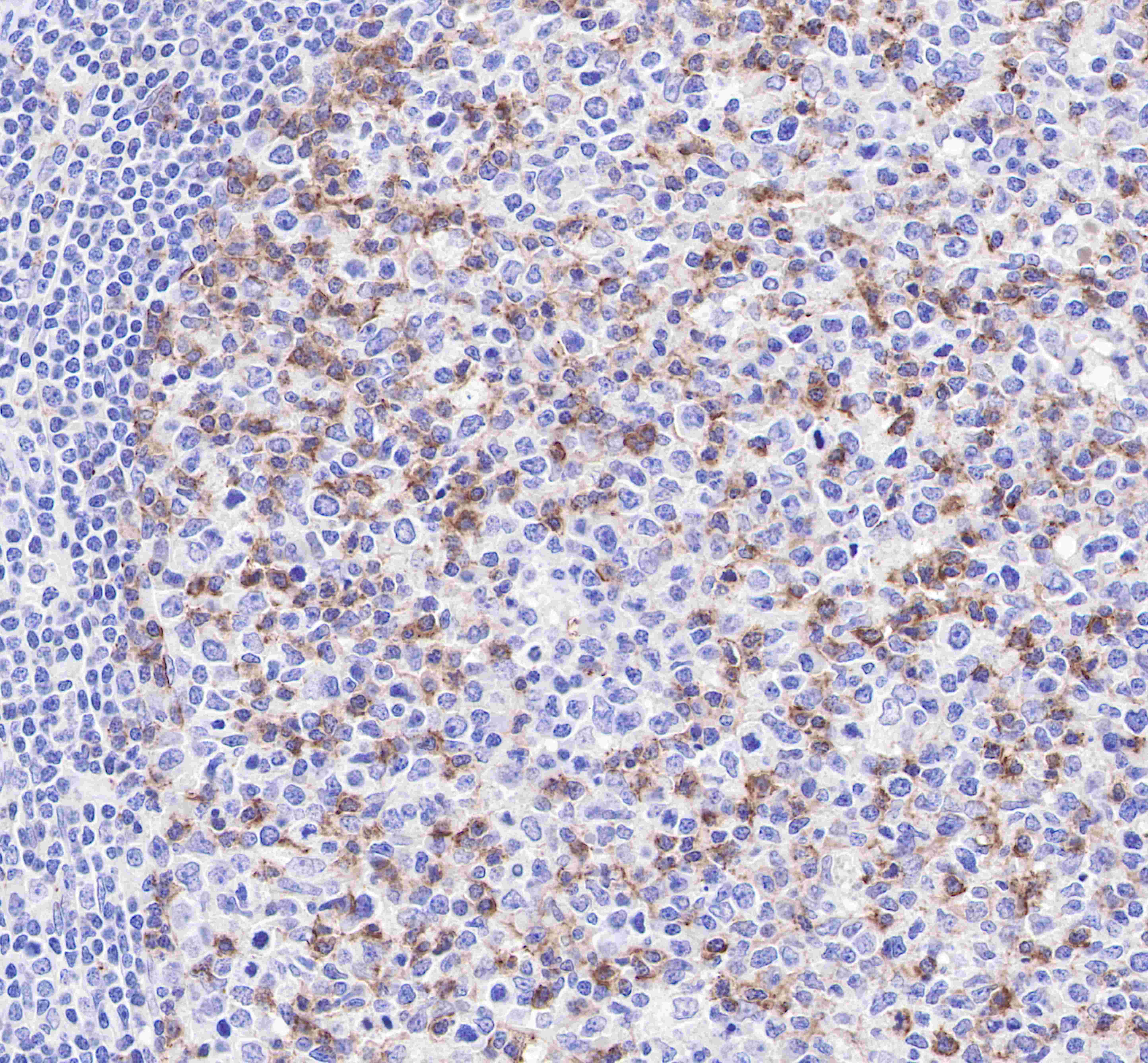
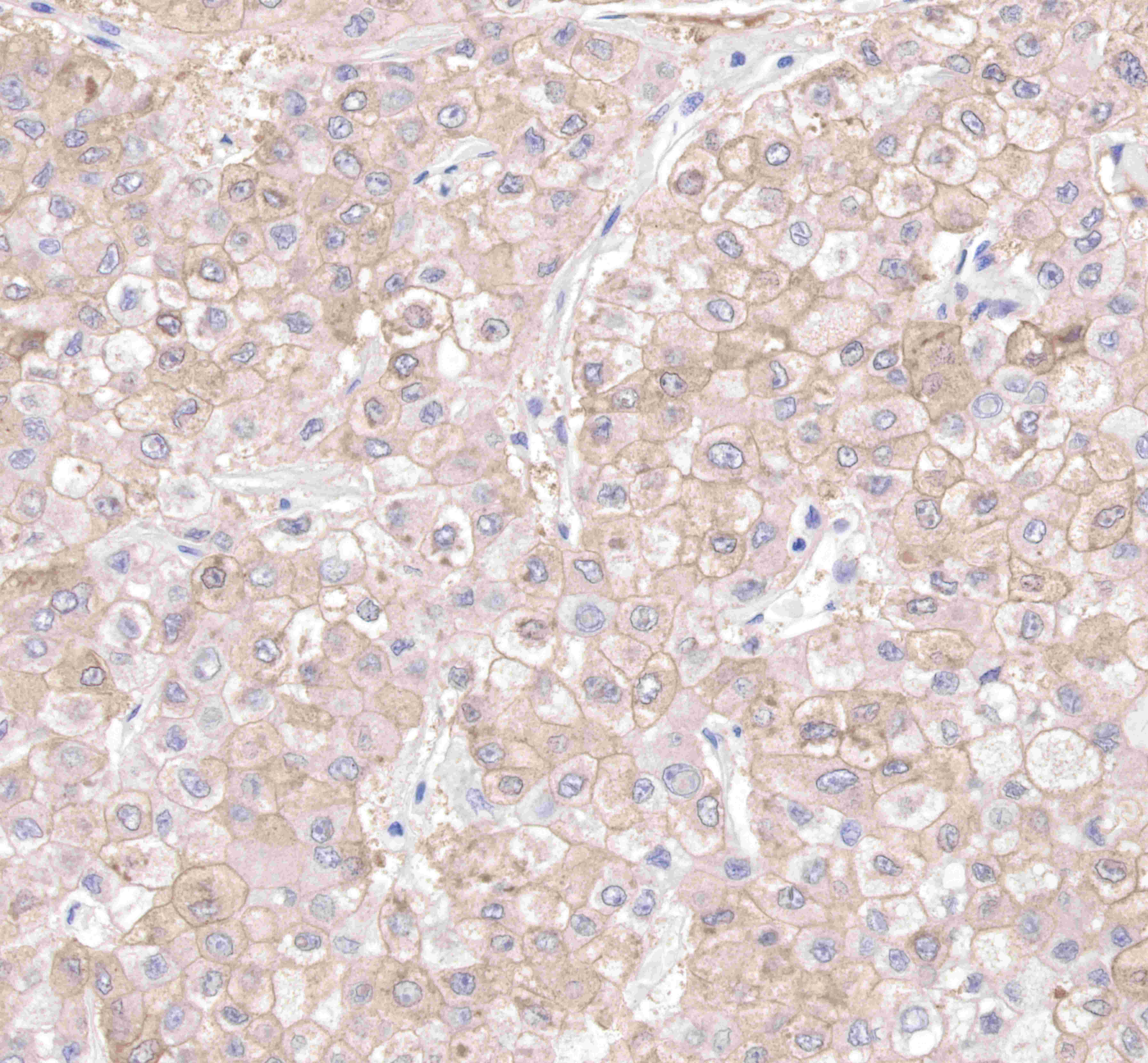
Product Specification


Host Rabbit
Antigen PD-1
Synonyms CD279, PDCD1
Immunogen Synthetic Peptide
Location Membrane
Accession Q15116
Clone Number SDT-035-25
Antibody Type Rabbit mAb
Application IHC-P
Reactivity Hu
Purification Protein A
Concentration 0.5mg/ml
Conjugation Unconjugated
Physical Appearance Liquid
Storage Buffer PBS, 40% Glycerol, 0.05%BSA, 0.03% Proclin 300
Stability & Storage 12 months from date of receipt / reconstitution, -20 °C as supplied

Dilution


application dilution species
IHC-P 1:500

Background

Programmed cell death protein 1, also known as PD-1 and CD279 (cluster of differentiation 279), is a protein on the surface of T and B cells that has a role in regulating the immune system's response to the cells of the human body by down-regulating the immune system and promoting self-tolerance by suppressing T cell inflammatory activity. This prevents autoimmune diseases, but it can also prevent the immune system from killing cancer cells. PD-1 is an immune checkpoint and guards against autoimmunity through two mechanisms. First, it promotes apoptosis (programmed cell death) of antigen-specific T-cells in lymph nodes. Second, it reduces apoptosis in regulatory T cells (anti-inflammatory, suppressive T cells).

Picture

Immunohistochemistry

IHC shows positive staining in paraffin-embedded human tonsil. Anti-PD1 antibody was used at 1/500 dilution, followed by a Goat Anti-Rabbit IgG H&L (HRP) ready to use.

Counterstained with hematoxylin.              

Heat mediated antigen retrieval with Tris/EDTA buffer pH9.0 was performed before commencing with IHC staining protocol.

IHC shows positive staining in paraffin-embedded human tonsil. Anti-PD1 antibody was used at 1/500 dilution, followed by a Goat Anti-Rabbit IgG H&L (HRP) ready to use.

Counterstained with hematoxylin.            

 Heat mediated antigen retrieval with Tris/EDTA buffer pH9.0 was performed before commencing with IHC staining protocol.

IHC shows positive staining in paraffin-embedded human Hepatocellular carcinoma.

Anti-PD1 antibody was used at 1/500 dilution, followed by a Goat Anti-Rabbit IgG H&L (HRP) ready to use.

Counterstained with hematoxylin.              

Heat mediated antigen retrieval with Tris/EDTA buffer pH9.0 was performed before commencing with IHC staining protocol.

Customer Reviews

Be the first to write a review
0%
(0)
0%
(0)
0%
(0)
0%
(0)
0%
(0)