WB result of PARP1 Rabbit mAb
Primary antibody: PARP1 Rabbit mAb at 1/1000 dilution
Lane 1: Untreated Jurkat whole cell lysate 20 µg
Lane 2: Jurkat treated with 1 µM staurosporine for 4 hours whole cell lysate 20 µg
Secondary antibody: Goat Anti-Rabbit IgG, (H+L), HRP conjugated at 1/10000 dilution
Predicted MW: 113 kDa
Observed MW: 116, 85, 71, 55, 42 kDa
PARP1 Recombinant Rabbit mAb (S-R299)
PARP1 Recombinant Rabbit mAb (S-R299)
Price:
Regular price
$100 USD
Regular price
Sale price
$100 USD
Unit price
per
For shipping services or bulk orders, you may request a quotation.
Secure checkout with

View full details
Product Details
Product Details
Product Specification
| Host | Rabbit |
| Synonyms | Poly [ADP-ribose] polymerase 1, ADP-ribosyltransferase diphtheria toxin-like 1 (ARTD1), DNA ADP-ribosyltransferase PARP1, NAD(+) ADP-ribosyltransferase 1 (ADPRT 1), Poly[ADP-ribose] synthase 1, Protein poly-ADP-ribosyltransferase PARP1, ADPRT, PPOL |
| Location | Cytoplasm, Nucleus |
| Accession | P09874 |
| Clone Number | S-R299 |
| Antibody Type | Recombinant mAb |
| Isotype | IgG |
| Application | WB, IHC-P, ICC, ICFCM, IP |
| Reactivity | Hu, Ms |
| Purification | Protein A |
| Concentration | 0.5 mg/ml |
| Conjugation | Unconjugated |
| Physical Appearance | Liquid |
| Storage Buffer | PBS, 40% Glycerol, 0.05% BSA, 0.03% Proclin 300 |
| Stability & Storage | 12 months from date of receipt / reconstitution, -20 °C as supplied. |
Dilution
| application | dilution | species |
| WB | 1:1000 | null |
| IHC-P | 1:500 | null |
| ICC | 1:500 | null |
| ICFCM | 1:50 | null |
| IP | 1:50 | null |
Background
Poly [ADP-ribose] polymerase 1 (PARP-1) also known as NAD+ ADP-ribosyltransferase 1 or poly [ADP-ribose] synthase 1 is an enzyme that in humans is encoded by the PARP1 gene. It is the most abundant of the PARP family of enzymes, accounting for 90% of the NAD+ used by the family. PARP1 is mostly present in cell nucleus, but cytosolic fraction of this protein was also reported. PARP1 acts as a first responder that detects DNA damage and then facilitates choice of repair pathway. PARP1 contributes to repair efficiency by ADP-ribosylation of histones leading to decompaction of chromatin structure, and by interacting with and modifying multiple DNA repair factors. PARP1 is implicated in the regulation of several DNA repair processes including the pathways of nucleotide excision repair, non-homologous end joining, microhomology-mediated end joining, homologous recombinational repair, and DNA mismatch repair.
Picture
Picture
Western Blot
FC
Flow cytometric analysis of 4% PFA fixed 90% methanol permeabilized HeLa (Human cervix adenocarcinoma epithelial cell) cells labelling PARP1 antibody at 1/50 dilution (1 μg)/ (Red) compared with a Rabbit monoclonal IgG (Black) isotype control and an unlabelled control (cells without incubation with primary antibody and secondary antibody) (Blue). Goat Anti - Rabbit IgG Alexa Fluor® 488 was used as the secondary antibody.
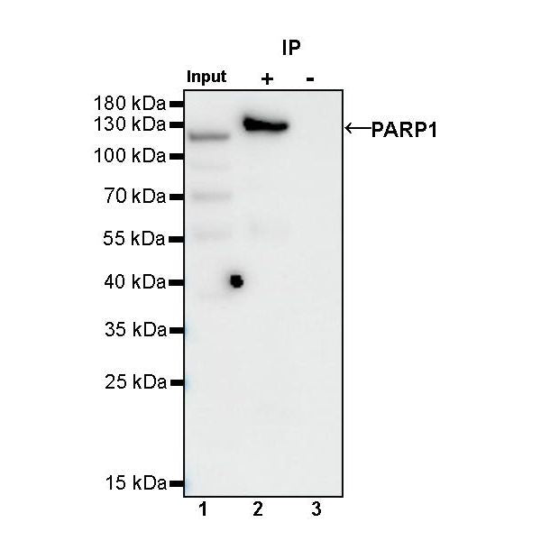
IP
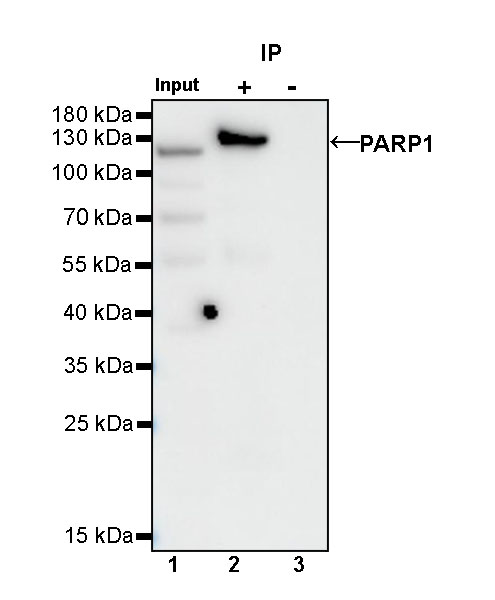
PARP1 Rabbit mAb at 1/50 dilution (1 µg) immunoprecipitating PARP1 in 0.4 mg Jurkat whole cell lysate.
Western blot was performed on the immunoprecipitate using PARP1 Rabbit mAb at 1/1000 dilution.
Secondary antibody (HRP) for IP was used at 1/1000 dilution.
Lane 1: Jurkat whole cell lysate 10 µg (Input)
Lane 2: PARP1 Rabbit mAb IP in Jurkat whole cell lysate
Lane 3: Rabbit monoclonal IgG IP in Jurkat whole cell lysate
Predicted MW: 113 kDa
Observed MW: 116, 85, 71, 55, 42 kDa
This blot was developed with high sensitivity substrate
Immunohistochemistry
IHC shows positive staining in paraffin-embedded human tonsil. Anti-PARP1 antibody was used at 1/500 dilution, followed by a HRP Polymer for Mouse & Rabbit IgG (ready to use). Counterstained with hematoxylin. Heat mediated antigen retrieval with Tris/EDTA buffer pH9.0 was performed before commencing with IHC staining protocol.
IHC shows positive staining in paraffin-embedded human testis. Anti-PARP1 antibody was used at 1/500 dilution, followed by a HRP Polymer for Mouse & Rabbit IgG (ready to use). Counterstained with hematoxylin. Heat mediated antigen retrieval with Tris/EDTA buffer pH9.0 was performed before commencing with IHC staining protocol.
IHC shows positive staining in paraffin-embedded human breast cancer. Anti-PARP1 antibody was used at 1/500 dilution, followed by a HRP Polymer for Mouse & Rabbit IgG (ready to use). Counterstained with hematoxylin. Heat mediated antigen retrieval with Tris/EDTA buffer pH9.0 was performed before commencing with IHC staining protocol.
IHC shows positive staining in paraffin-embedded human cervical squamous cell carcinoma. Anti-PARP1 antibody was used at 1/500 dilution, followed by a HRP Polymer for Mouse & Rabbit IgG (ready to use). Counterstained with hematoxylin. Heat mediated antigen retrieval with Tris/EDTA buffer pH9.0 was performed before commencing with IHC staining protocol.
IHC shows positive staining in paraffin-embedded mouse testis. Anti-PARP1 antibody was used at 1/500 dilution, followed by a HRP Polymer for Mouse & Rabbit IgG (ready to use). Counterstained with hematoxylin. Heat mediated antigen retrieval with Tris/EDTA buffer pH9.0 was performed before commencing with IHC staining protocol.
Immunocytochemistry
ICC shows positive staining in HeLa cells. Anti-PARP1 antibody was used at 1/500 dilution (Green) and incubated overnight at 4°C. Goat polyclonal Antibody to Rabbit IgG - H&L (Alexa Fluor® 488) was used as secondary antibody at 1/1000 dilution. The cells were fixed with 100% ice-cold methanol and permeabilized with 0.1% PBS-Triton X-100. Nuclei were counterstained with DAPI (Blue). Counterstain with tubulin (Red).