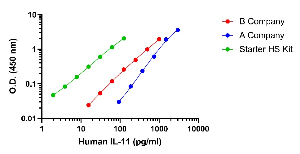
Leading Competitor comparison
IL-11 HS ELISA Kit
IL-11 HS ELISA Kit
Price:
Regular price
$855 USD
Regular price
Sale price
$855 USD
Unit price
per
For shipping services or bulk orders, you may request a quotation.
Secure checkout with

View full details
Product Details
Product Details
Product Specification
| Antigen | IL-11 |
| Immunogen | Recombinant Protein |
| Antibody Type | Recombinant mAb |
| Reactivity | Hu |
| Purification | Protein A |
| Stability & Storage |
2 to 8 °C as supplied.
|
Kit
| Range |
1.95 pg/ml - 125 pg/ml
|
| Sensitivity |
<0.7 pg/ml
|
| Assay type |
Sandwich (quantitative)
|
| Sample type |
Cell culture supernatant, Serum, Plasma
|
| Species reactivity |
human
|
| Precision |
Intra-assay: 2.8%; Inter-assay: 3.8% |
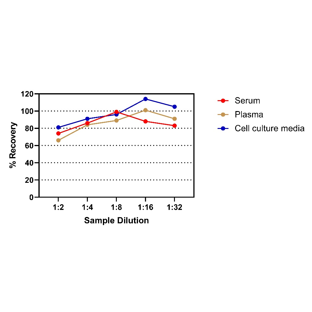
| Recovery |
Cell culture supernatant 91% (80-97%) Serum 102% (88-120%) Plasma 89% (86-91%) |
| Assay time | 3 hours |
Background
IL-11 was discovered in 1990 and first described as a hematopoietic factor that stimulates plasmacytoma proliferation and megakaryocyte formation. The dependence of IL-11 on the ubiquitously expressed receptor gp130 designates IL-11 as a member of the IL-6 family of cytokines. IL-6 family cytokines have been shown to bind to gp130 alone, although the accepted mode of signaling requires the interaction of cytokines with a specific, high-affinity receptor subunit (alpha chain) prior to binding to gp130. The gp130 receptor subsequently triggers a number of different pathways, including canonical JAK/STAT signaling, as well as ERK and AKT Loss-of-function mutations in IL-6 family cytokines or receptors result in a diverse set of genetic disorders of varying severity, which suggests nonredundant and distinct biological functions of members of this cytokine family. In the lung, IL-11 receptor subunit alpha (IL11RA) is highly expressed on cells such as fibroblasts, vascular smooth muscle cells, and epithelial cells. These cell types are central for both acute and chronic diseases of the lung/respiratory tract. This receptor expression pattern differentiates IL11RA from the IL-6 receptor, which is most highly expressed on immune cells, including monocytes and tissue-resident macrophages.
Picture
Picture
ELISA
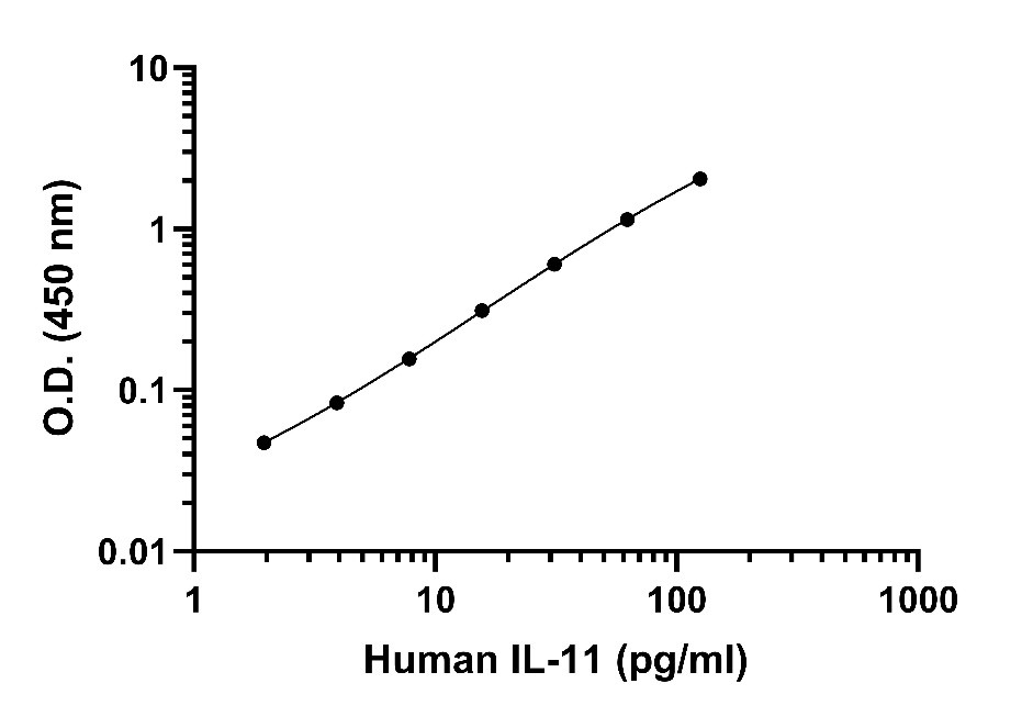
Standard curve
Example of human IL-11 standard curve in Assay Diluent #1.
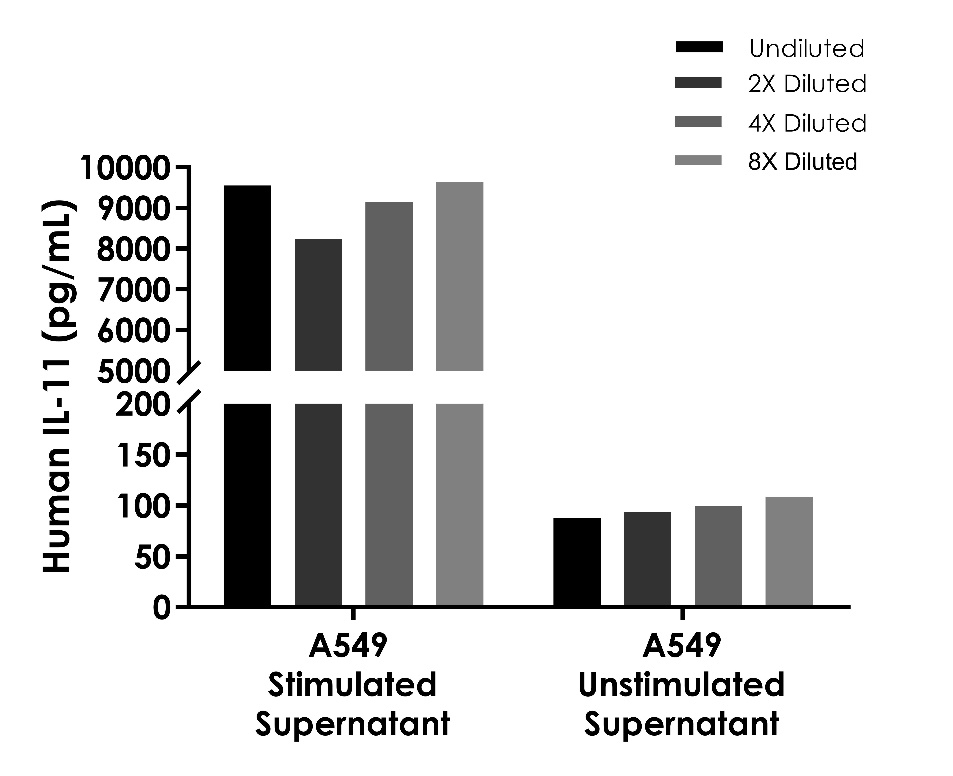
Linearity
Linearity
The concentrations of IL-11 were measured and interpolated from the target standard curves and corrected for sample dilution.
#1 sample is undiluted samples are as follows: human A549 cells stimulated with 2ng/ml TGF-β for 2days (0.16%). The interpolated dilution factor corrected values are plotted. The mean target concentration was determined to be 9,139 pg/mL in stimulated human A549 supernatant.
#2 sample is undiluted samples are as follows: human A549 cells culture for 2days (100%). The interpolated dilution factor corrected values are plotted. The mean target concentration was determined to be 97 pg/mL in unstimulated human A549 supernatant.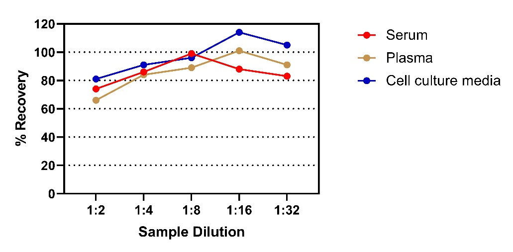
To assess the linearity of the assay, three samples were spiked with high concentrations of Human IL-11 in various matrices and diluted with the appropriate Calibrator Diluent to produce samples with values within the dynamic range of the assay.