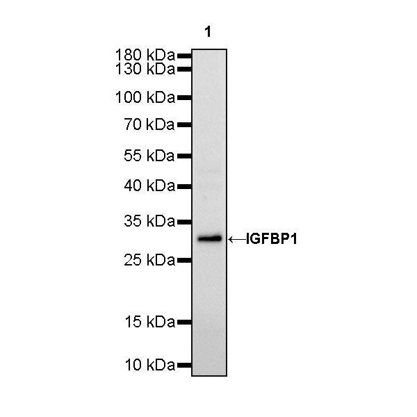
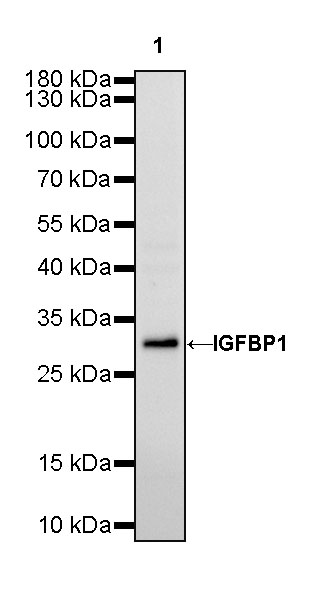
WB result of IGFBP1 Rabbit mAb
Primary antibody: IGFBP1 Rabbit mAb at 1/1000 dilution
Lane 1: HepG2 whole cell lysate 20 µg
Secondary antibody: Goat Anti-Rabbit IgG, (H+L), HRP conjugated at 1/10000 dilution
Predicted MW: 28 kDa
Observed MW: 30 kDa
IGFBP1 Recombinant Rabbit mAb (SDT-196-47)
IGFBP1 Recombinant Rabbit mAb (SDT-196-47)
Price:
Regular price
$100 USD
Regular price
Sale price
$100 USD
Unit price
per
For shipping services or bulk orders, you may request a quotation.
Secure checkout with

View full details
Product Details
Product Details
Product Specification
| Host | Rabbit |
| Antigen | IGFBP1 |
| Synonyms | Insulin-like growth factor-binding protein 1, IBP-1; IGF-binding protein 1, Placental protein 12, PP12 |
| Immunogen | Recombinant Protein |
| Location | Secreted |
| Accession | P08833 |
| Clone Number | SDT-196-47 |
| Antibody Type | Recombinant mAb |
| Application | WB, ICC, ICFCM |
| Reactivity | Hu |
| Purification | Protein A |
| Concentration | 0.5 mg/ml |
| Conjugation | Unconjugated |
| Physical Appearance | Liquid |
| Storage Buffer | PBS, 40% Glycerol, 0.05% BSA, 0.03% Proclin 300 |
| Stability & Storage | 12 months from date of receipt / reconstitution, -20 °C as supplied |
Dilution
| application | dilution | species |
| WB | 1:1000 | null |
| ICC | 1:250 | null |
Background
Insulin-like growth factor binding proteins (IGFBPs) are a family of proteins binding to Insulin-like growth factors (IGFs), generally including IGFBP1, IGFBP2, IGFBP3, IGFBP4, IGFBP5, and IGFBP6 [PMID: 30214426]. IGFBP1, a 40 to 50 kDa protein, which plays an indispensable role in normal growth and development, and in the pathophysiology of various tumors. IGFBP-1 has been shown to be associated with the risk of various tumors, and has a vital function in regulating tumor behaviors such as proliferation, migration, invasion and adhesion through different molecular mechanisms. The biological actions of IGFBP-1 in cancer are found to be related to its phosphorylation state, and the IGF-dependent and -independent mechanisms. [PMID: 1648311, PMID: 33841624].
Picture
Picture
Western Blot
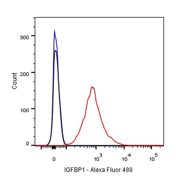
FC
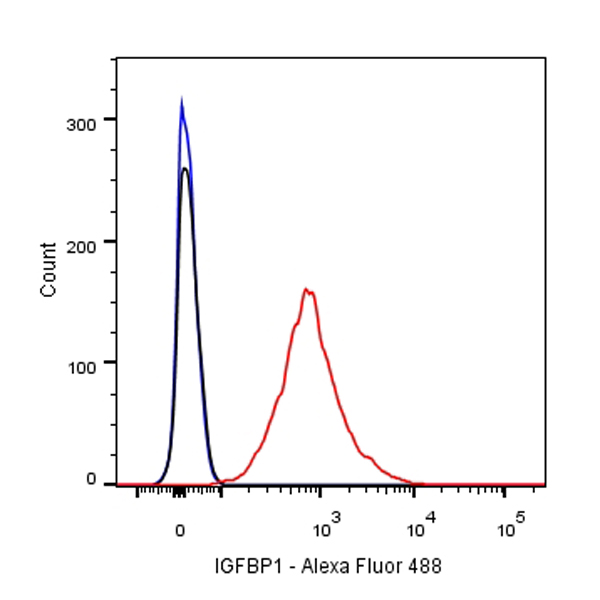
Flow cytometric analysis of HepG2 cells labelling IGFBP1 antibody at 1/500 (0.1 μg) dilution/ (red) compared with a Rabbit monoclonal IgG (Black) isotype control and an unlabelled control (cells without incubation with primary antibody and secondary antibody) (Blue). Goat Anti-Rabbit IgG Alexa Fluor® 488 was used as the secondary antibody.
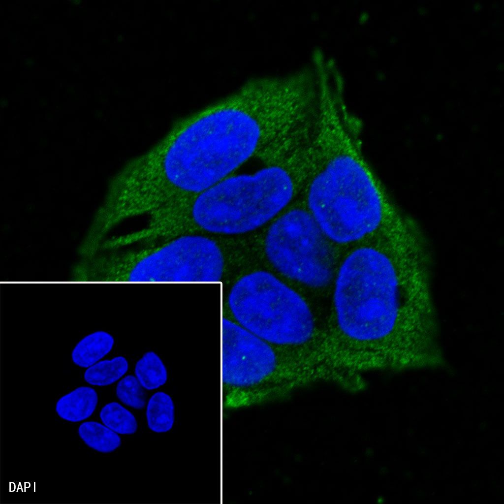
Immunocytochemistry
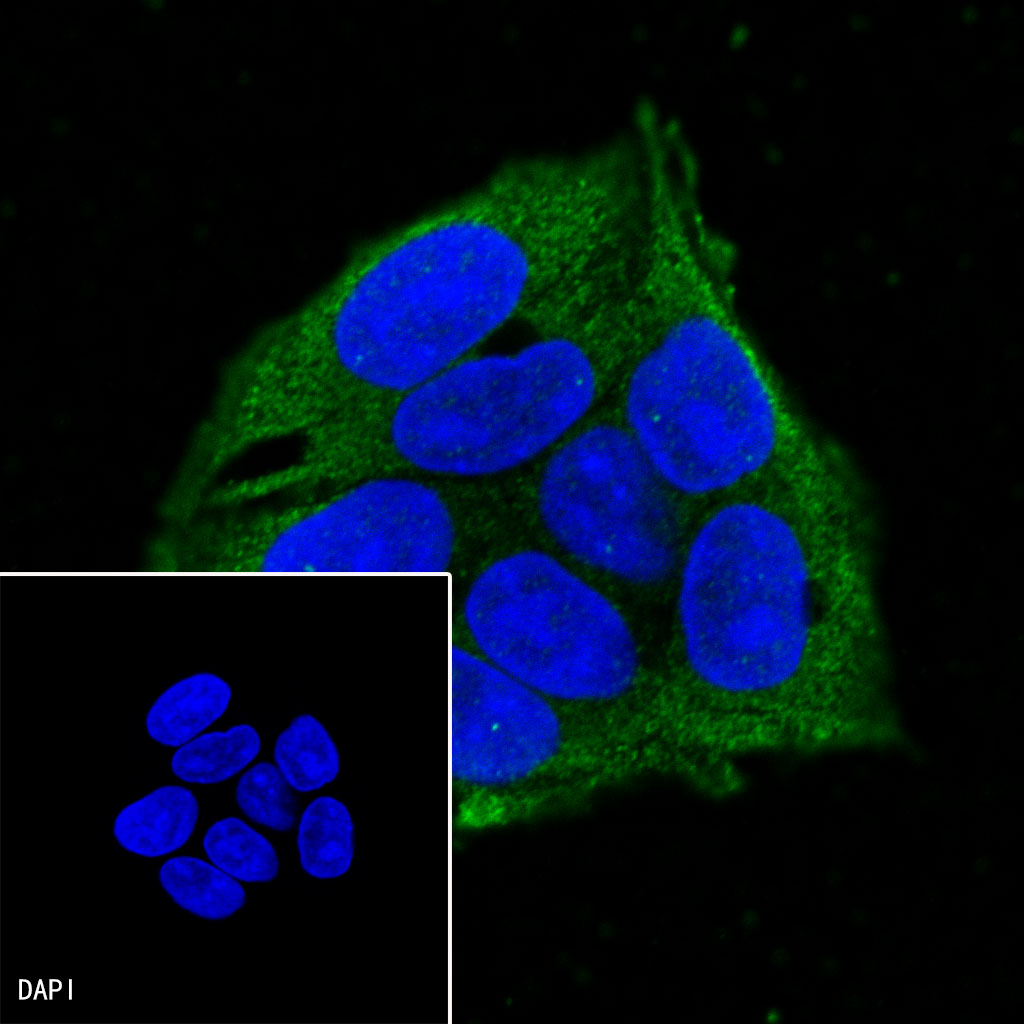
ICC shows positive staining in HepG2 cells. Anti-IGFBP1 antibody was used at 1/250 dilution (Green) and incubated overnight at 4°C. Goat polyclonal Antibody to Mouse IgG - H&L (Alexa Fluor® 488) was used as secondary antibody at 1/1000 dilution. The cells were fixed with 100% ice-cold methanol and permeabilized with 0.1% PBS-Triton X-100. Nuclei were counterstained with DAPI (Blue).