Product Specification
| Host |
Rabbit |
| Antigen |
Glypican-3 |
| Synonyms |
GTR2-2, MXR7,GPC3,OCI5 |
| Immunogen |
N/A |
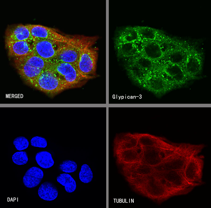
| Location |
Cytoplasm, Cell membrane |
| Accession |
P51654 |
| Clone Number |
SDT-R032 |
| Antibody Type |
Rabbit mAb |
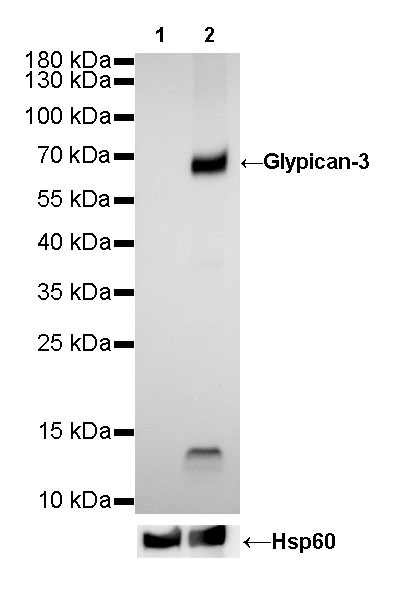
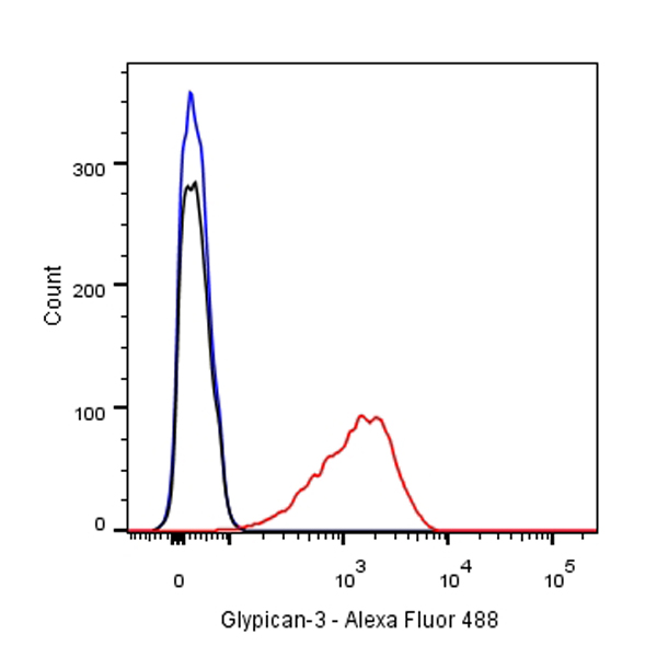
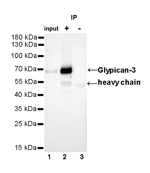
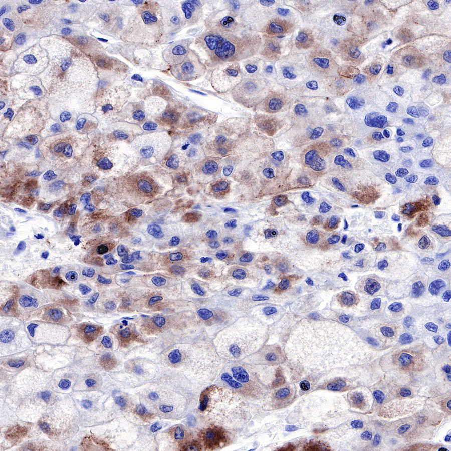
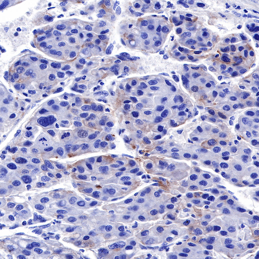
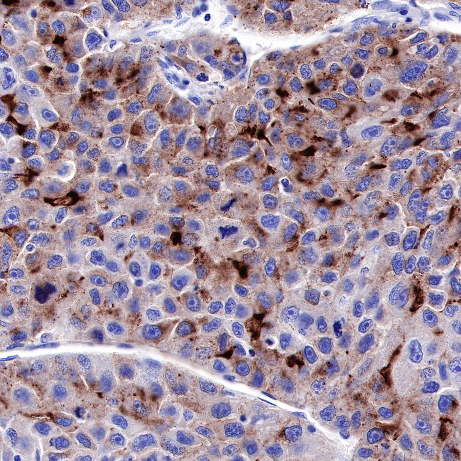
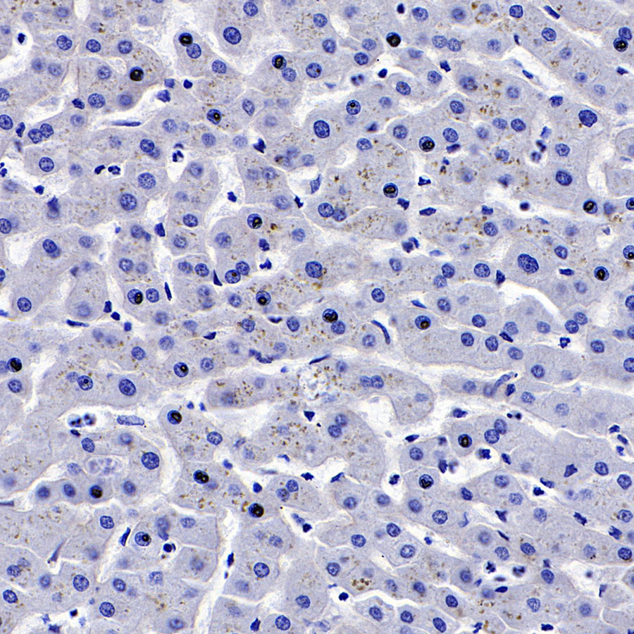
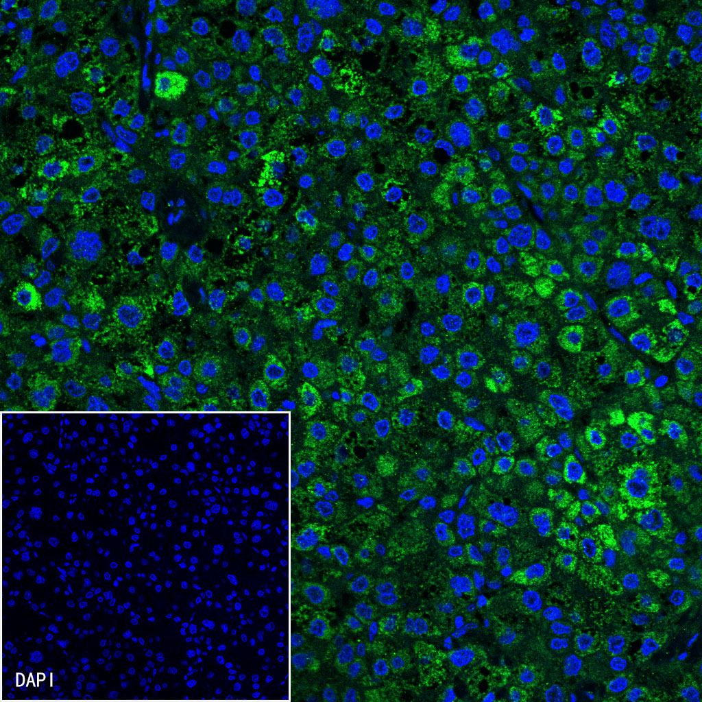
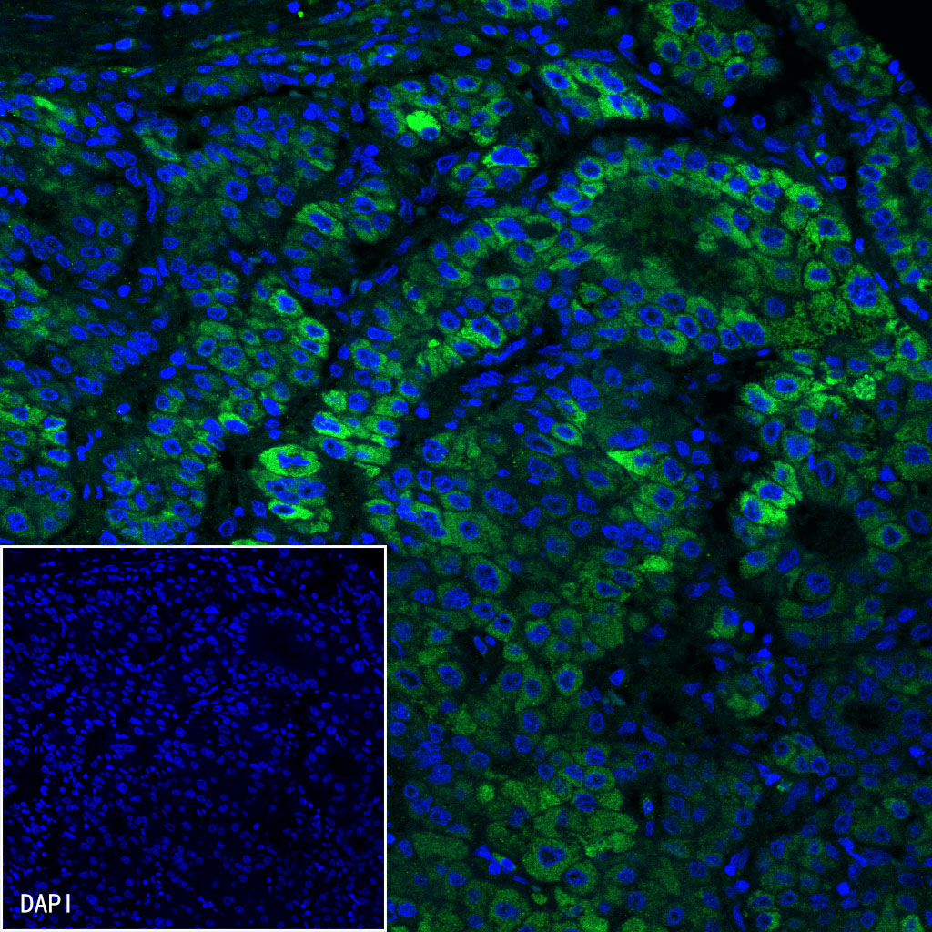
| Application |
WB, IHC-P, ICC, FCM, IP, IF |
| Reactivity |
Hu |
| Purification |
Protein A |
| Concentration |
0.5 mg/ml |
| Physical Appearance |
Liquid |
| Storage Buffer |
PBS, 40% Glycerol, 0.05%BSA, 0.03% Proclin 300 |
| Stability & Storage |
12 months from date of receipt / reconstitution, -20 °C as supplied |
Dilution
| application |
dilution |
species |
| FCM |
1:500 |
null |
| IP |
1:25 |
null |
| IHC-P |
1:500 |
null |
| WB |
1:1000 |
null |
| ICC |
1:500 |
null |
| IF |
1:200 |
null |
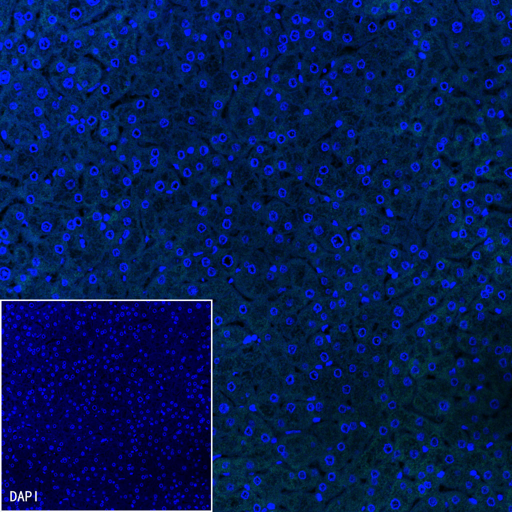
Background
Glypican-3 (GPC3) is a cell-surface glyco phosphatidylinositol (GPI)-anchored protein that belongs to the heparan sulfate (HS) proteoglycan family, which plays important roles in cell growth, differentiation and migration. Many studies have shown that GPC3 is highly expressed in HCC, while its expression is absent in most nonmalignant adult tissues. GPC3 is currently used as an informative immunohistochemical biomarker for HCC, and it is believed to be an attractive target for HCC therapy.