WB result of FGF-19 Recombinant Rabbit mAb
Primary antibody: FGF-19 Recombinant Rabbit mAb at 1/1000 dilution
Lane 1: 293T whole cell lysate 20 µg
Lane 2: HT-29 whole cell lysate 20 µg
Lane 3: SW480 whole cell lysate 20 µg
Lane 4: COLO 205 whole cell lysate 20 µg
Negative control: 293T whole cell lysate
Secondary antibody: Goat Anti-rabbit IgG, (H+L), HRP conjugated at 1/10000 dilution
Predicted MW: 24 kDa
Observed MW: 24 kDa
FGF-19 Recombinant Rabbit mAb (SDT-1070-17)
FGF-19 Recombinant Rabbit mAb (SDT-1070-17)
Price:
Regular price
$100 USD
Regular price
Sale price
$100 USD
Unit price
per
For shipping services or bulk orders, you may request a quotation.
Secure checkout with

View full details
Product Details
Product Details
Product Specification
| Host | Rabbit |
| Antigen | FGF-19 |
| Synonyms | Fibroblast growth factor 19; FGF19 |
| Immunogen | Recombinant Protein |
| Location | Secreted |
| Accession | O95750 |
| Clone Number | SDT-1070-17 |
| Antibody Type | Recombinant mAb |
| Isotype | IgG |
| Application | WB, IHC-P, IP |
| Reactivity | Hu |
| Purification | Protein A |
| Concentration | 0.5 mg/ml |
| Conjugation | Unconjugated |
| Physical Appearance | Liquid |
| Storage Buffer | PBS, 40% Glycerol, 0.05% BSA, 0.03% Proclin 300 |
| Stability & Storage | 12 months from date of receipt / reconstitution, -20 °C as supplied |
Dilution
| application | dilution | species |
| WB | 1:1000 | |
| IHC | 1:100 | |
| IP | 1:50 |
Background
FGF-19 is a member of the fibroblast growth factor (FGF) family. FGF family members possess broad mitogenic and cell survival activities, and are involved in a variety of biological processes including embryonic development cell growth, morphogenesis, tissue repair, tumor growth and invasion. This growth factor is a high affinity, heparin dependent ligand for FGFR4. FGF19 has important roles as a hormone produced in the ileum in response to bile acid absorption. Bile acids bind to the farnesoid X receptor (FXR), stimulating FGF19 transcription. FGF19 regulates new bile acid synthesis, acting through the FGFR4/Klotho-β receptor complexes in the liver to inhibit CYP7A1. FGF19 also has metabolic effects, affecting glucose and lipid metabolism when used in experimental mouse models. Furthermore, FGF19 is frequently amplified in human cancers. Amplification of the FGF19 genomic locus was found in liver cancer, breast cancer, lung cancer, prostate cancer, bladder cancer, and esophageal cancer, among others. Targeting FGF19 inhibits tumor growth in colon cancer cells and hepatocellar carcinoma. Increase in FGF19 correlates with tumor progression and poorer prognosis of hepatocellular carcinoma.
Picture
Picture
Western Blot
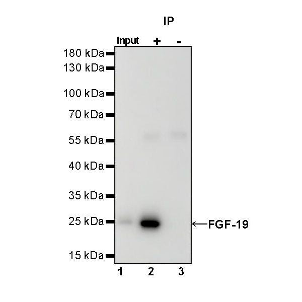
IP
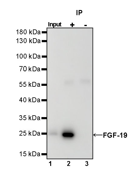
FGF-19 Rabbit mAb at 1/50 dilution (1 µg) immunoprecipitating FGF-19 in 0.4 mg SW480 whole cell lysate.
Western blot was performed on the immunoprecipitate using FGF-19 Rabbit mAb at 1/1000 dilution.
Secondary antibody (HRP) for IP was used at 1/1000 dilution.
Lane 1: SW480 whole cell lysate 20 µg (Input)
Lane 2: FGF-19 Rabbit mAb IP in SW480 whole cell lysate
Lane 3: Rabbit monoclonal IgG IP in SW480 whole cell lysate
Predicted MW: 24 kDa
Observed MW: 24 kDa
Immunohistochemistry
IHC shows positive staining in paraffin-embedded human gall bladder. Anti- FGF-19 antibody was used at 1/100 dilution, followed by a HRP Polymer for Mouse & Rabbit IgG (ready to use). Counterstained with hematoxylin. Heat mediated antigen retrieval with Tris/EDTA buffer pH9.0 was performed before commencing with IHC staining protocol.
IHC shows positive staining in paraffin-embedded human hepatocellular carcinoma. Anti- FGF-19 antibody was used at 1/100 dilution, followed by a HRP Polymer for Mouse & Rabbit IgG (ready to use). Counterstained with hematoxylin. Heat mediated antigen retrieval with Tris/EDTA buffer pH9.0 was performed before commencing with IHC staining protocol.
Negative control: IHC shows negative staining in paraffin-embedded human liver. Anti- FGF-19 antibody was used at 1/100 dilution, followed by a HRP Polymer for Mouse & Rabbit IgG (ready to use). Counterstained with hematoxylin. Heat mediated antigen retrieval with Tris/EDTA buffer pH9.0 was performed before commencing with IHC staining protocol.