WB result of EGFR Rabbit pAb
Primary antibody: EGFR Rabbit pAb at 1/1000 dilution
Lane 1: A431 whole cell lysate 20 µg
Secondary antibody: Goat Anti-rabbit IgG, (H+L), HRP conjugated at 1/10000 dilution
Predicted MW: 134 kDa
Observed MW: 180 kDa
EGFR Rabbit Polyclonal Antibody
EGFR Rabbit Polyclonal Antibody
Price:
Regular price
$100 USD
Regular price
Sale price
$100 USD
Unit price
per
For shipping services or bulk orders, you may request a quotation.
Secure checkout with

View full details
Product Details
Product Details
Product Specification
| Host | Rabbit |
| Antigen | EGFR |
| Synonyms | Epidermal growth factor receptor; Proto-oncogene c-ErbB-1; Receptor tyrosine-protein kinase erbB-1; ERBB; ERBB1; HER1; EGF Receptor |
| Immunogen | Synthetic Peptide |
| Location | Nucleus, Secreted, Endosome, Cell membrane, Endoplasmic reticulum |
| Accession | P00533 |
| Antibody Type | Polyclonal antibody |
| Isotype | IgG |
| Application | WB, ICC |
| Reactivity | Hu |
| Positive Sample | A431 |
| Predicted Reactivity | Mq |
| Purification | Immunogen Affinity |
| Concentration | 2 mg/ml |
| Conjugation | Unconjugated |
| Physical Appearance | Liquid |
| Storage Buffer | PBS, 40% Glycerol, 0.05% BSA, 0.03% Proclin 300 |
| Stability & Storage | 12 months from date of receipt / reconstitution, -20 °C as supplied |
Dilution
| application | dilution | species |
| WB | 1:1000 | Hu |
| ICC | 1:100 |
Background
The EGFR (Epidermal Growth Factor Receptor) is a transmembrane glycoprotein located on the cell membrane and belongs to the ErbB receptor tyrosine kinase family (HER1/ErbB1). Its structure consists of three main domains: an extracellular region, a transmembrane region, and an intracellular region. The extracellular region contains four domains (I-IV), with domains II and IV being cysteine-rich and responsible for binding ligands (such as EGF and TGF-α), leading to receptor dimerization. The transmembrane region is a single α-helix that anchors the receptor in the cell membrane. The intracellular region possesses tyrosine kinase activity and includes a juxtamembrane domain, a kinase domain (with an ATP-binding site), and a C-terminal regulatory tail containing multiple autophosphorylation sites (e.g., Y992, Y1045, Y1068). EGFR activates downstream signaling pathways such as RAS/RAF/MEK/ERK and PI3K/AKT, regulating cell proliferation, differentiation, migration, and survival. Mutations or overexpression of EGFR are strongly associated with various cancers, including lung and colorectal cancers, making it a key target for drugs like gefitinib and osimertinib.
Picture
Picture
Western Blot
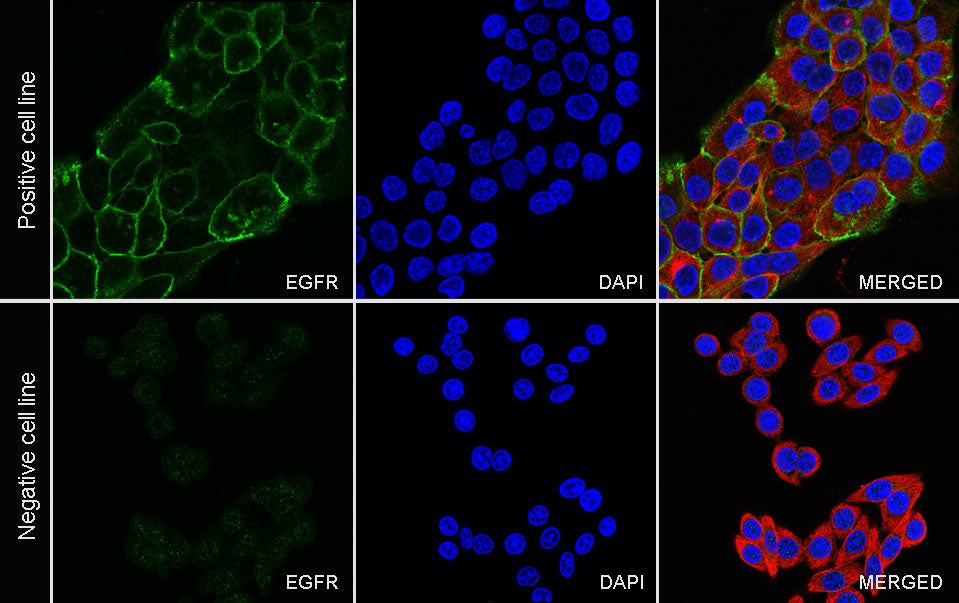
Immunocytochemistry

ICC shows positive staining in A431 cells (top panel) and negative staining in SW480 cells (below panel). Anti-EGFR antibody was used at 1/100 dilution (Green) and incubated overnight at 4°C. Goat polyclonal Antibody to Rabbit IgG - H&L (Alexa Fluor® 488) was used as secondary antibod at 1/1000 dilution. The cells were fixed with 100% ice-cold methanol and permeabilized with 0.1% PBS-Triton X-100. Nuclei were counterstained with DAPI (Blue). Counterstain with tubulin (Red).