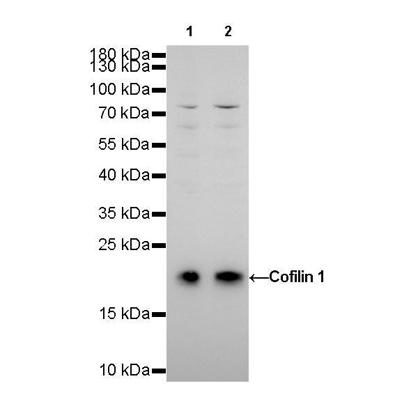
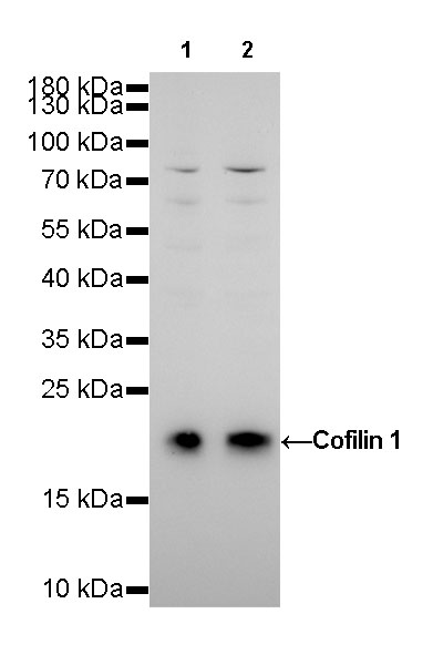
WB result of Cofilin 1 Rabbit mAb
Primary antibody: Cofilin 1 Rabbit mAb at 1/1000 dilution
Lane 1: HeLa whole cell lysate 20 µg
Lane 2: MCF7 whole cell lysate 20 µg
Secondary antibody: Goat Anti-Rabbit IgG, (H+L), HRP conjugated at 1/10000 dilution
Predicted MW: 19 kDa
Observed MW: 19 kDa
Exposure time: 180s
Cofilin 1 Recombinant Rabbit mAb (SDT-R090)
Cofilin 1 Recombinant Rabbit mAb (SDT-R090)
Price:
Regular price
$100 USD
Regular price
Sale price
$100 USD
Unit price
per
For shipping services or bulk orders, you may request a quotation.
Secure checkout with

View full details
Product Details
Product Details
Product Specification
| Host | Rabbit |
| Antigen | Cofilin 1 |
| Synonyms | p18, Cofilin, non-muscle isoform |
| Immunogen | N/A |
| Location | Cytoplasm, Nucleus, Membrane |
| Accession | P23528 |
| Clone Number | SDT-R090 |
| Antibody Type | Rabbit mAb |
| Application | WB, ICC, ICFCM |
| Reactivity | Hu, Ms, Rt |
| Purification | Protein A |
| Concentration | 0.5 mg/ml |
| Physical Appearance | Liquid |
| Storage Buffer | PBS, 40% Glycerol, 0.05%BSA, 0.03% Proclin 300 |
| Stability & Storage | 12 months from date of receipt / reconstitution, -20 °C as supplied. |
Dilution
| application | dilution | species |
| ICFCM | 1:500 | null |
| ICC | 1:1000 | null |
| WB | 1:1000-1:20000 | null |
Background
Cofilin 1 (non-muscle; n-cofilin), also known as CFL1, is a human gene, part of the ADF/cofilin family. Cofilin is a widely distributed intracellular actin-modulating protein that binds and depolymerizes filamentous F-actin and inhibits the polymerization of monomeric G-actin in a pH-dependent manner. It is involved in the translocation of actin-cofilin complex from cytoplasm to nucleus.
Picture
Picture
Western Blot
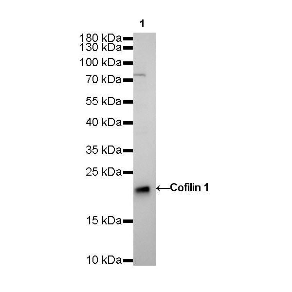
WB result of Cofilin 1 Rabbit mAb
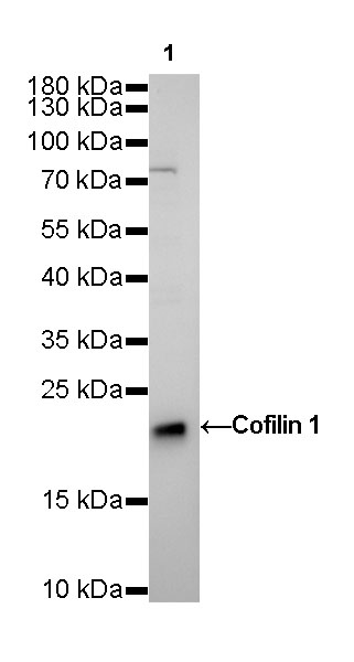
Primary antibody: Cofilin 1 Rabbit mAb at 1/5000 dilution
Lane 1: NIH/3T3 whole cell lysate 20 µg
Secondary antibody: Goat Anti-Rabbit IgG, (H+L), HRP conjugated at 1/10000 dilution
Predicted MW: 19 kDa
Observed MW: 19 kDa
Exposure time: 60s
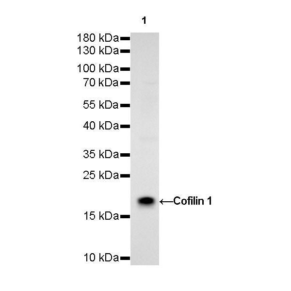
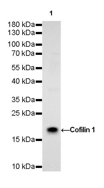
WB result of Cofilin 1 Rabbit mAb
Primary antibody: Cofilin 1 Rabbit mAb at 1/1000 dilution
Lane 1: PC-12 whole cell lysate 20 µg
Secondary antibody: Goat Anti-Rabbit IgG, (H+L), HRP conjugated at 1/10000 dilution
Predicted MW: 19 kDa
Observed MW: 19 kDa
Exposure time: 180s
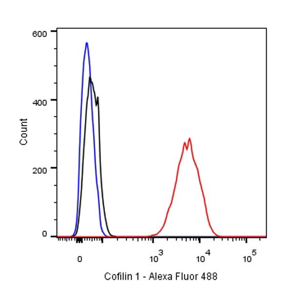
FC
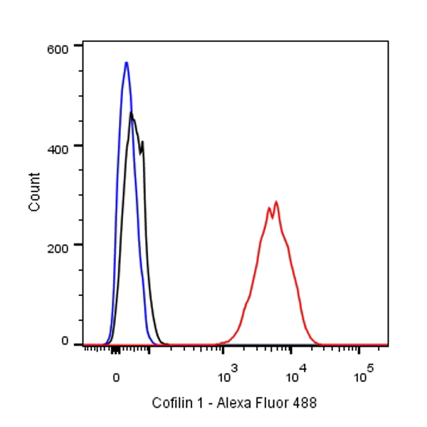
Flow cytometric analysis of HeLa cells labelling Cofilin 1 antibody at 1/500(0.1 μg) dilution/ (red) compared with a Rabbit monoclonal IgG (Black) isotype control and an unlabelled control (cells without incubation with primary antibody and secondary antibody) (Blue). Goat Anti-Rabbit IgG Alexa Fluor® 488 was used as the secondary antibody.
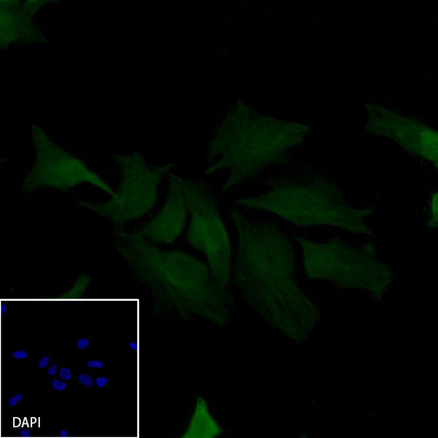
Immunocytochemistry
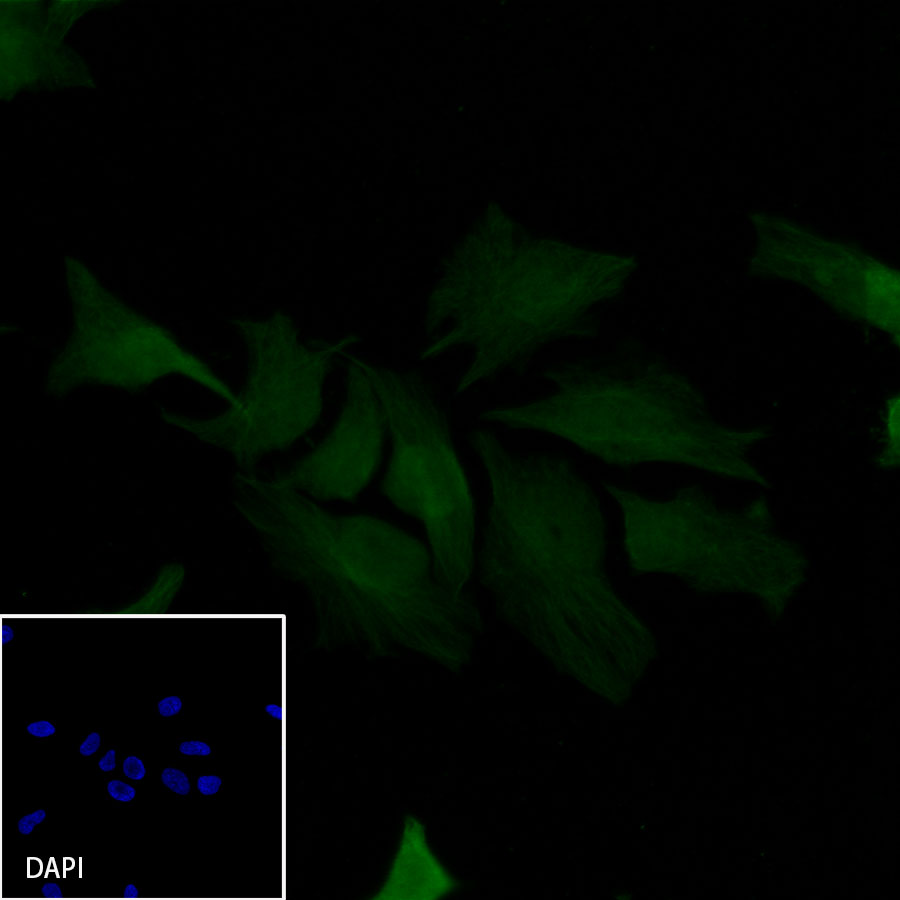
ICC shows positive staining in HeLa cells. Anti-Cofilin 1 antibody was used at 1/1000 dilution and incubated overnight at 4°C. Goat polyclonal Antibody to Rabbit IgG - H&L (Alexa Fluor® 488) was used as secondary antibody at 1/1000 dilution.The cells were fixed with 4% PFA and permeabilized with 0.1% PBS-Triton X-100. Nuclei were counterstained with DAPI.