CD42b Recombinant Rabbit mAb (SDT-R147)
CD42b Recombinant Rabbit mAb (SDT-R147)
Product Details
Product Details
Product Specification
| Host | Rabbit |
| Antigen | CD42b |
| Synonyms | GPIb, Platelet glycoprotein Ib alpha chain, GPIb-alpha, GPIbA, Glycoprotein Ibalpha, Antigen CD42b-alpha |
| Immunogen | N/A |
| Location | Membrane |
| Accession | P07359 |
| Clone Number | SDT-R147 |
| Antibody Type | Recombinant mAb |
| Application | IHC-P, IF |
| Reactivity | Hu, Ms, Rt |
| Purification | Protein A |
| Concentration | 0.25 mg/ml |
| Physical Appearance | Liquid |
| Storage Buffer | PBS, 40% Glycerol, 0.05% BSA, 0.03% Proclin 300 |
| Stability & Storage | 12 months from date of receipt / reconstitution, -20 °C as supplied |
Dilution
| application | dilution | species |
| IHC-P | 1:1000 | null |
| IF | 1:250 | null |
Background
Platelet glycoprotein Ib alpha chain also known as glycoprotein Ib (platelet), alpha polypeptide or CD42b (Cluster of Differentiation 42b), is a protein that in humans is encoded by the GP1BA gene. Glycoprotein Ib (GPIb) is a platelet surface membrane glycoprotein composed of a heterodimer, an alpha chain and a beta chain, that are linked by disulfide bonds. The GpIb functions as a receptor for von Willebrand factor (VWF). The complete receptor complex includes noncovalent association of the alpha and beta subunits with platelet glycoprotein IX and platelet glycoprotein V to form the glycoprotein Ib-IX-V complex. The binding of the GP Ib-IX-V complex to VWF facilitates initial platelet adhesion to vascular subendothelium after vascular injury, and also initiates signaling events within the platelet that lead to enhanced platelet activation, thrombosis, and hemostasis.
Picture
Picture
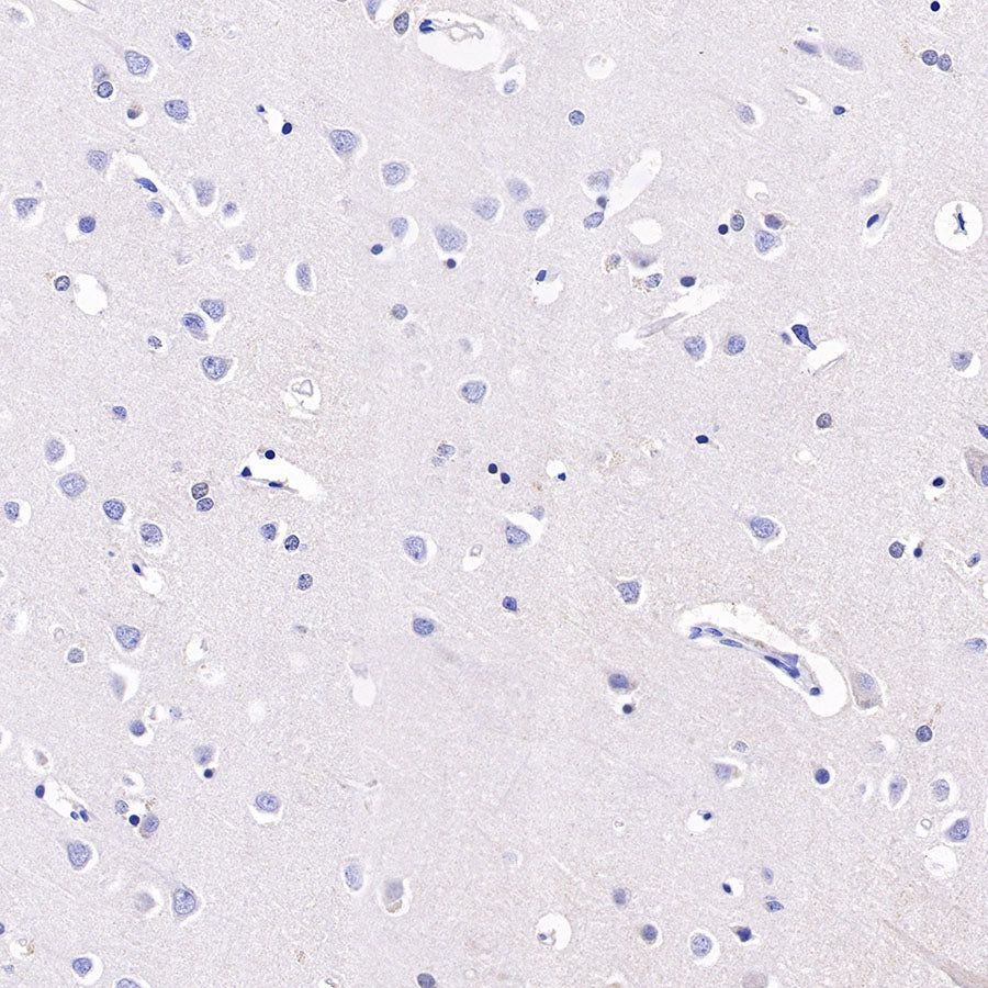
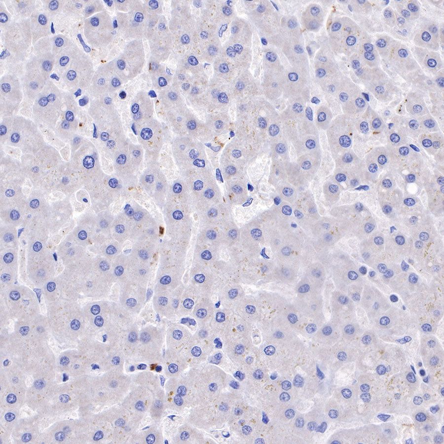
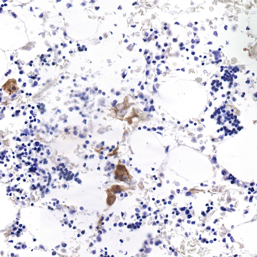
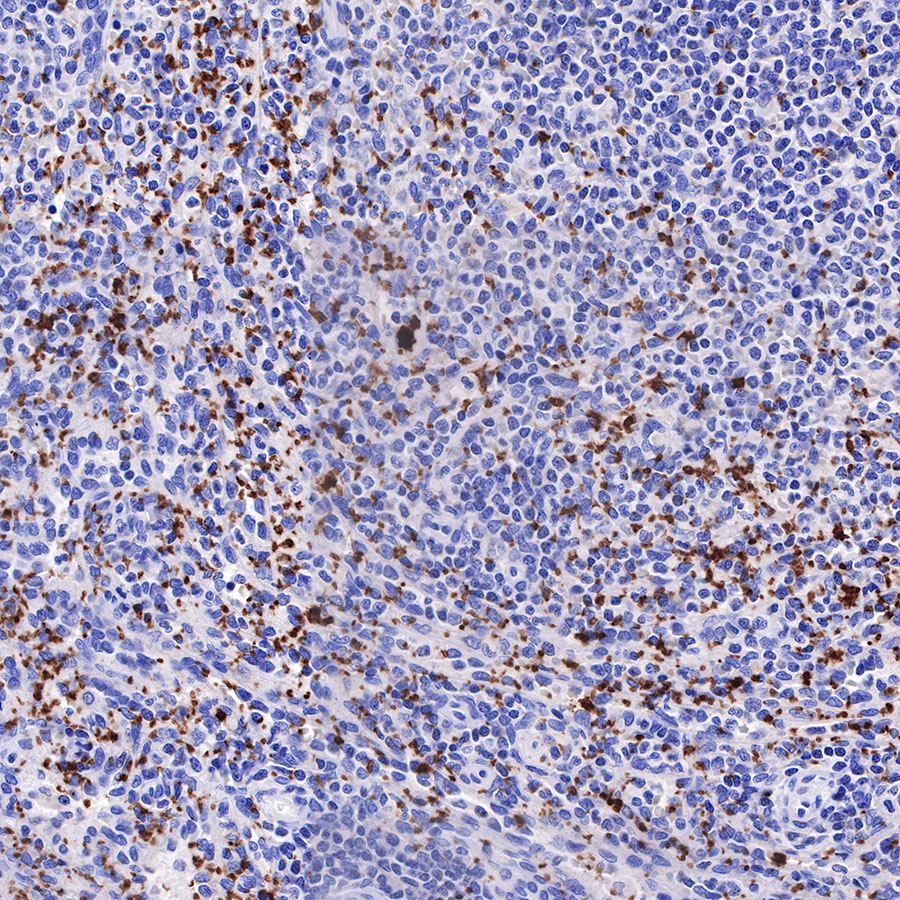
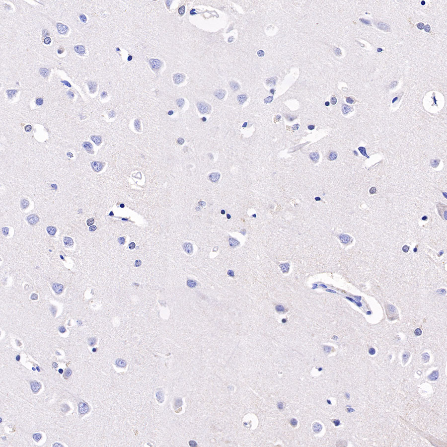
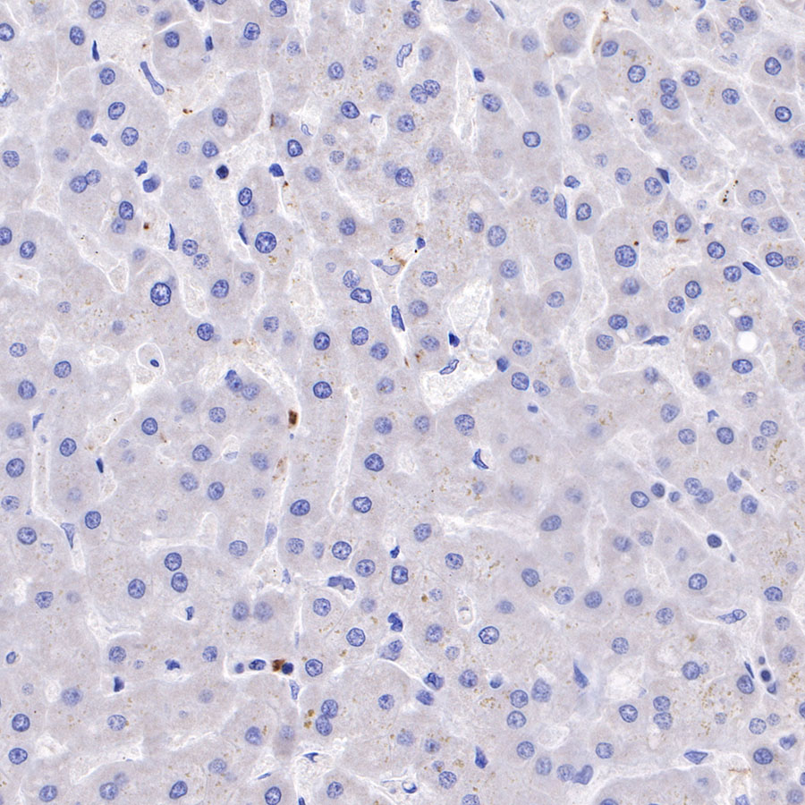
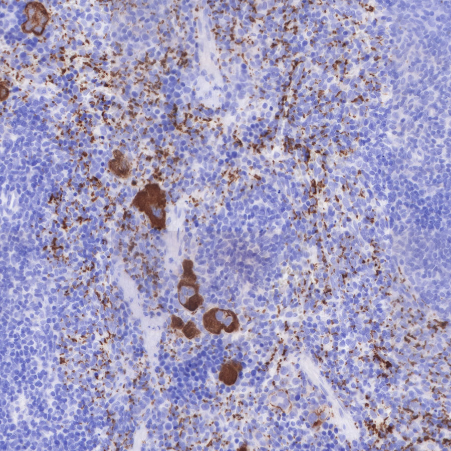
Immunohistochemistry

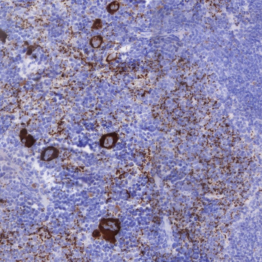
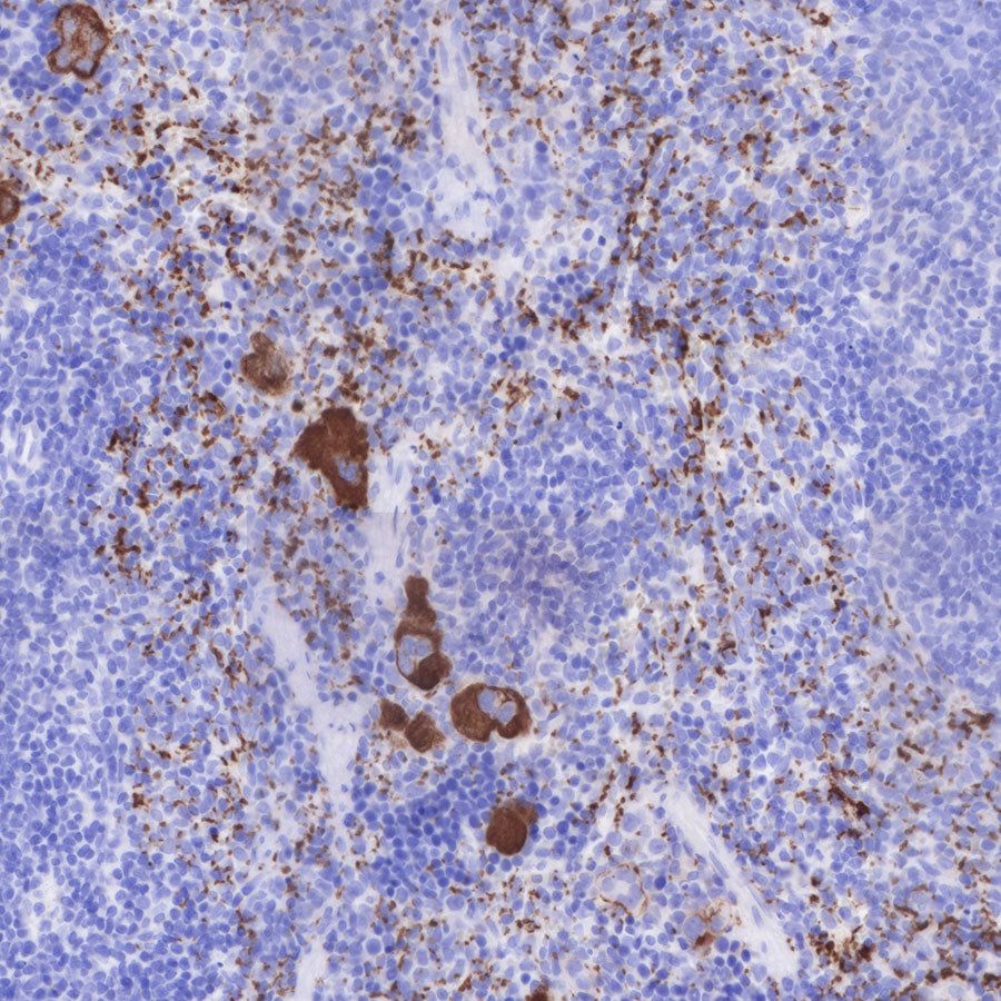
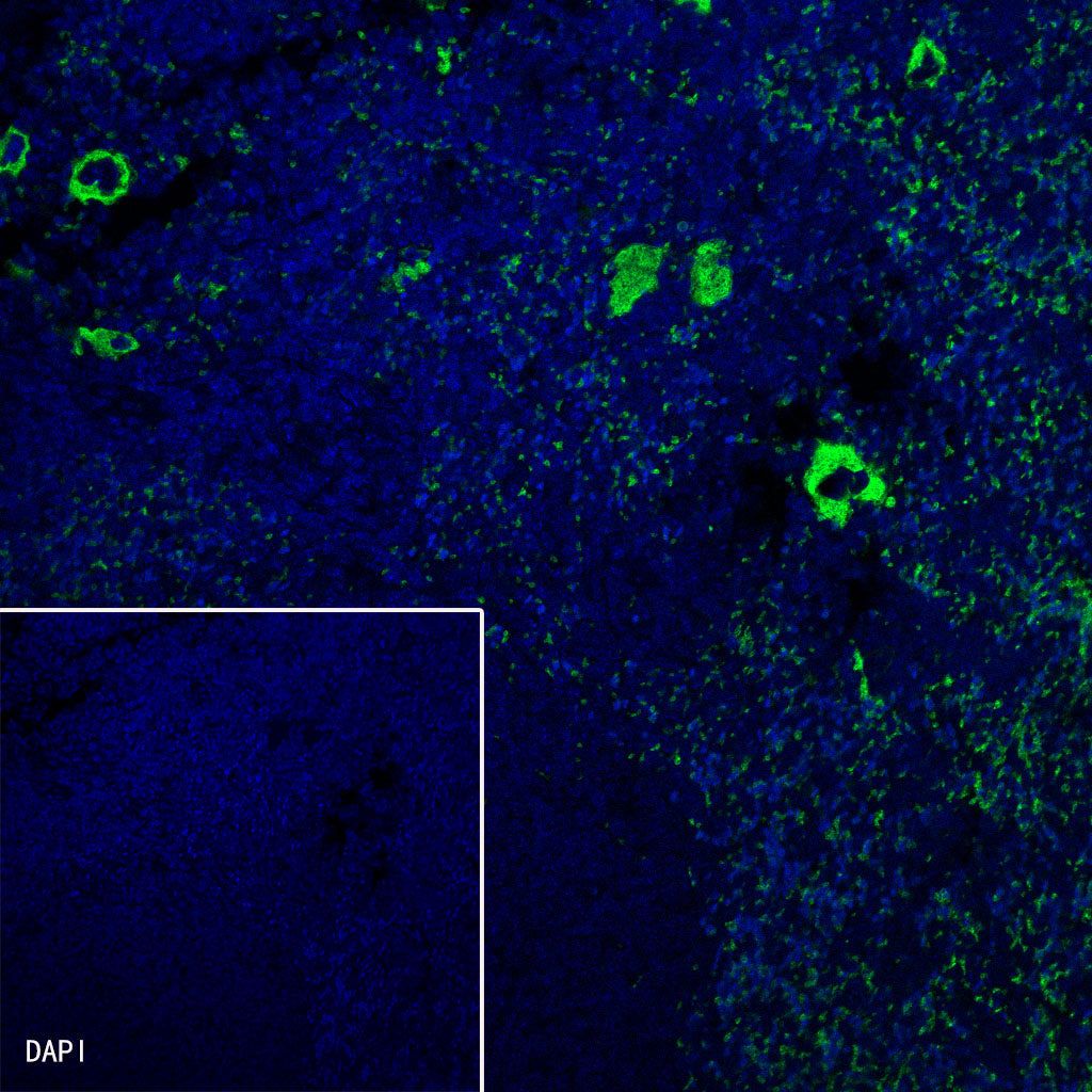
Immunofluorescence
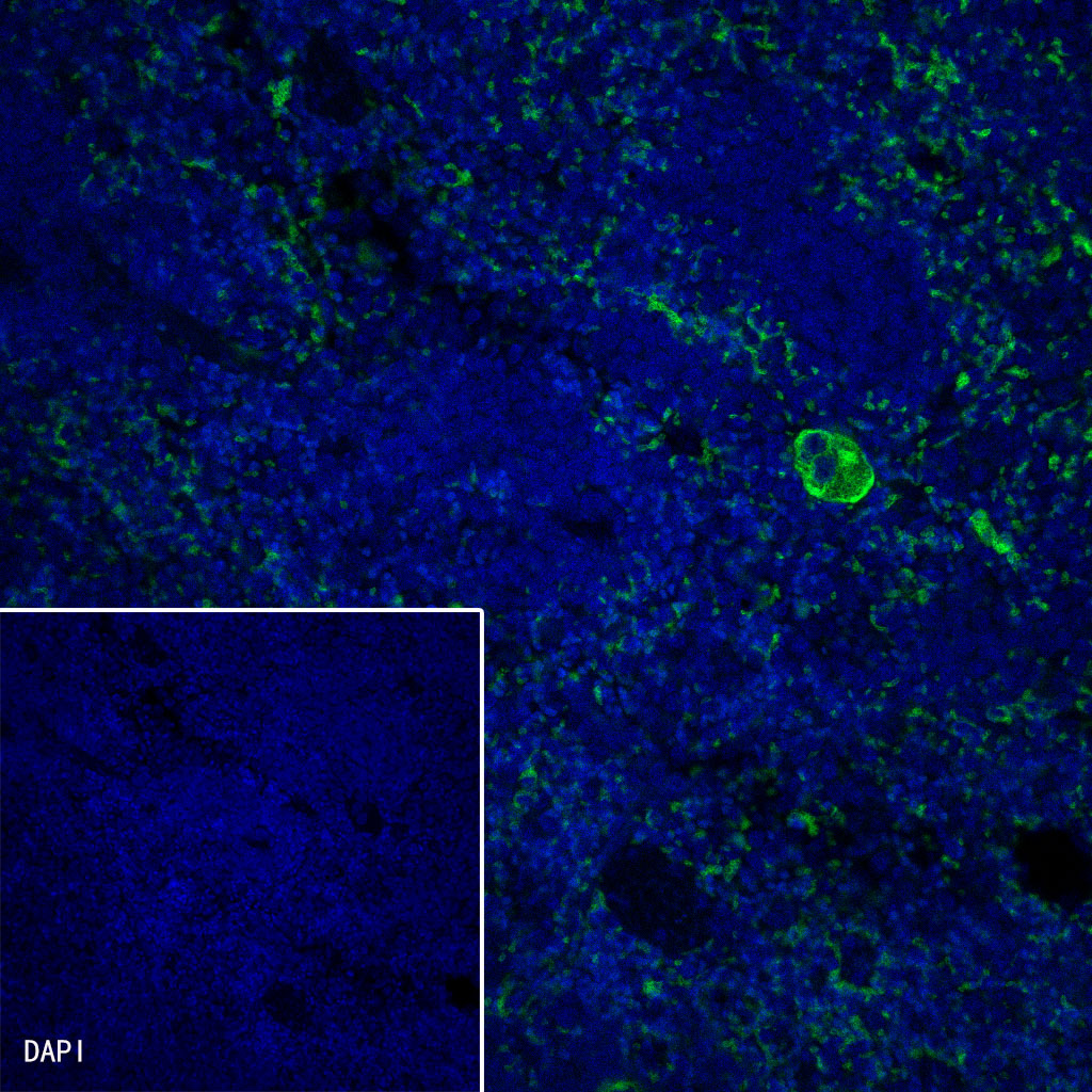
IF shows positive staining in paraffin-embedded rat spleen. Anti-CD42b antibody was used at 1/250 dilution (Green). Goat polyclonal Antibody to Rabbit IgG - H&L (Alexa Fluor® 488) was used as secondary antibody at 1/1000 dilution. Counterstained with DAPI (Blue). Heat mediated antigen retrieval with Tris/EDTA buffer pH9.0 was performed before commencing with IF staining protocol.
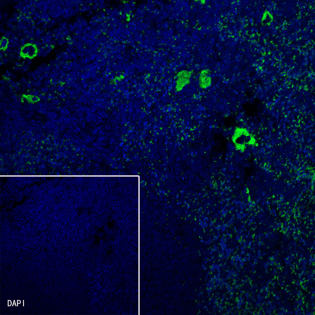
IF shows positive staining in paraffin-embedded mouse spleen. Anti-CD42b antibody was used at 1/250 dilution (Green). Goat polyclonal Antibody to Rabbit IgG - H&L (Alexa Fluor® 488) was used as secondary antibody at 1/1000 dilution. Counterstained with DAPI (Blue). Heat mediated antigen retrieval with Tris/EDTA buffer pH9.0 was performed before commencing with IF staining protocol.