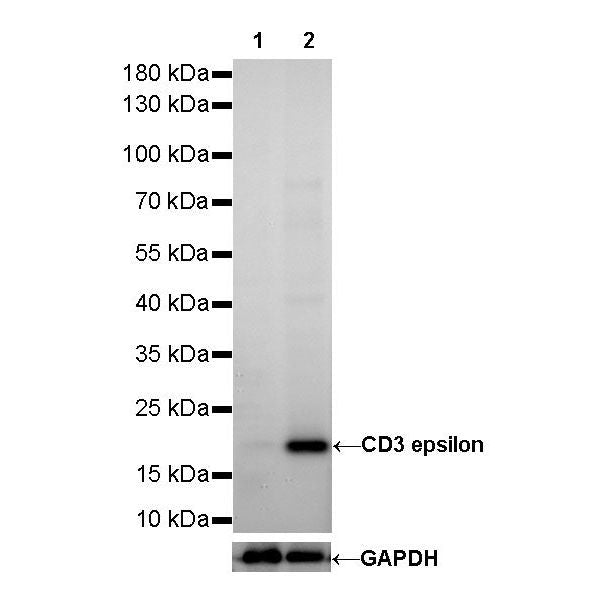
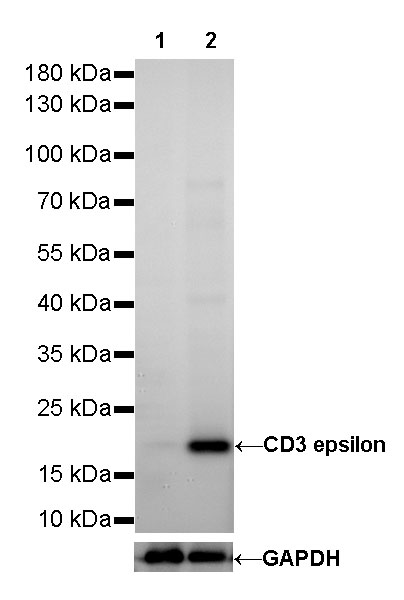
WB result of CD3 epsilon Rabbit mAb
Primary antibody: CD3 epsilon Rabbit mAb at 1/500 dilution
Lane 1: Raji whole cell lysate 20 µg
Lane 2: Jurkat whole cell lysate 20 µg
Negative control: Raji whole cell lysate
Secondary antibody: Goat Anti-Rabbit IgG, (H+L), HRP conjugated at 1/10000 dilution
Predicted MW: 20 kDa
Observed MW: 20 kDa
CD3 epsilon Recombinant Rabbit mAb (SDT-241-49)
CD3 epsilon Recombinant Rabbit mAb (SDT-241-49)
Price:
Regular price
$100 USD
Regular price
Sale price
$100 USD
Unit price
per
For shipping services or bulk orders, you may request a quotation.
Secure checkout with

View full details
Product Details
Product Details
Product Specification
| Host | Rabbit |
| Antigen | CD3 EPSILON |
| Synonyms | T-cell surface antigen T3/Leu-4 epsilon chain, CD3e, T3E, CD3E |
| Immunogen | Synthetic Peptide |
| Location | Cell membrane |
| Accession | P07766 |
| Clone Number | SDT-241-49 |
| Antibody Type | Rabbit mAb |
| Application | WB, IHC-P, ICC, IP, IF |
| Reactivity | Hu, Ms, Rt |
| Predicted Reactivity | Rb, Mq, Pg, Sh |
| Purification | Protein A |
| Concentration | 0.25 mg/ml |
| Physical Appearance | Liquid |
| Storage Buffer | PBS, 40% Glycerol, 0.05% BSA, 0.03% Proclin 300 |
| Stability & Storage | 12 months from date of receipt / reconstitution, -20 °C as supplied |
Dilution
| application | dilution | species |
| WB | 1:500 | |
| IHC-P | 1:250-1:500 | |
| IP | 1:25 | |
| ICC | 1:250 | |
| IF | 1:1000 |
Background
CD3 (cluster of differentiation 3) is a protein complex and T cell co-receptor that is involved in activating both the cytotoxic T cell (CD8+ naive T cells) and T helper cells (CD4+ naive T cells). It is composed of four distinct chains. In mammals, the complex contains a CD3γ chain, a CD3δ chain, and two CD3ε chains. These chains associate with the T-cell receptor (TCR) and the CD3-zeta (ζ-chain) to generate an activation signal in T lymphocytes. The TCR, CD3-zeta, and the other CD3 molecules together constitute the TCR complex. The CD3–T cell receptor (TCR) complex plays a central role in the T-cell-mediated immunoresponse as it is involved in the recognition of antigens and subsequent signal transduction and activation of immunocompetent T lymphocytes. Because CD3 is required for T cell activation, drugs (often monoclonal antibodies) that target it are being investigated as immunosuppressant therapies (e.g., otelixizumab, teplizumab) for type 1 diabetes and other autoimmune diseases.
Picture
Picture
Western Blot
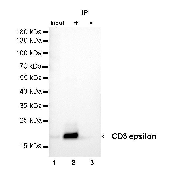
IP
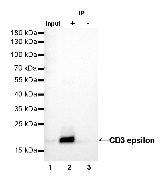
CD3 epsilon Rabbit mAb at 1/25 dilution (1µg) immunoprecipitating CD3 epsilon in 0.4mg Jurkat whole cell lysate.
Western blot was performed on the immunoprecipitate using CD3 epsilon Rabbit mAb at 1/1000 dilution.
Secondary antibody (HRP) for IP was used at 1/400 dilution.
Lane 1 : Jurkat whole cell lysate 10µg(input)
Lane 2 : CD3 epsilon Rabbit mAb IP in Jurkat whole cell lysate
Lane 3 : Rabbit monoclonal IgG IP in Jurkat whole cell lysate
Predicted MW: 20 kDa
Observed MW: 20 kDa
Immunohistochemistry
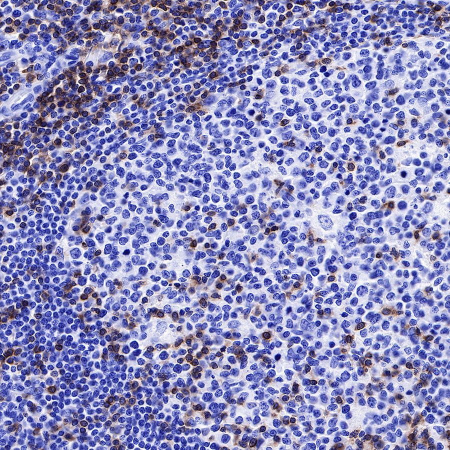
IHC shows positive staining in paraffin-embedded human spleen. Anti-CD3 epsilon antibody was used at 1/2000 dilution, followed by a HRP Polymer for Mouse & Rabbit IgG (ready to use). Counterstained with hematoxylin. Heat mediated antigen retrieval with Tris/EDTA buffer pH9.0 was performed before commencing with IHC staining protocol.
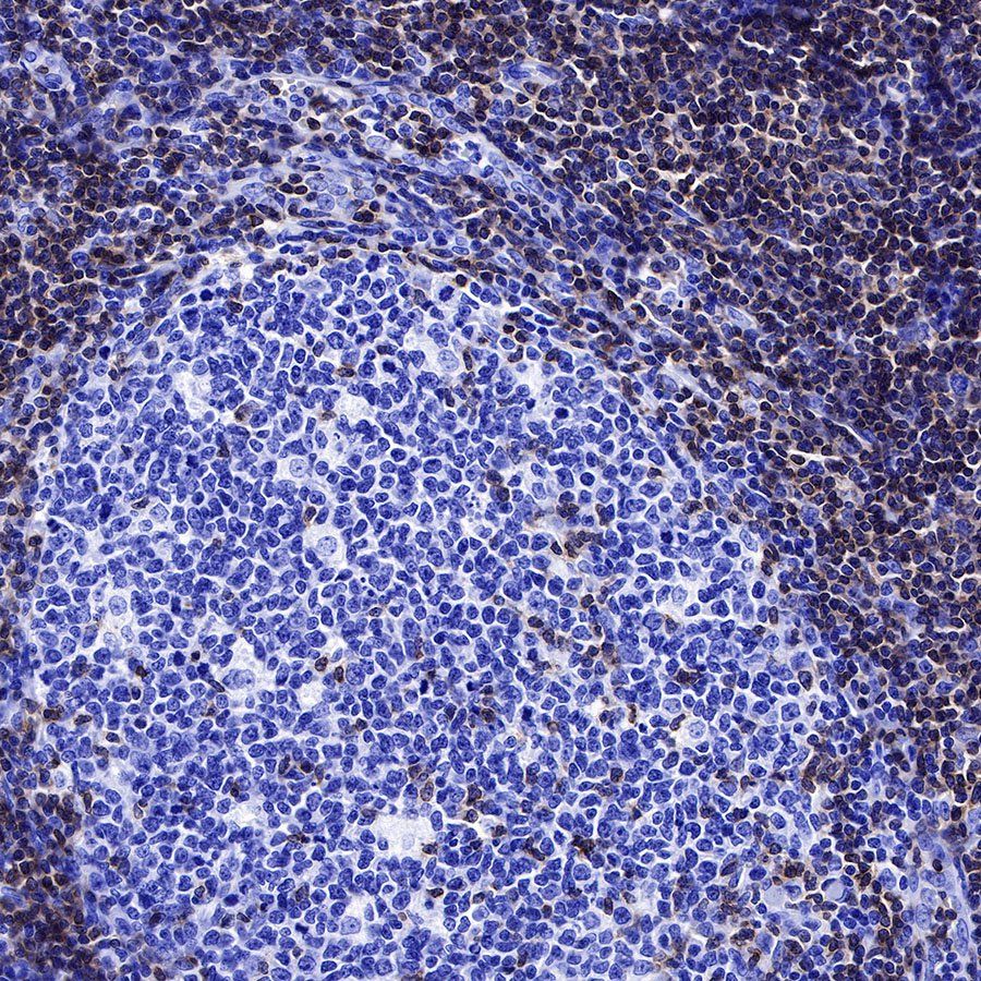
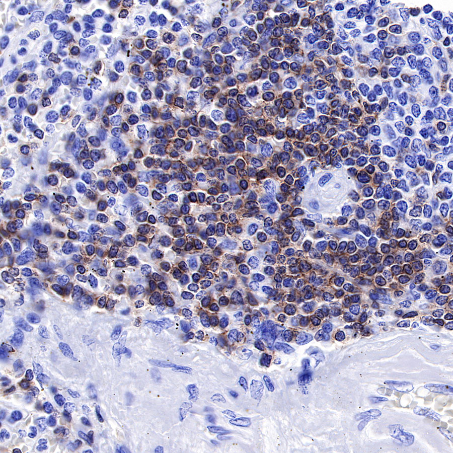
IHC shows positive staining in paraffin-embedded human tonsil. Anti-CD3 epsilon antibody was used at 1/2000 dilution, followed by a HRP Polymer for Mouse & Rabbit IgG (ready to use). Counterstained with hematoxylin. Heat mediated antigen retrieval with Tris/EDTA buffer pH9.0 was performed before commencing with IHC staining protocol.

IHC shows positive staining in paraffin-embedded human tonsil. Anti-CD3 epsilon antibody was used at 1/2000 dilution, followed by a HRP Polymer for Mouse & Rabbit IgG (ready to use). Counterstained with hematoxylin. Heat mediated antigen retrieval with Tris/EDTA buffer pH9.0 was performed before commencing with IHC staining protocol.
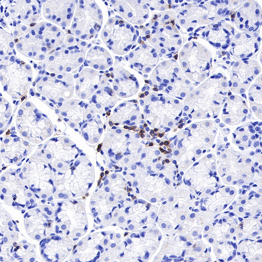
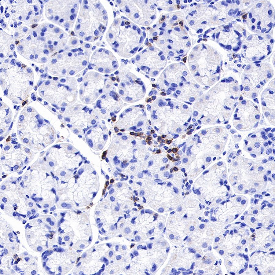
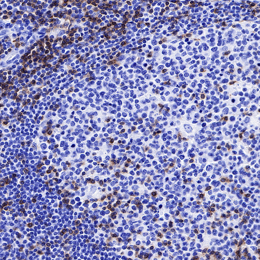
IHC shows positive staining in paraffin-embedded human stomach. Anti-CD3 epsilon antibody was used at 1/2000 dilution, followed by a HRP Polymer for Mouse & Rabbit IgG (ready to use). Counterstained with hematoxylin. Heat mediated antigen retrieval with Tris/EDTA buffer pH9.0 was performed before commencing with IHC staining protocol.

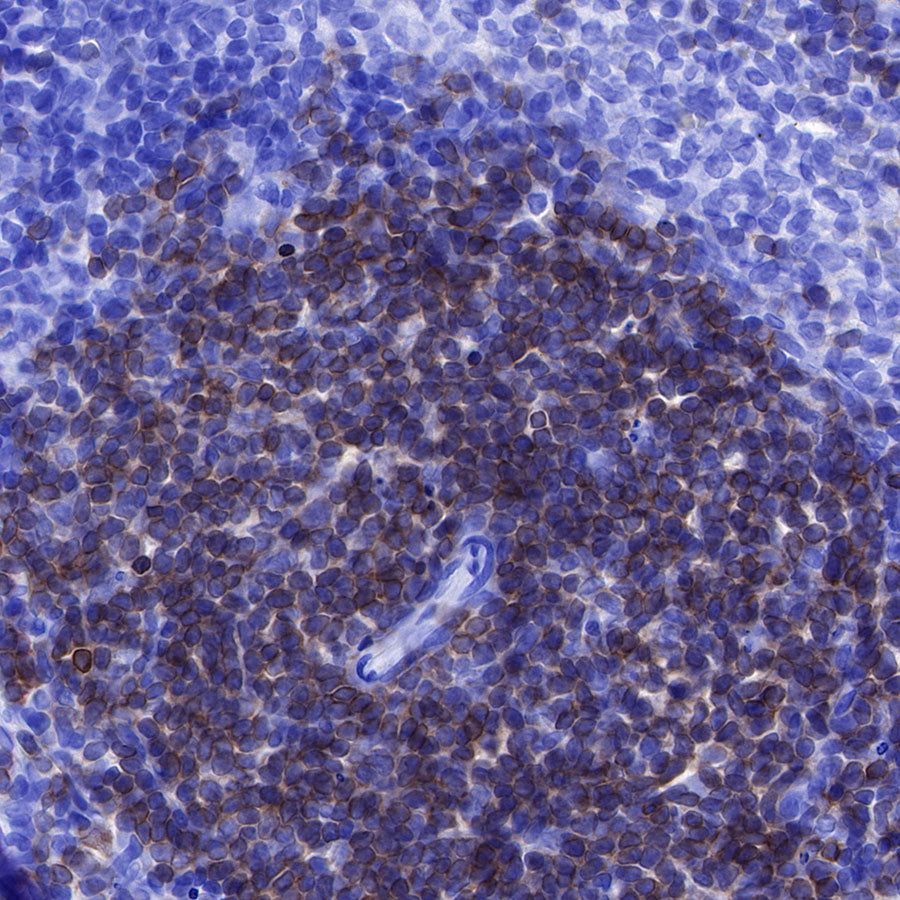
IHC shows positive staining in paraffin-embedded mouse spleen. Anti-CD3 epsilon antibody was used at 1/4000 dilution, followed by a HRP Polymer for Mouse & Rabbit IgG (ready to use). Counterstained with hematoxylin. Heat mediated antigen retrieval with Tris/EDTA buffer pH9.0 was performed before commencing with IHC staining protocol.

IHC shows positive staining in paraffin-embedded rat spleen. Anti-CD3 epsilon antibody was used at 1/4000 dilution, followed by a HRP Polymer for Mouse & Rabbit IgG (ready to use). Counterstained with hematoxylin. Heat mediated antigen retrieval with Tris/EDTA buffer pH9.0 was performed before commencing with IHC staining protocol.
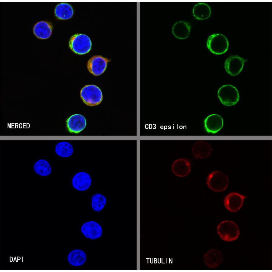
Immunocytochemistry
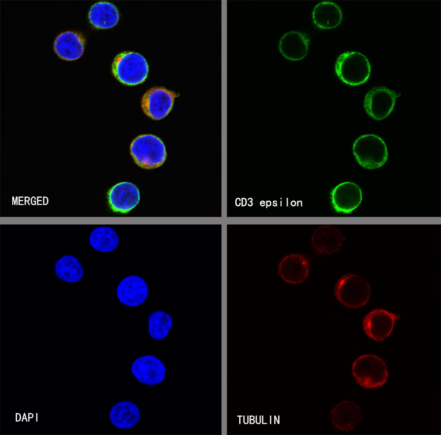
ICC shows positive staining in Jurkat cells. Anti-CD3 epsilon antibody was used at 1/250 dilution (Green) and incubated overnight at 4°C. Goat polyclonal Antibody to Rabbit IgG - H&L (Alexa Fluor® 488) was used as secondary antibody at 1/1000 dilution. The cells were fixed with 4% PFA and permeabilized with 0.1% PBS-Triton X-100. Nuclei were counterstained with DAPI (Blue). Counterstain with tubulin (red).
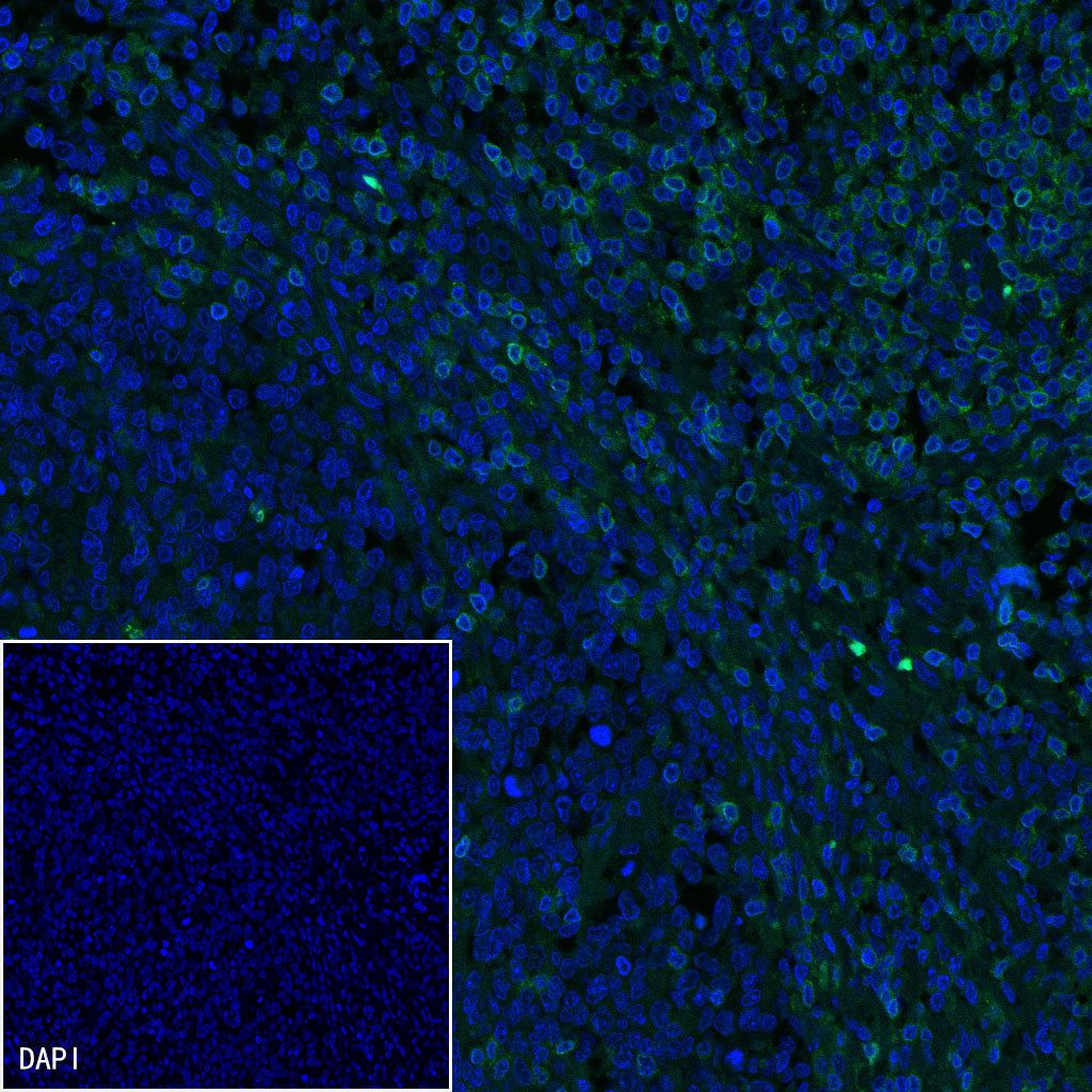
Immunofluorescence
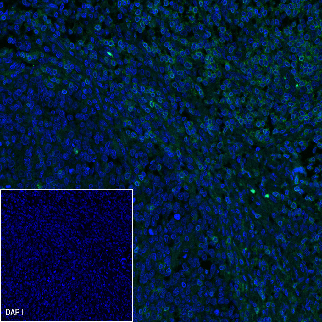
IF shows positive staining in paraffin-embedded human tonsil. Anti-CD3 epsilon antibody was used at 1/1000 dilution (Green) and incubated overnight at 4°C. Goat polyclonal Antibody to Rabbit IgG - H&L (Alexa Fluor® 488) was used as secondary antibody at 1/1000 dilution. Counterstained with DAPI (Blue). Heat mediated antigen retrieval with EDTA buffer pH9.0 was performed before commencing with IF staining protocol.