WB result of CD11c Rabbit mAb
Primary antibody: CD11c Rabbit mAb at 1/1000 dilution
Lane 1: THP-1 whole cell lysate 20 µg
Lane 2: THP-1 serum starvation 2 hour then treated with Phorbol-12-myristate-13-acetate (100ng/ml, 96 h) whole cell lysate 20 µg
Secondary antibody: Goat Anti-Rabbit IgG, (H+L), HRP conjugated at 1/10000 dilution
Predicted MW: 128 kDa
Observed MW: 145kDa
(This blot was developed with high sensitivity substrate)
CD11c Recombinant Rabbit mAb (SDT-396-48)
CD11c Recombinant Rabbit mAb (SDT-396-48)
Price:
Regular price
$100 USD
Regular price
Sale price
$100 USD
Unit price
per
For shipping services or bulk orders, you may request a quotation.
Secure checkout with

View full details
Product Details
Product Details
Product Specification
| Host | Rabbit |
| Antigen | CD11c |
| Synonyms | Integrin alpha-X, CD11 antigen-like family member C, Leu M5, Leukocyte adhesion glycoprotein p150/95 alpha chain, Leukocyte adhesion receptor p150/95, ITGAX |
| Immunogen | Synthetic Peptide |
| Location | Membrane |
| Accession | P20702 |
| Clone Number | SDT-396-48 |
| Antibody Type | Recombinant mAb |
| Application | WB, IHC-P, IP |
| Reactivity | Hu |
| Purification | Protein A |
| Concentration | 0.5 mg/ml |
| Conjugation | Unconjugated |
| Physical Appearance | Liquid |
| Storage Buffer | PBS, 40% Glycerol, 0.05%BSA, 0.03% Proclin 300 |
| Stability & Storage | 12 months from date of receipt / reconstitution, -20 °C as supplied |
Dilution
| application | dilution | species |
| WB | 1:1000 | |
| IHC | 1:500 | |
| IP | 1:50 |
Background
CD11c, also known as Integrin, is an integrin alpha X chain protein. Integrins are heterodimeric integral membrane proteins composed of an alpha chain and a beta chain. This protein combines with the beta 2 chain (ITGB2) to form a leukocyte-specific integrin referred to as inactivated-C3b (iC3b) receptor 4 (CR4). The alpha X beta 2 complex seems to overlap the properties of the alpha M beta 2 integrin in the adherence of neutrophils and monocytes to stimulated endothelium cells, and in the phagocytosis of complement coated particles. CD11c is a type I transmembrane protein found at high levels on most human dendritic cells, but also on monocytes, macrophages, neutrophils, and some B cells that induces cellular activation and helps trigger neutrophil respiratory burst; expressed in hairy cell leukemias, acute nonlymphocytic leukemias, and some B-cell chronic lymphocytic leukemias.
Picture
Picture
Western Blot
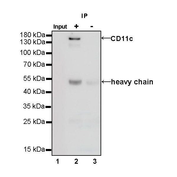
IP
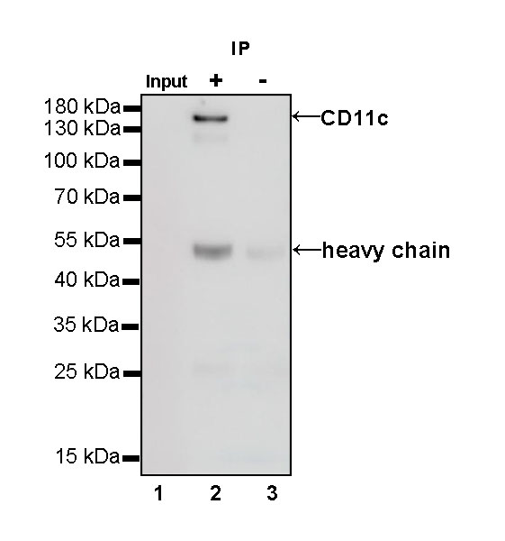
CD11c Rabbit mAb at 1/50 dilution (1 µg) immunoprecipitating CD11c in 0.4 mg THP-1 serum starvation 2 hours then treated with Phorbol-12-myristate-13-acetate (100ng/ml, 96 h) whole cell lysate.
Western blot was performed on the immunoprecipitate using CD11c Rabbit mAb at 1/1000 dilution.
Secondary antibody (HRP) for IP was used at 1/400 dilution.
Lane 1: THP-1 serum starvation 2 hours then treated with Phorbol-12-myristate-13-acetate (100ng/ml, 96 h) whole cell lysate 20 µg (Input)
Lane 2: CD11c Rabbit mAb IP in THP-1 serum starvation 2 hours then treated with Phorbol-12-myristate-13-acetate (100ng/ml, 96 h) whole cell lysate
Lane 3: Rabbit monoclonal IgG IP in THP-1 serum starvation 2 hours then treated with Phorbol-12-myristate-13-acetate (100ng/ml, 96 h) whole cell lysate
Predicted MW: 128 kDa
Observed MW: 145 kDa
Immunohistochemistry
IHC shows positive staining in paraffin-embedded human tonsil. Anti-CD11c antibody was used at 1/500 dilution, followed by a HRP Polymer for Mouse & Rabbit IgG (ready to use). Counterstained with hematoxylin. Heat mediated antigen retrieval with Tris/EDTA buffer pH9.0 was performed before commencing with IHC staining protocol.
IHC shows positive staining in paraffin-embedded human spleen. Anti-CD11c antibody was used at 1/500 dilution, followed by a HRP Polymer for Mouse & Rabbit IgG (ready to use). Counterstained with hematoxylin. Heat mediated antigen retrieval with Tris/EDTA buffer pH9.0 was performed before commencing with IHC staining protocol.
IHC shows positive staining in paraffin-embedded human Hodgkin’s lymphoma. Anti-CD11c antibody was used at 1/500 dilution, followed by a HRP Polymer for Mouse & Rabbit IgG (ready to use). Counterstained with hematoxylin. Heat mediated antigen retrieval with Tris/EDTA buffer pH9.0 was performed before commencing with IHC staining protocol.
IHC shows positive staining in paraffin-embedded human colon cancer. Anti-CD11c antibody was used at 1/500 dilution, followed by a HRP Polymer for Mouse & Rabbit IgG (ready to use). Counterstained with hematoxylin. Heat mediated antigen retrieval with Tris/EDTA buffer pH9.0 was performed before commencing with IHC staining protocol.
IHC shows positive staining in paraffin-embedded human lung cancer. Anti-CD11c antibody was used at 1/500 dilution, followed by a HRP Polymer for Mouse & Rabbit IgG (ready to use). Counterstained with hematoxylin. Heat mediated antigen retrieval with Tris/EDTA buffer pH9.0 was performed before commencing with IHC staining protocol.