Product Specification
| Host |
Rabbit |
| Antigen |
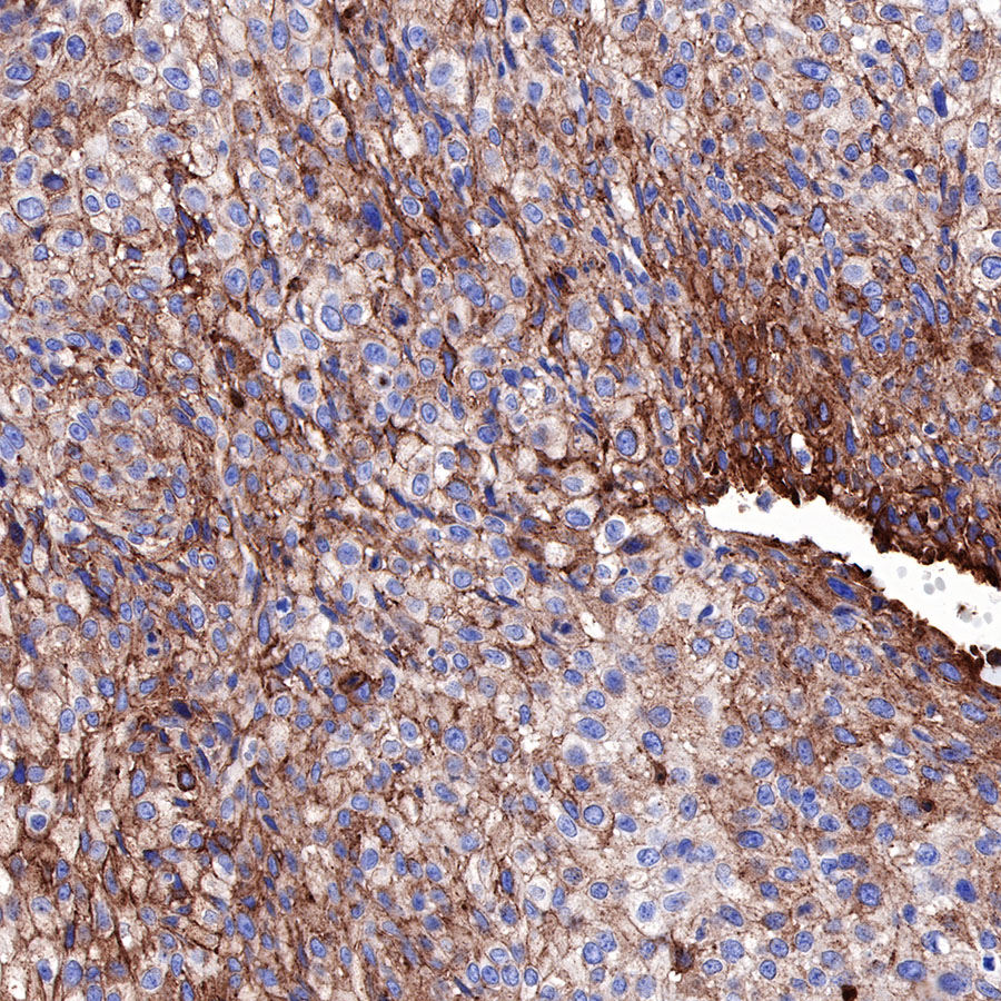
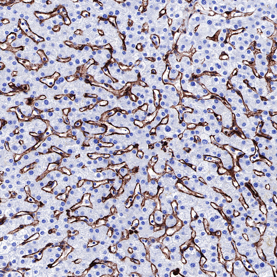
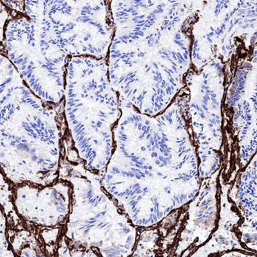
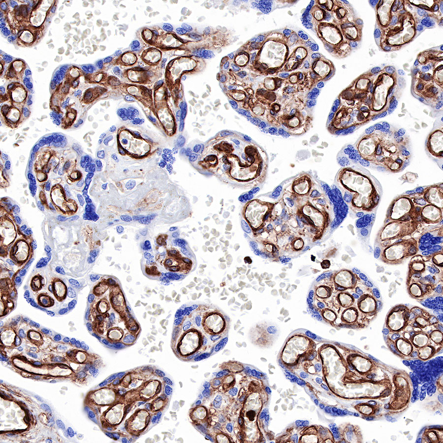
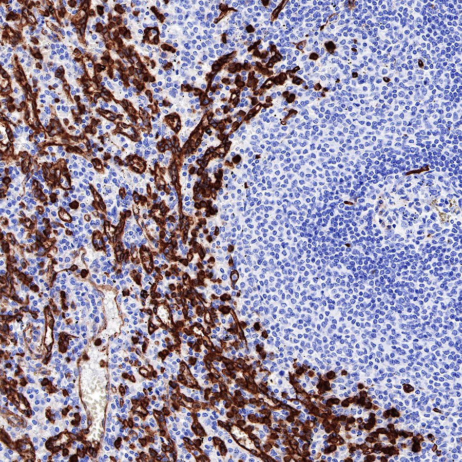
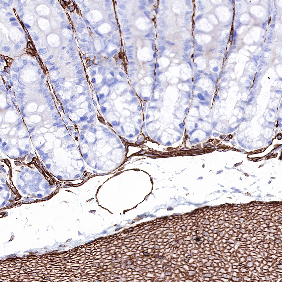
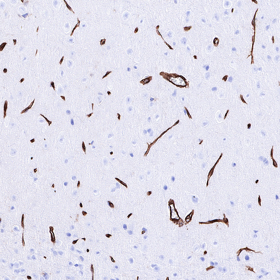
Caveolin-1 |
| Synonyms |
CAV1 |
| Immunogen |
Synthetic Peptide |
| Location |
Membrane, Caveola |
| Accession |
Q03135 |
| Clone Number |
SDT-065-20 |
| Antibody Type |
Rabbit mAb |
| Application |
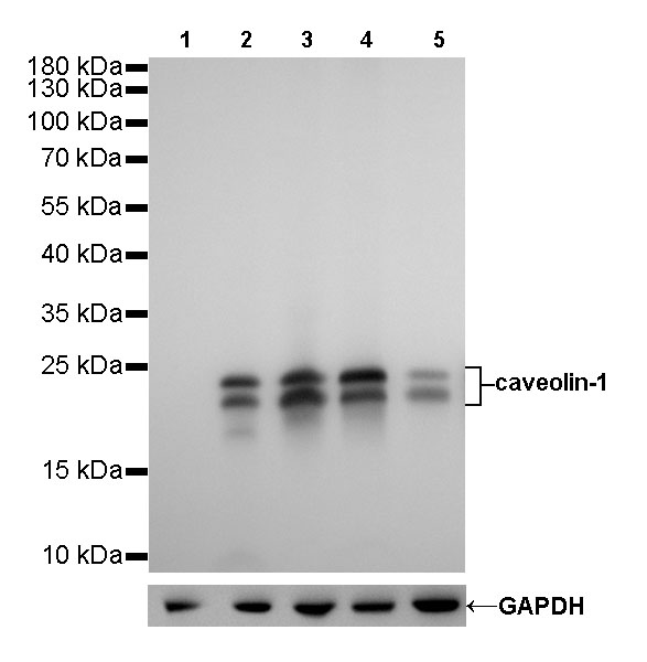
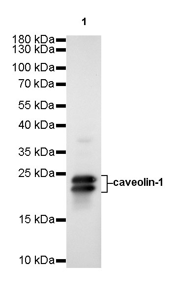
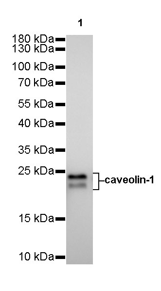
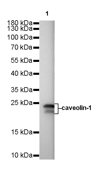
WB, IHC-P |
| Reactivity |
Hu, Ms, Rt |
| Predicted Reactivity |
Or, Bb |
| Purification |
Protein A |
| Concentration |
0.25 mg/ml |
| Physical Appearance |
Liquid |
| Storage Buffer |
PBS, 40% Glycerol, 0.05%BSA, 0.03% Proclin 300 |
| Stability & Storage |
12 months from date of receipt / reconstitution, -20 °C as supplied |
Dilution
| application |
dilution |
species |
| WB |
1:500-1:10000 |
|
| IHC-P |
1:1000 |
|
Background
Caveolin-1 is a 22 kDa protein encoded by CAV1 gene, and occupies flask-shaped plasma membrane invaginations called caveolae. It is one of three known caveolins (CAV1, 2 and 3) and is ubiquitously expressed in all cell types as is CAV2; CAV3 is mostly found in skeletal muscles. Besides earlier studies that implicated CAV1 in endocytosis, signaling and lipid disorders, research activities in the last two decades also focused on clarifying its relevance in cancer. Caveolin-1 (Cav-1) has been considered as a master regulator among the various signaling molecules. It has been emerging as a chief protein regulating cellular events associated with homeostasis, caveolae formation, and caveolae trafficking. In addition to the physiological role of cav-1, it has a complex role in the progression of various diseases. Caveolin-1 has been identified as a prognosticator in patients with cancer and has a dual role in tumorigenesis. The expression of Cav-1 in hippocampal neurons and synapses is related to neurodegeneration, cognitive decline, and aging.