Product Specification
| Host |
Rabbit |
| Antigen |
Caldesmon |
| Synonyms |
CDM, CALD1, CAD |
| Immunogen |
Synthetic Peptide |
| Location |
Cytoskeleton, Intracellular |
| Accession |
Q05682 |
| Clone Number |
SDT-056-48 |
| Antibody Type |
Rabbit mAb |
| Application |
WB, IHC-P, ICC, ICFCM, IP |
| Reactivity |
Hu, Ms, Rt |
| Purification |
Protein A |
| Concentration |
0.5mg/ml |
| Conjugation |
Unconjugated |
| Physical Appearance |
Liquid |
| Storage Buffer |
PBS, 40% Glycerol, 0.05%BSA, 0.03% Proclin 300 |
| Stability & Storage |
12 months from date of receipt / reconstitution, -20 °C as supplied |
Dilution
| application |
dilution |
species |
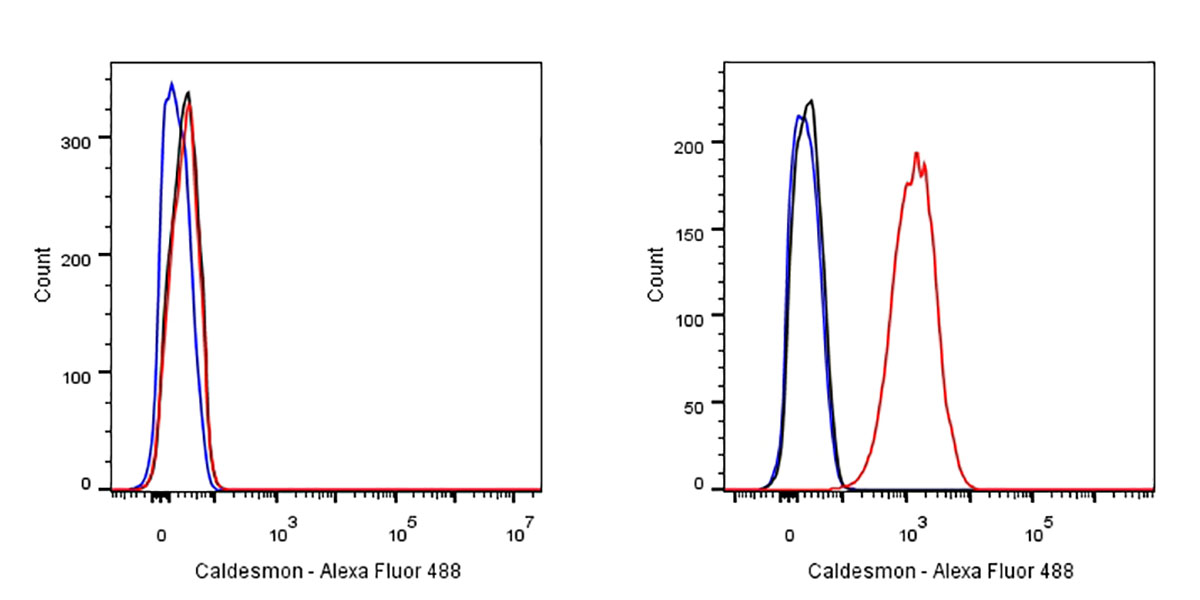
| ICFCM |
1:500 |
|
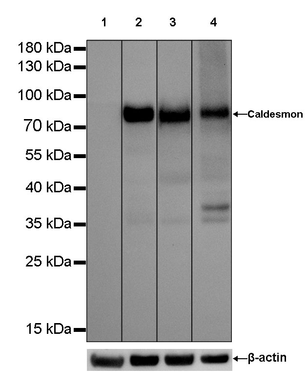
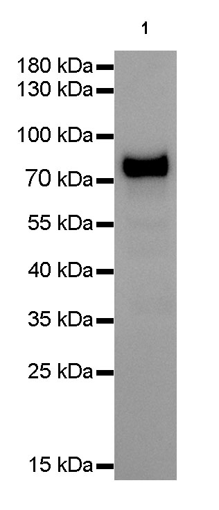
| WB |
1:1000 |
|
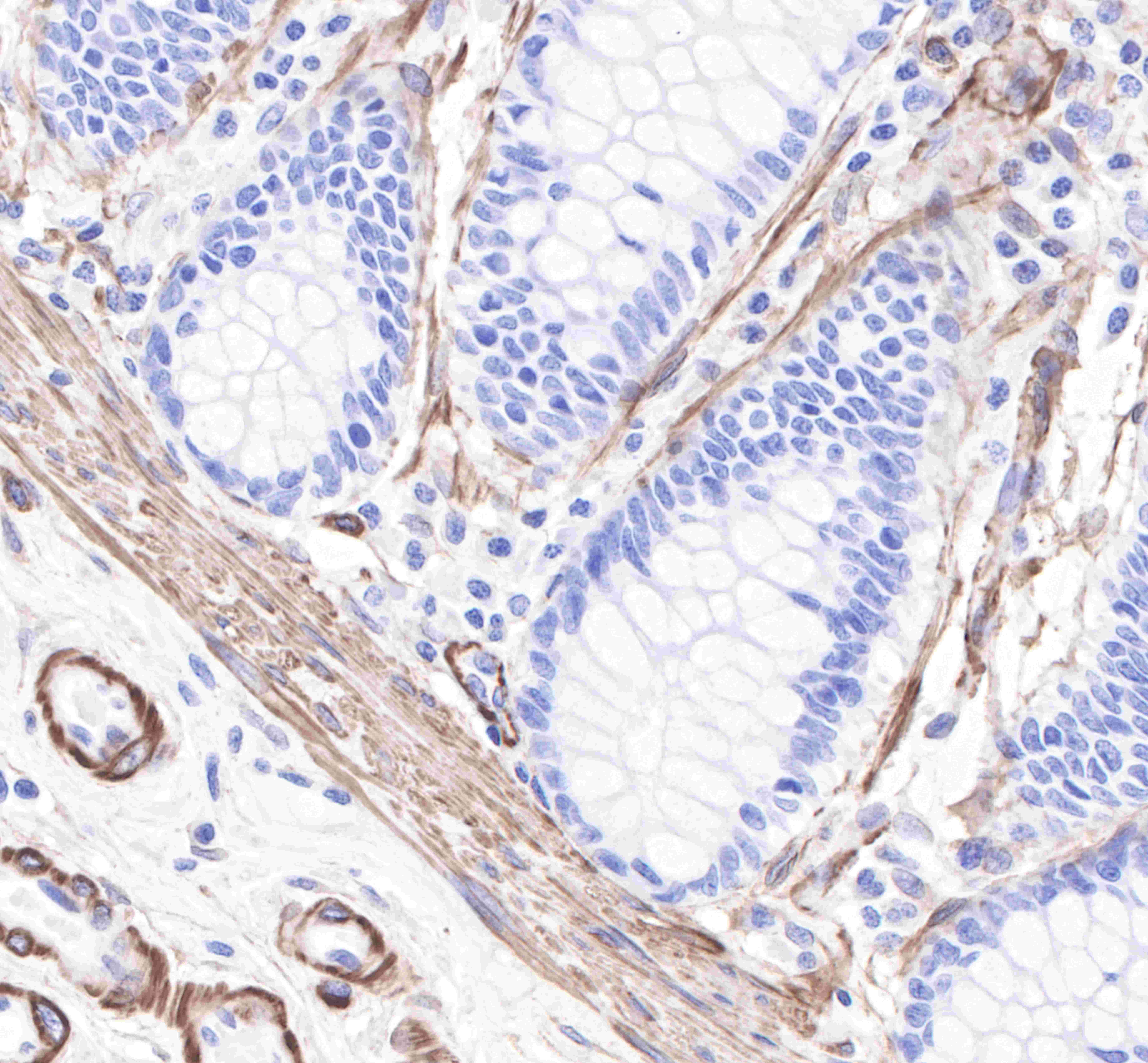
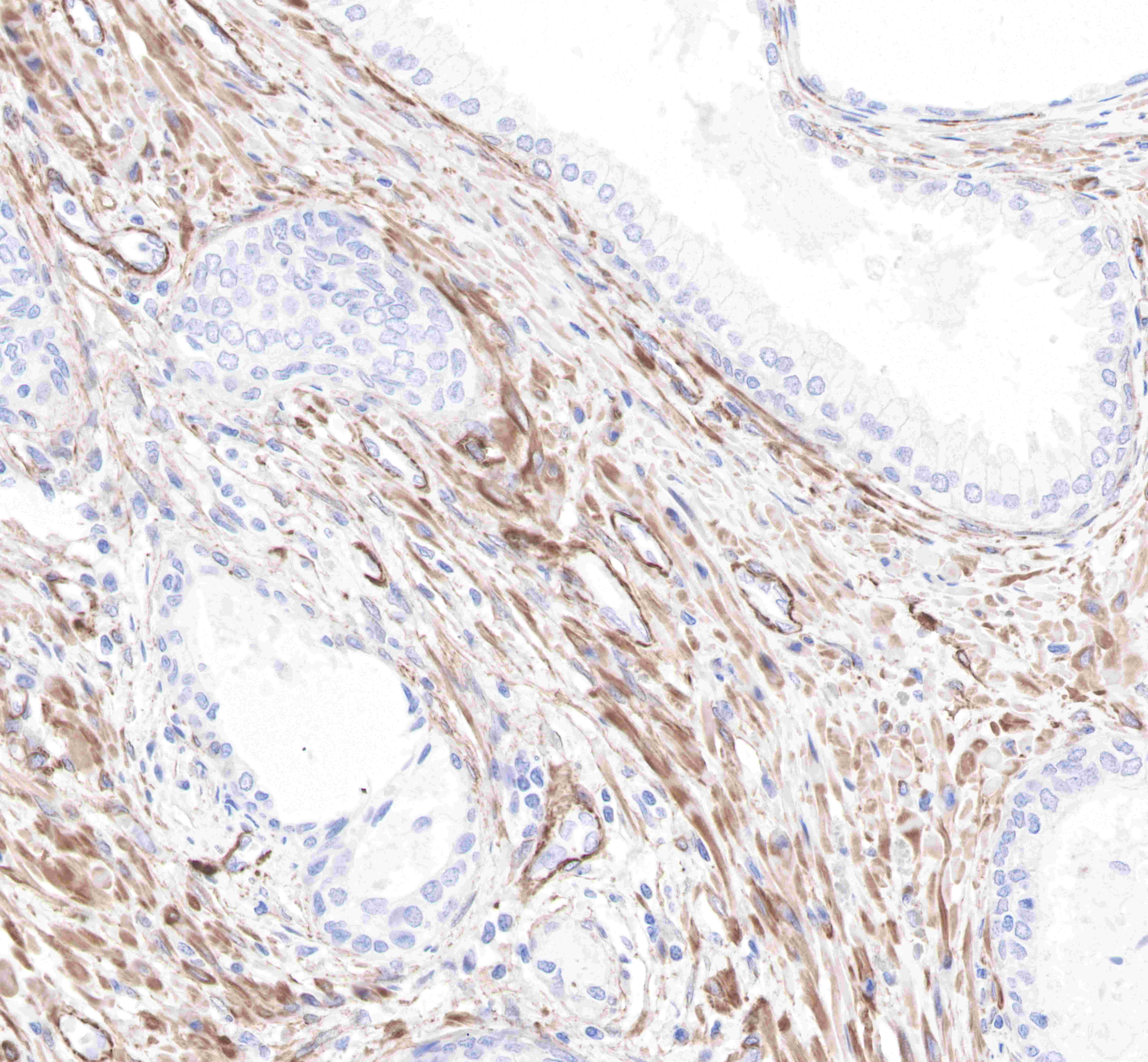
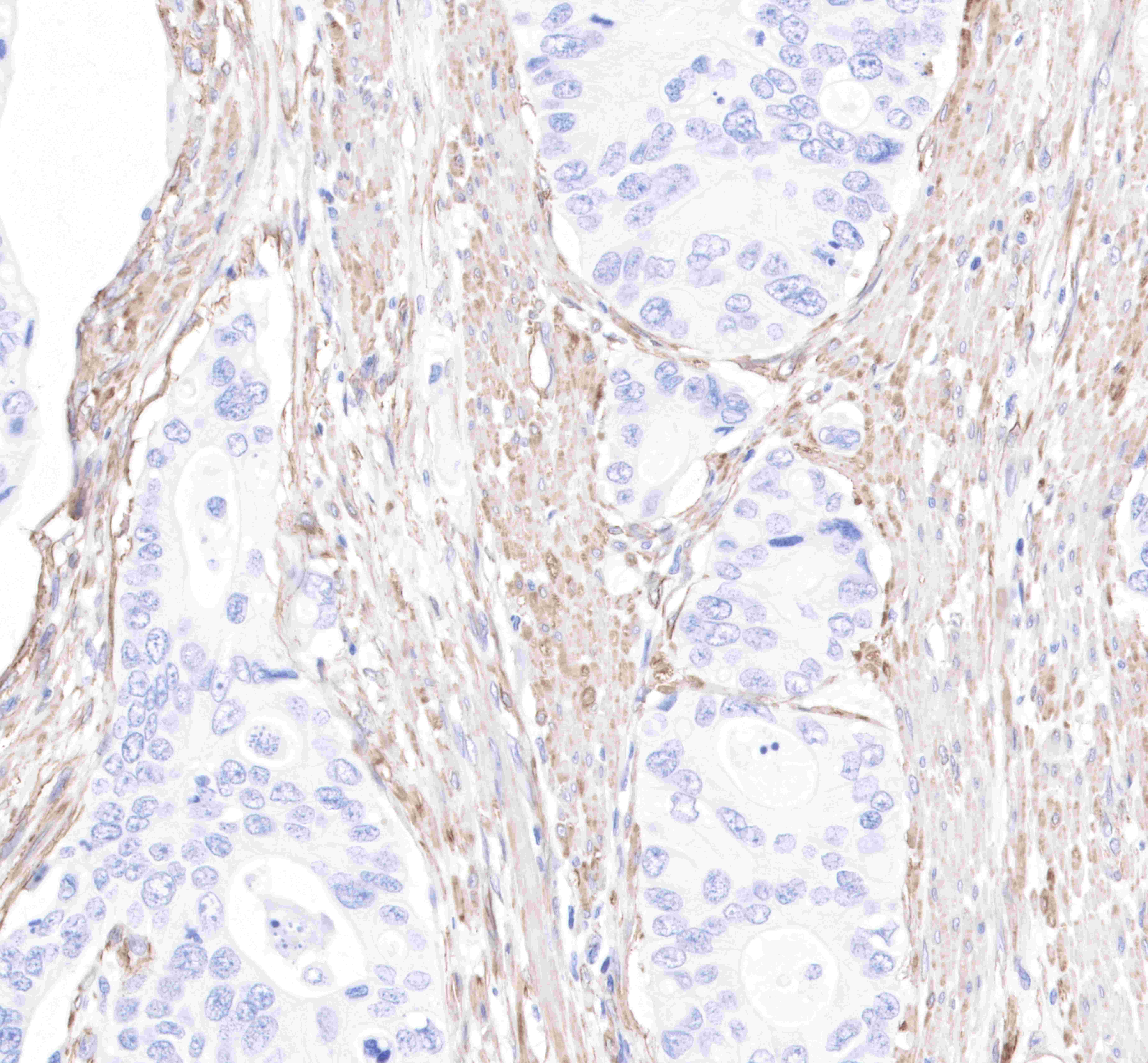
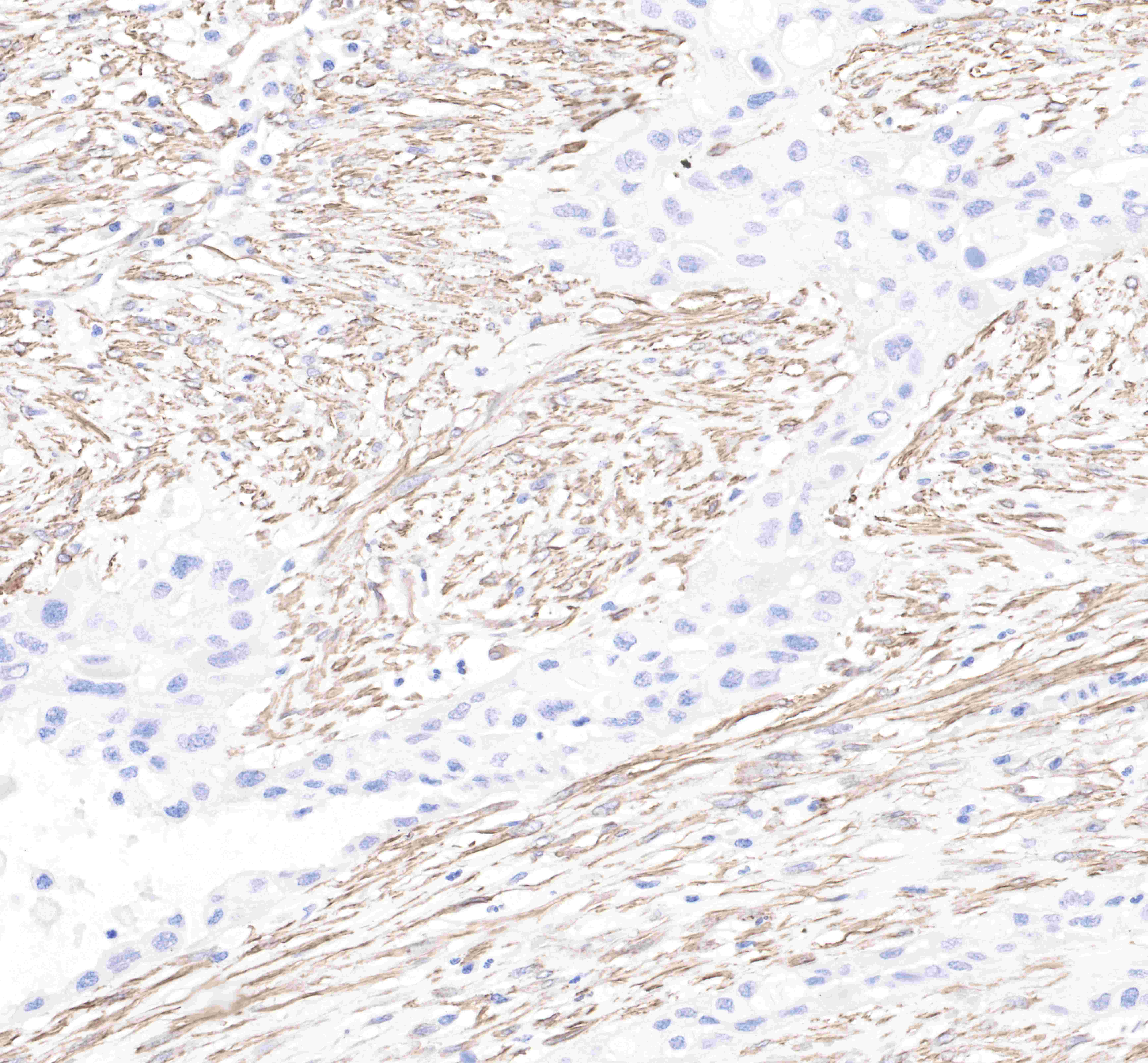
| IHC-P |
1:2000 |
|
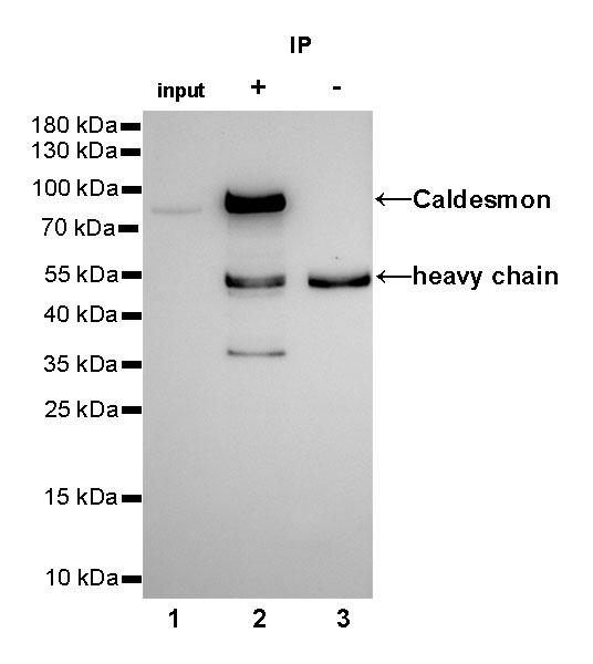
| IP |
1:25 |
|
| ICC |
1:500 |
|
Background
Caldesmon is a binding protein of smooth actin and calmodulin, a cytoskeleton-associated protein present in thin filaments (smooth and striated muscles) that regulates the interaction of actin and myosin. It is available in low molecular weight (l-Caldesmon) and high molecular weight (h-Caldesmon). The latter is thought to be present only in visceral and vascular smooth muscle and myoepithelium. Caldesmon is commonly used in the diagnosis of smooth muscle differentiation tumors.