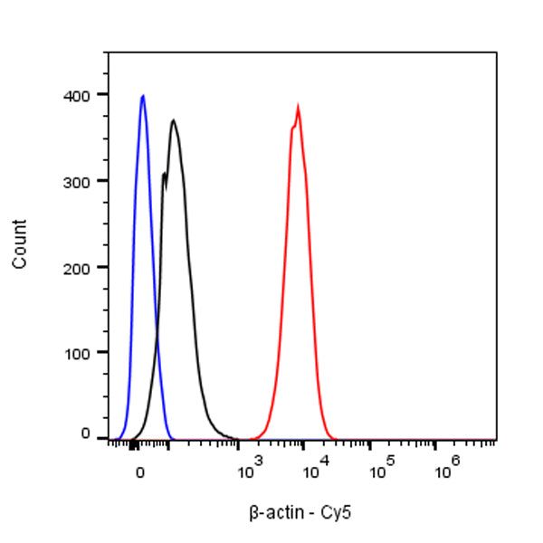
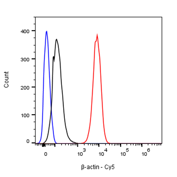
Flow cytometric analysis of Human CD326 expression on 4% PFA fixed 90% methanol permeabilized HeLa cells. Cells from the Hela (Human cervix adenocarcinoma epithelial cells) were stained with either Cy5 Rabbit IgG Isotype Control (Black line histogram) or SDT β-actin Recombinant Rabbit mAb (Cy5 Conjugate) (Red line histogram) at 0.1 μg/test, cells without incubation with primary antibody and secondary antibody (Blue line histogram) was used as unlabelled control. Flow cytometry and data analysis were performed using BD FACSymphony™ A1 and FlowJo™ software.
β-actin Recombinant Rabbit mAb (Cy5 Conjugate) (SDT-R156)
β-actin Recombinant Rabbit mAb (Cy5 Conjugate) (SDT-R156)
Price:
Regular price
$40 USD
Regular price
Sale price
$40 USD
Unit price
per
For shipping services or bulk orders, you may request a quotation.
Secure checkout with

View full details
Product Details
Product Details
Product Specification
| Host | Rabbit |
| Antigen | β-Actin |
| Synonyms | ACTB |
| Immunogen | N/A |
| Location | Cytoplasm, Cytoskeleton |
| Accession | P60709 |
| Clone Number | SDT-R156 |
| Antibody Type | Recombinant mAb |
| Application | ICC, ICFCM |
| Reactivity | Hu, Ms, Rt |
| Purification | Protein A |
| Concentration | 2 mg/ml |
| Conjugation | Cy5 |
| Physical Appearance | Liquid |
| Storage Buffer | PBS, 0.1% BSA, 0.01% Proclin 300 |
| Stability & Storage | 12 months from date of receipt / reconstitution, 2 to 8 °C as supplied. |
Dilution
| application | dilution | species |
| ICC | 1:200 | |
| ICFCM | 1:2000 |
Background
Beta-actin (human gene and protein abbreviation ACTB/ACTB) is one of six different actin isoforms which have been identified in humans. This is one of the two non-muscle cytoskeletal actins. Actins are highly conserved proteins that are involved in cell motility, structure and integrity. Beta actin is often used in Western blotting as a loading control, to normalize total protein amounts and check for eventual protein degradation in the samples. Its transcript is also commonly used as a housekeeping gene standard in qPCR. Its molecular weight is approximately 42 kDa.
Picture
Picture
FC
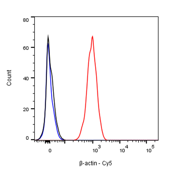
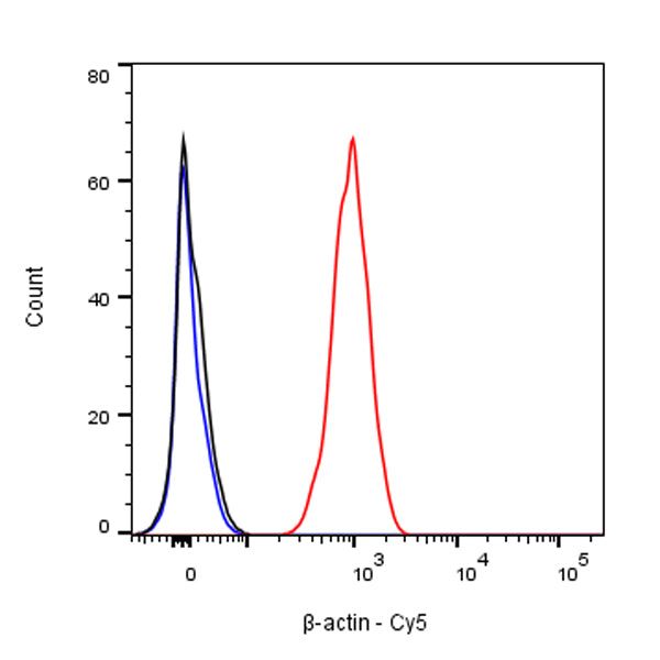
Flow cytometric analysis of Human CD326 expression on 4% PFA fixed 90% methanol permeabilized NIH/3T3 cells. Cells from the NIH/3T3 (Mouse embryonic fibroblast) were stained with either Cy5 Rabbit IgG Isotype Control (Black line histogram) or SDT β-actin Recombinant Rabbit mAb (Cy5 Conjugate) (Red line histogram) at 0.1 μg/test, cells without incubation with primary antibody and secondary antibody (Blue line histogram) was used as unlabelled control. Flow cytometry and data analysis were performed using BD FACSymphony™ A1 and FlowJo™ software.
Immunocytochemistry
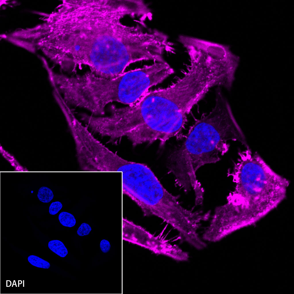
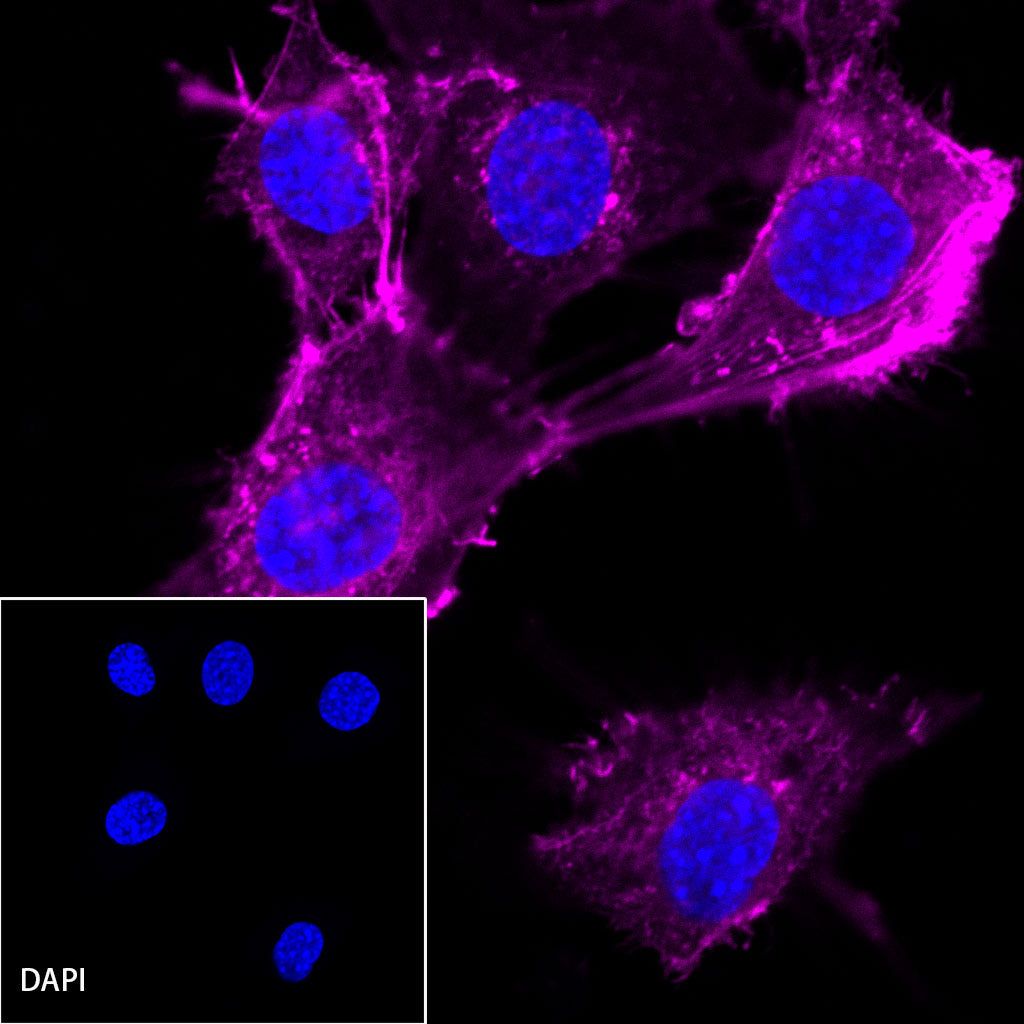
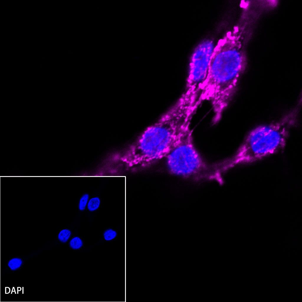
ICC shows positive staining in HeLa cells. Anti-β-actin (Cy5 Conjugate) antibody was used at 1/200 dilution (magenta) and incubated overnight at 4°C. The cells were fixed with 4% PFA and permeabilized with 0.1% PBS-Triton X-100. Nuclei were counterstained with DAPI (Blue).

ICC shows positive staining in NIH/3T3 cells. Anti-β-actin (Cy5 Conjugate) antibody was used at 1/200 dilution (magenta) and incubated overnight at 4°C. The cells were fixed with 4% PFA and permeabilized with 0.1% PBS-Triton X-100. Nuclei were counterstained with DAPI (Blue).

ICC shows positive staining in C6 cells. Anti-β-actin (Cy5 Conjugate) antibody was used at 1/200 dilution (magenta) and incubated overnight at 4°C. The cells were fixed with 4% PFA and permeabilized with 0.1% PBS-Triton X-100. Nuclei were counterstained with DAPI (Blue).