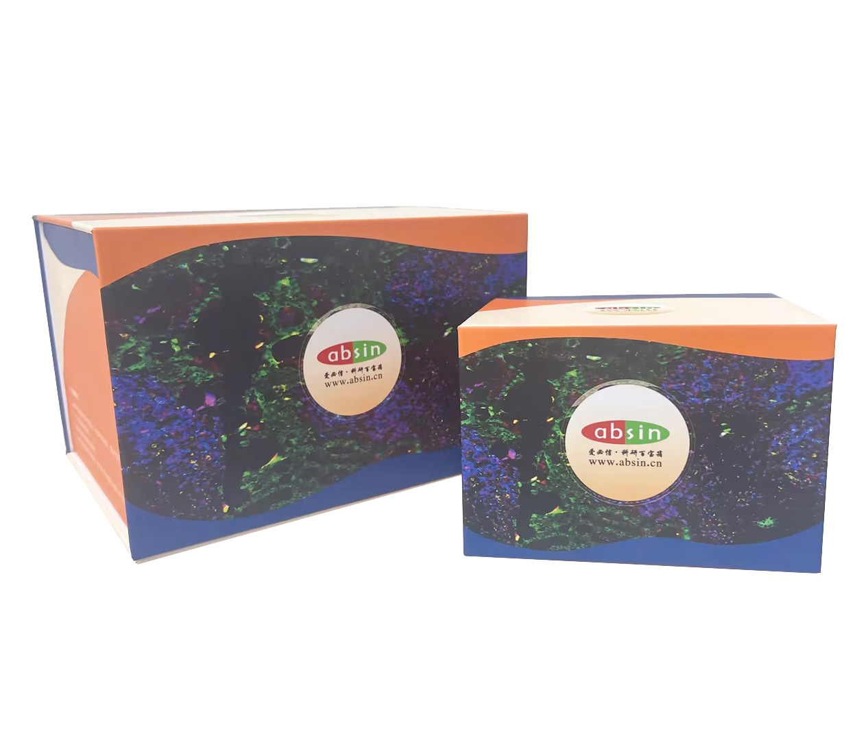
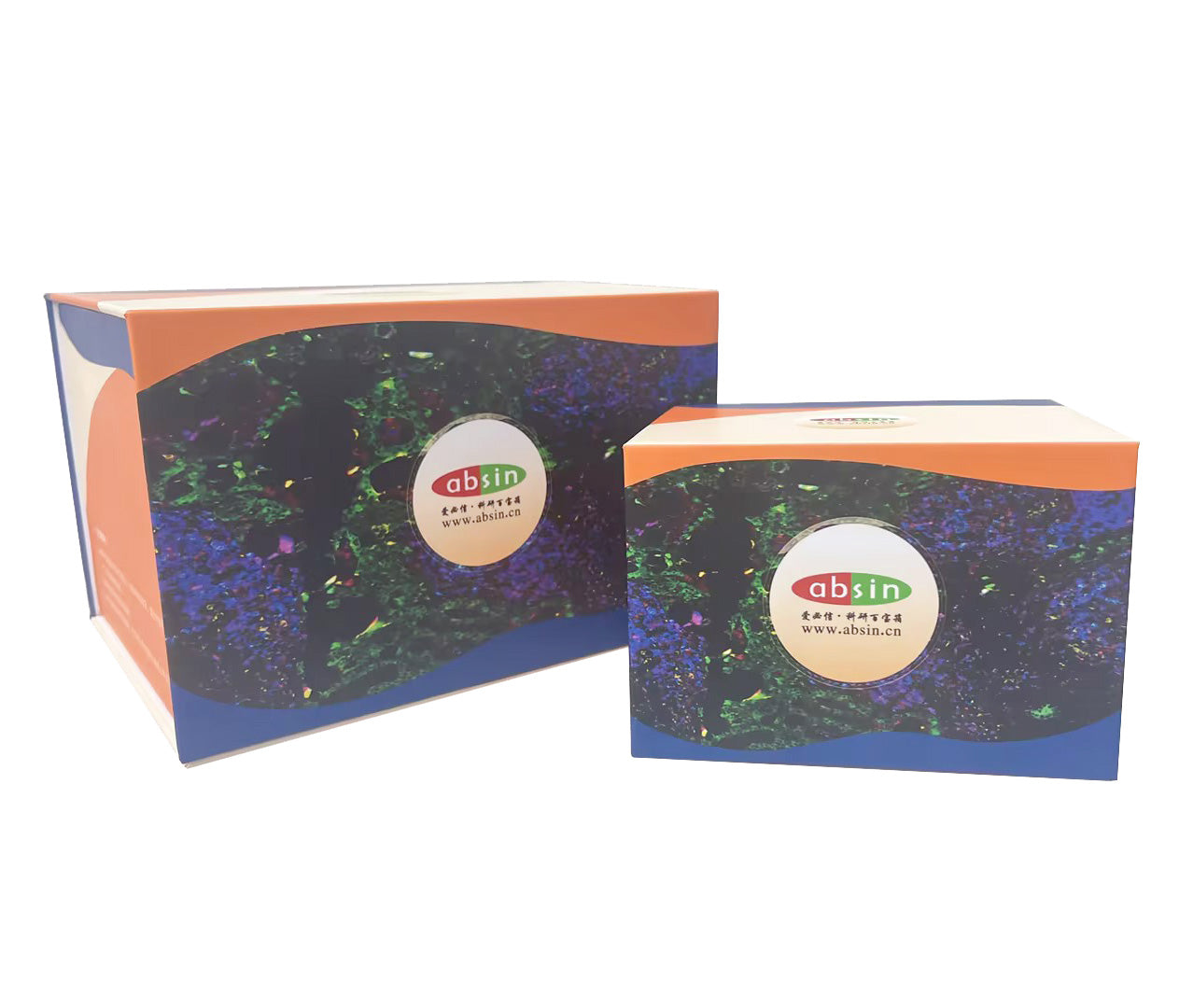
Product Details
Product Details
Product Specification
| References | 1. Mingzhu Yang, et al., Chemical-induced chromatin remodeling reprograms mouse ESCs to totipotent-like stem cells. Cell Stem Cell 2022 Mar 3 Xin Guan, et al., Nanoparticle-enhanced radiotherapy synergizes with PD-L1 blockade to limit post-surgical cancer recurrence and metastasis. Nature Communication. May 20, 2022 Qian-Yue Zhang, et al., Lymphocyte infiltration and thyrocyte destruction are driven by stromal and immune cell components in Hashimoto's thyroiditis. Nature Communications Feb 9, 2022 4. Dabbs David J. Diagnostic immunohistochemistry (M). Beijing: Peking University Medical Press, September 2008. Ed Harlow, David Lane. Technical guidelines for antibodies (M). Beijing: Science Press, 2002: 79-80, 105. Stack, E. C., et al., Multiplexed immunohistochemistry, imaging, and quantitation: a review, with an assessment of Tyramide signal amplification, multispectral imaging and multiplex analysis. Methods, 2014 70 (1): 46-58. 7. Money helps the country. Jiao Lei. Application of multi-labeled immunofluorescence staining and Doppler imaging in histological research. Chinese Journal of Histochemistry and Cytochemistry, 2017 (4): 373-382. |
| Usage |
1. For TSA dye, please use the signal amplification reaction solution to dilute it at 1: 100 to prepare the dye working solution (ready-to-use); DAPI prepares the working solution using sterile water dilution at 1: 100. |
| Theory | Multiplex fluorescence immunohistochemical staining is based on the principle of specific binding of antigen and antibody. The secondary antibody labeled with horseradish peroxidase (HRP) is selected to activate the fluorescent dye in the kit and covalently bind the signal to the antigen. In situ multi-target staining of tissues or cells is achieved by multiple rounds of staining cycles with the aid of different fluorescent dye labels. |
| Synonym | Multicolor kit |
| Description | There are complex cells in tissue microenvironment, and the phenotype, state, abundance and distribution of these cells have important biological significance and clinical value. It can be rendered in situ in tissue by means of antibody staining. Immunohistochemical staining is a common technique to study tissue morphology and protein expression in situ. Routine IHC detection can only show a single indicator, and it is difficult to present the cell composition, state and relationship in the complex tissue microenvironment. This information is crucial to the diagnosis and treatment of diseases! |
| Composition | TSA monochromatic fluorescent dye 520, TSA monochromatic fluorescent dye 570, TSA monochromatic fluorescent dye 620, TSA monochromatic fluorescent dye 690, TSA monochromatic fluorescent dye 770, signal amplification reaction solution, pika universal HRP labeled secondary antibody, anti-fluorescence quenching encapsulation tablet, DAPI. |
| General Notes | 1. This kit is only used for immunohistochemistry and is not used for other purposes. 2. This kit is restricted to professional use only. 3. Appropriate protective measures should be taken to avoid contact of the reagent with the skin and eyes. 4. The activity of reagents beyond the expiration date may be reduced, so reagents beyond the expiration date should not be used. 5. If the dyeing components of this kit are mixed with products of other companies, abnormalities may occur during the dyeing process. 6. Incomplete dewaxing will easily affect the dyeing effect. 7. In order to prevent possible false negative and false positive results, positive control and negative control should be carried out at the same time during the experiment. 8. All kinds of wastes generated in the use of this kit should be disposed of in accordance with the Regulations on Medical Waste Management. |
| Storage Temp. | Fluorescent dyes should be stored in the dark from light at 2-8 ℃, and the validity period is 12 months from receipt of the goods. |
| Applications | It is mainly used for immunohistochemical staining of tissues, paraffin sections and TMA chips. It can also be used for frozen sections and cell crawling sections, and needs to be matched with abs994 antibody eluate (specific for mIHC). |
Picture
Picture
Immunohistochemistry


