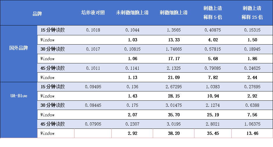
SEAP Detection Kit Reaction Kinetics: THP1-Dual cells at a concentration of 5×10⁵/ml were seeded into a 12-well cell culture plate. After stimulation with Pam3CSK4 to a final concentration of 3mg/ml for 24 hours, the supernatant was collected for enzyme activity analysis. Detection was carried out according to the kit instructions, with 180µl of detection reagent added to 20µl of supernatant. After mixing, absorbance was measured at different time points. The sample in the figure represents the results of undiluted supernatant directly detected after stimulation, showing the average of two replicates, with a detection wavelength of 650nm.
UA-Blue SEAP Detection Kit
UA-Blue SEAP Detection Kit
Price:
Regular price
$195 USD
Regular price
Sale price
$195 USD
Unit price
per
For shipping services or bulk orders, you may request a quotation.
Secure checkout with

View full details
Product Details
Product Details
Product Specification
| Synonyms | Alkaline phosphatase detection reagent |
| Stability & Storage |
Store reagents at -20°C or below, protected from light . Shelf Life: 12 months when stored as recommended. For research use only. |
Background
Secreted Alkaline Phosphatase (SEAP) is widely used in reporter gene analysis studies. Its advantage is that it only requires the analysis of cell culture supernatant, without the need to lyse cells. Moreover, SEAP is relatively heat-resistant and has a high tolerance for specific alkaline phosphatase inhibitors. Therefore, samples can be subjected to specific pre-treatment to inactivate endogenous alkaline phosphatase and eliminate interference.
The UA-Blue SEAP Detection Kit is a homogeneous ready-for-use reagent that completes the detection through the simple steps of “add-mix-detect.” This kit is used to detect alkaline phosphatase activity in the culture medium of cells transfected with the SEAP reporter gene. There is no need to lyse the cells, which can be further cultured or used for other multiplex analyses. Compared with other similar products, the detection sensitivity is at least one time higher, and the reaction time is shortened by at least one time. It is particularly suitable for the analysis of cells with low enzyme activity and the screening of inhibitory compounds.
Components
|
Size |
Buffer (40x) |
Substrate (250x) |
Tests in 96-well plates |
Tests in 384-well plates |
|
2500 tests |
5x 2.5 ml |
5x 400 ml |
2,500 |
12,500 |
|
25000 tests |
5x 25 ml |
5 x 4ml |
25,000 |
125,000 |
Protocol
1). Cell Seeding & Preparation
Plate cells expressing SEAP reporter gene in 96-well or 384-well plates.
If using FBS-containing medium: Heat-inactivate FBS at 56°C for 30 min to eliminate endogenous alkaline phosphatase (AP) activity.
2). Cell Treatment & Stimulation
Treat cells with test compounds and stimulate reporter gene expression as required.
3). Sample Collection & Pre-Treatment
Collect cell culture supernatant at the desired timepoint post-stimulation. For high endogenous AP background: Heat samples at 65°C for 5-10 min to inactivate interfering phosphatases (optional).
Note: The QT-Blue™ SEAP Assay Kit reagent suppresses low levels of endogenous AP without affecting SEAP activity.
4). Reagent Preparation
Equilibrate UA-Blue detection reagent to room temperature (RT) before use.
Buffer Handling (40× Buffer)
May appear slightly turbid (no precipitate) - vortex well before dilution.
Storage: 4°C: Stable for 2 weeks; -20°C: Long-term storage
Substrate Handling (250× Substrate)
Appearance: Red-brown solution; precipitate may form during storage.
To resolubilize: Vortex vigorously for 1–2 min, OR Incubate at 37°C for 5–10 min.
Storage: 4°C: Stable for 4 weeks; -20°C: Long-term storage
5). 1× Working Solution Preparation
Example (40 mL 1× detection reagent):
- Dilute 1 mL 40× Buffer in 39 mL sterile water → 1× QB Buffer.
- Add 167 µL 250× Substrate → immediate flocculation occurs. Vortex immediately until fully dissolved (clear solution). Delay in mixing causes irreversible precipitation.
6). HTS Optimization (e.g., 1536-well plates):
Prepare 3× or 6× concentrated detection reagent to minimize pipetting steps. Example:
3× reagent (2 µL) + 4 µL sample → 1× final concentration.
Storage for 1x/3x/6x detection reagents: 4°C, stable for 1 week (may form slight precipitate; vortex before use).
7). Assay Setup (96-Well Plate Example)
- Add 20 µL sample/control per well.
- Add 180 µL 1× detection reagent.
- Mix on plate shaker (15 sec).
8). Incubation: 37°C for 30 min-6 hrs (protect from light).
9). Detection: Measure absorbance at 600-650 nm (OD) using a plate reader.
Guidelines
Signal Stability: Readings should be taken within 6 hr of reagent addition.
HTS Adaptation: For 384/1536-well plates, scale down volumes proportionally.
Interference Control: Heat pretreatment (65°C) is recommended for high-background samples.
Picture
Picture
Bioactivity