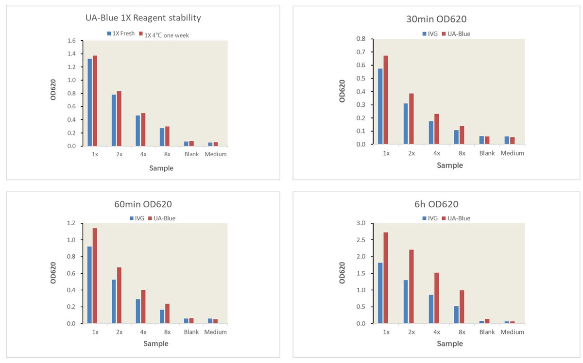
SEAP Detection Kit Reaction Kinetics: THP1-Dual cells at a concentration of 5×10⁵/ml were seeded into a 12-well cell culture plate. After stimulation with Pam3CSK4 to a final concentration of 3mg/ml for 24 hours, the supernatant was collected for enzyme activity analysis. Detection was carried out according to the kit instructions, with 180µl of detection reagent added to 20µl of supernatant. After mixing, absorbance was measured at different time points. The sample in the figure represents the results of undiluted supernatant directly detected after stimulation, showing the average of two replicates, with a detection wavelength of 650nm.
UA-Blue SEAP Detection Kit
UA-Blue SEAP Detection Kit
Price:
Regular price
$120 USD
Regular price
Sale price
$120 USD
Unit price
per
For shipping services or bulk orders, you may request a quotation.
Secure checkout with

View full details
Product Details
Product Details
Product Specification
| Synonyms | Alkaline phosphatase detection reagent (secreted alkaline phosphatase assay kit) |
| Stability & Storage | Dry ice transportation. Store at -20°C or below, protected from light. The expiration date is indicated on the reagent bottle label. |
Background
Secreted Alkaline Phosphatase (SEAP) is widely used in reporter gene analysis studies. Its advantage is that it only requires the analysis of cell culture supernatant, without the need to lyse cells. Moreover, SEAP is relatively heat-resistant and has a high tolerance for specific alkaline phosphatase inhibitors. Therefore, samples can be subjected to specific pre-treatment to inactivate endogenous alkaline phosphatase and eliminate interference.
The UA-Blue SEAP Detection Kit is a homogeneous ready-for-use reagent that completes the detection through the simple steps of “add-mix-detect.” This kit is used to detect alkaline phosphatase activity in the culture medium of cells transfected with the SEAP reporter gene. There is no need to lyse the cells, which can be further cultured or used for other multiplex analyses. Compared with other similar products, the detection sensitivity is at least one time higher, and the reaction time is shortened by at least one time. It is particularly suitable for the analysis of cells with low enzyme activity and the screening of inhibitory compounds.
Components
Size |
Buffer (40x) |
Substrate (250x) |
Tests in 96-well plates |
Tests in 384-well plates |
500 tests |
2.5 ml |
400 μl |
500 |
2,500 |
2500 tests |
5x 2.5 ml |
5x 400 μl |
2,500 |
12,500 |
25000 tests |
5x 25 ml |
5 x 4ml |
25,000 |
125,000 |
Protocol
1.According to the experimental design, plate cells expressing the SEAP reporter gene in 96-well or 384-well reaction plates.If the cell culture medium contains FBS, it should be heat-inactivated at 56°C for 30 minutes to neutralize any potential alkaline phosphatase activity in the FBS.
2.Treat the cells with compounds and stimulate the reporter gene as required by the experiment.
3.After an appropriate incubation period following reporter gene stimulation, collect the cell supernatant for SEAP activity detection. The QT-BlueTM Secreted Alkaline Phosphatase Assay Kit can inhibit a certain level of endogenous alkaline phosphatase activity without affecting SEAP activity.If the SEAP-expressing cells secrete high levels of endogenous alkaline phosphatase, the samples can be heat-inactivated at 65°C for 5-10 minutes before SEAP detection to neutralize the endogenous enzyme.
4.Before the experiment, equilibrate the QT-BlueTM Secreted Alkaline Phosphatase Assay Kit to room temperature.
5.The QB buffer (40x) may appear slightly turbid but without precipitation. Mix by vortexing before dilution and use. The QB buffer (40x) can be stored at 4°C for 2 weeks or at -20°C for long-term storage.
6.The QB substrate (250x) solution is reddish-brown. Long-term storage at 4°C or -20°C may cause precipitation. Vortex vigorously for 1-2 minutes to dissolve the precipitate, or incubate at 37°C for 5-10 minutes to aid dissolution. The QB substrate (250x) can be stored at 4°C for 4 weeks or at -20°C for long-term storage.
7.Prepare the required volume of 1x reaction solution according to the experiment. The substrate in the kit is 250x, and the buffer is 40x. First, dilute the buffer to 1x with sterile pure water, then use the 1x buffer to dilute the substrate to 1x reaction solution.Follow the steps below strictly for preparation:
For example, to prepare 40 mL of 1x detection reagent (1x buffer, 1x substrate), add 1 mL of QB buffer (40x) to 39 mL of sterile pure water and mix well (1x QB buffer). Then add 167 µL of QB substrate (250x). The substrate will immediately form flocculent precipitates.Vortex immediately until the substrate is completely dissolved.This is the 1x detection reagent, which should be a clear solution.If the substrate is not vortexed immediately after addition, precipitation may form and become difficult to dissolve.
Preparation of high-concentration detection reagent for HTS experiments:For HTS experiments, high-concentration detection reagent can be added directly to the cell culture plate without pipetting. For example, for a 1536-well plate with 4 µL of cell culture medium per well, prepare 3x detection reagent (3x buffer, 3x substrate) and add 2 µL of 3x detection reagent to 4 µL of cell culture medium, resulting in a final concentration of 1x. To enhance signal detection, higher concentrations such as 6x detection reagent can be prepared and added to the cell culture plate at a 1:2 ratio.
The 1x detection reagent (or higher concentrations like 3x, 6x) can be stored at 4°C for 1 week. Long-term storage at 4°C may result in minor precipitation, but vortexing will dissolve it without affecting performance.
8.Add 20 µL of detection sample or control sample to each well of a 96-well transparent detection plate, followed by 180 µL of 1x detection reagent. Shake the plate for 15 seconds to mix.
9.Incubate at 37°C and read the plate within 30 minutes to 6 hours.
10.Measure the absorbance (OD) at 620 nm using a multifunctional plate reader. Other wavelengths between 600-650 nm can also be used.
Guidelines
1. Different batches are not recommended for mixed use 2. Without strict validation, it is not recommended to change the dosage of detection reagents 3. For research use only
Picture
Picture
Bioactivity
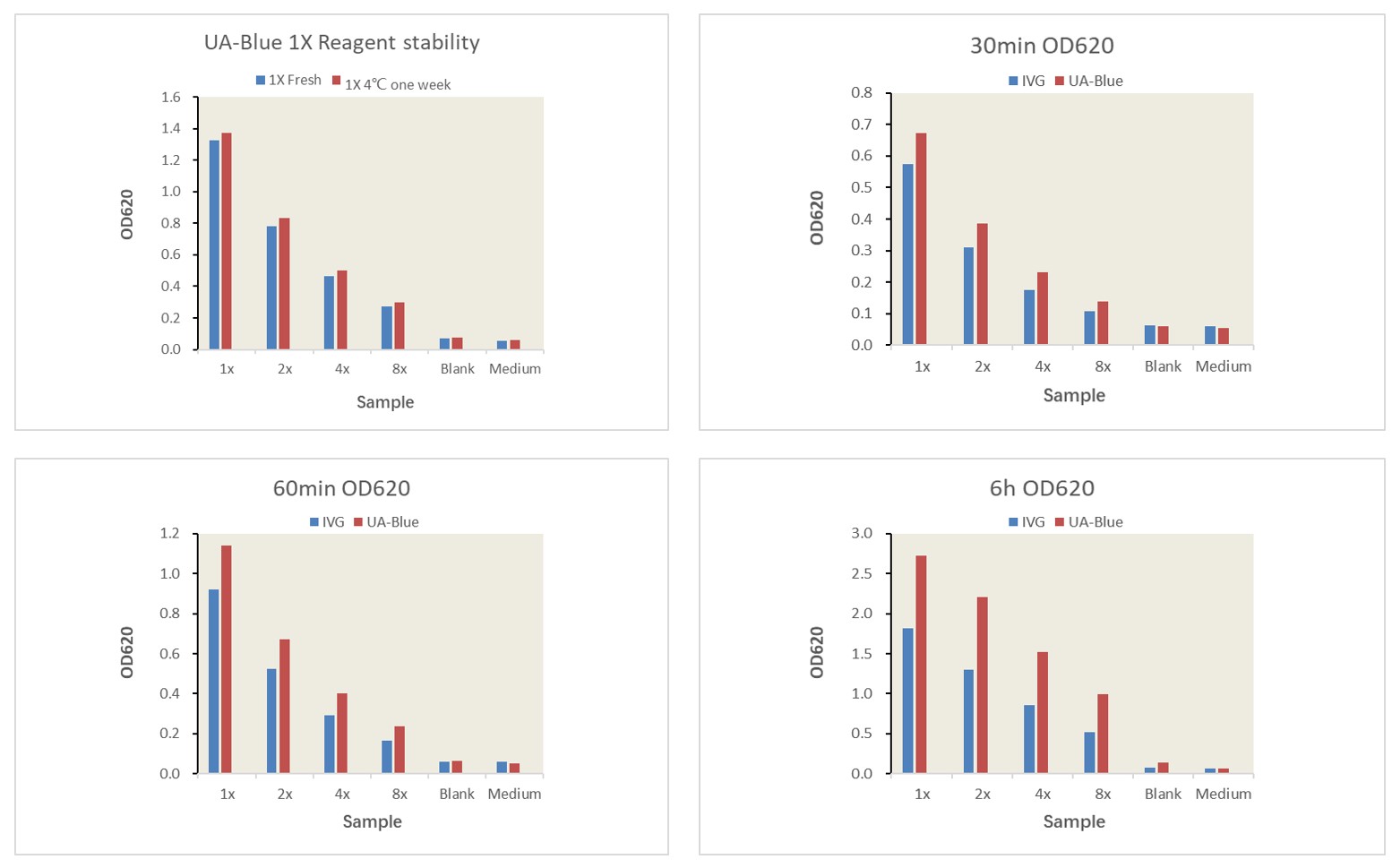
Signal window and stability comparison