New Product Express | Glass gel phosphorylated protein gels

● What is protein phosphorylation?
Protein phosphorylation is an important post-translational modification (PTM) of proteins, which refers to the process of adding phosphate groups to specific amino acid residues of proteins. This process is typically catalyzed by a class of enzymes called protein kinases, while the removal of phosphate groups is catalyzed by phosphatases. Phosphorylation occurs mainly on Serine, Threonine, and Tyrosine residues.

Fig.1 Schematic diagram of protein phosphorylation
● Common protein kinases
The International Union of Biochemistry classifies protein kinases into the following categories based on the specificity of receptor amino acids:
1) Phosphotransferases that use protein ethanol groups as acceptors are called protein serine or threonine kinases;
2) phosphotransferases with phenyl as phosphoreceptors are called protein tyrosine kinases;
3) Phosphotransferases with His, Arg, or Lys as their receptors are called protein His kinases;
4) Phosphotransferases with Cys residues as acceptors are called protein Cys kinases;
5) Phosphotransferases that use acetyl groups as acceptors are called asparagus or glutamine kinases.
The first two classes of enzymes are the most common, and many protein filament/threonine or tyrosine kinases have been purified.
● What is the significance of studying protein phosphorylation?
1) Cell signaling and regulation: Protein phosphorylation plays a key role in intracellular signaling. Through phosphorylation modification, proteins can be activated or inhibited, thereby participating in the regulation of various intracellular signaling pathways, including cell proliferation, apoptosis, differentiation, etc. This modification mode plays an important role in biological processes such as cell signaling, gene expression regulation, and cell cycle regulation.
2) Cell cycle and proliferation: Protein phosphorylation plays an important role in various stages of the cell cycle. Phosphorylation modification can regulate the activity of cyclins, control the orderly division and proliferation of cells, and maintain the normal function of cells.
3) Disease initiation and treatment: The occurrence of many diseases is related to abnormal protein phosphorylation. Studying changes in protein phosphorylation can help us understand the pathogenesis of disease, discover potential therapeutic targets, and develop new drug treatment strategies. Aberrant protein phosphorylation has been implicated in many diseases, particularly cancer. For example, protein kinases encoded by certain oncogenes may cause inappropriate phosphorylation, which in turn promotes tumorigenesis and progression;
4) Drug development and screening: By analyzing the changes in protein phosphorylation, new drug targets and signaling pathways can be discovered to guide drug design and development. At the same time, protein phosphorylation analysis can also be used for drug screening to evaluate the regulatory effect of drugs on specific phosphorylation sites.
5) Personalized medicine: Protein phosphorylation analysis can provide important guidance for personalized medicine. By detecting the status of phosphorylation sites, it is possible to predict the patient's response to specific treatments and provide a basis for clinical treatment decisions.
6) Biomarker identification: Researchers have discovered a number of disease-related biomarkers by analyzing changes in protein phosphorylation. These biomarkers can be used for early diagnosis, prognosis assessment and treatment monitoring.
7) Protein function and stability: Phosphorylation can alter the conformation of proteins, thereby altering their activity and/or stability. For example, phosphorylation can regulate the activity of an enzyme by altering its active state. In addition, phosphorylation can also regulate protein stability by changing the rate of protein degradation;
8) Protein interactions: Phosphorylation can alter the interaction between proteins or proteins and other proteins. For example, phosphorylation can mediate the binding of proteins to ligands, which can affect the activation of signal transduction pathways. In addition, phosphorylation can also alter the affinity or antibodyiness between proteins and other proteins, affecting complex network interactions in cells.
● What are the commonly used protein phosphorylation detection methods?
Western blotting: This is a commonly used protein detection technique that can be used to detect protein phosphorylation levels. Key steps include protein extraction, protein isolation and transfer, antibody recognition, and signal detection. By selecting a specific phosphorylated antibody, the phosphorylation level of the target protein can be accurately detected.
Mass Spectrometry: Mass spectrometry is a highly sensitive and high-resolution protein analysis technique that can be used to detect protein phosphorylation sites and quantify phosphorylation levels. Commonly used methods include liquid chromatography-mass spectrometry coupled with LC-MS/MS and phosphorylated peptide enrichment. By analyzing mass spectrometry data, phosphorylated peptides can be identified and quantified, and phosphorylation sites can be identified.
Enzyme-linked immunosorbent assay (ELISA): Phosphorylation-specific antibodies can be used to quantify the level of phosphorylation in a protein sample. This microplate-based analysis typically utilizes a capture antibody specific to the protein of interest, independent of the phosphorylation state. The protein of interest is then bound to an antibody-coated assay plate, and a phosphorylation site-specific detection antibody to be analyzed is added.
Radioisotope labeling method: By using 32P isotope radiolabeled phosphate as a phosphate group donor, after phosphorylation enzymatic reaction, the phosphate group with 32P isotope radiolabeling is transferred to the corresponding reactive protein, which is separated by gel electrophoresis, and the phosphorylated protein can be detected by autoradiography or phosphorus storage screen.
Immunofluorescence and flow cytometry, among others, are also tools for protein phosphorylation studies. In addition, today I would like to introduce to you a new technology for protein phosphorylation detection - Phos-iso SDS-PAGE
● What is Phos-iso SDS-PAGE?
This is a phosphoaffinity electrophoresis technique that separates phosphorylated and non-phosphorylated proteins using the conventional SDS-PAGE procedure. The core ingredient of this technology, Phos-iso Acrylamide, is a binuclear metal complex that binds specifically to phosphate groups and enables the separation and detection of phosphorylated proteins. Simply add Phos-iso Acrylamide and metal ions (Mn2+ or Zn2+) to the regular SDS-PAGE gel. During electrophoresis, the Phos-iso complex immobilized on the gel can specifically bind to phosphorylated proteins, resulting in reduced electrophoretic mobility of phosphorylated proteins, resulting in the separation of phosphorylated and non-phosphorylated proteins. Phos-iso SDS-PAGE is simple to operate and can detect different forms of phosphorylated proteins, without being limited by phosphoantibodies, and can be used for western blotting, mass spectrometry, etc.
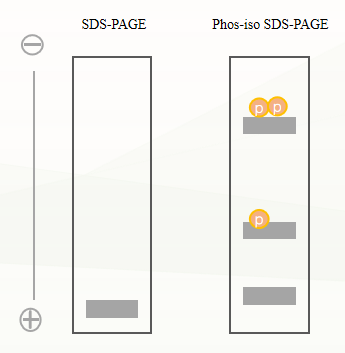
Fig.2 Schematic diagram of Phos-iso acrylamide compared to conventional SDS-PAGE
● Advantages of Phos-iso SDS-PAGE technology
1) There is no need to prepare antibodies against phosphorylated proteins, and both phosphorylated and non-phosphorylated proteins can be detected with total antibodies instead of anti-phosphoprotein antibodies;
2) It can be applied to subsequent operations such as western blotting and mass spectrometry;
3) Phosphorylation forms with different numbers and positions of phosphorylation sites can also be separated.
● Abison new product express - Glass gel phosphorylated protein precast gel
Glass gel phosphoprotein gel (with abs9941 phosphoprotein loading buffer (3×)) is pre-added with 100 μmol/L of Phos-iso Acrylamide, which can be used directly after opening the package. The gels contain zinc as a metal ion, which is stable in the central gel buffer and results in neat strips. After separation, the gels can be used for Coomassie brilliant blue staining, western blotting, and mass spectrometry experiments.
1) The use of automated glue filling production technology ensures the high stability and repeatability of product quality;
2) The use of glass plastic plate can effectively reduce the non-specific adsorption of protein, and make the protein band more acute and clear;
3) The glue clip is extremely easy to open, just use the blade to gently scratch the side of the glue clip to open;
4) Compatible with mainstream mini electrophoresis tanks on the market, such as Bio-Rad, Invitrogen, Tianneng and Junyi Oriental, etc.
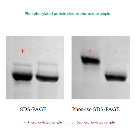
Fig.3. Example of phosphorylated protein electrophoresis
● Frequently Asked Questions
1. The strip is bent
1) The sample contains inorganic salts, surfactants, EDTA and vanadic acid (the sample can be treated with TCA precipitation or dialysis desalting);
2) Improper sample handling, such as the sample is viscous and acidic (full denaturation, the acidic sample solution is yellow-orange, add Tris buffer to neutralize to blue-purple);
3) The prestained Marker band bends adjacent bands due to bending due to the presence of chelating agents, etc., and blank lanes also cause bending of the bands (using loading buffer spacing between the sample and the Marker);
4) High electrophoresis temperature.
2. Unable to identify phosphorylated bands
Perform a regular SDS-PAGE to verify that no stripe migration has occurred.
3. If there is a corresponding phospho-antibody, how to choose between the phospho-antibody and the Phos-iso SDS-PAGE?
If phospho-antibodies are used, the phospho-antibody needs to be pressed, stripped off the membrane, and then pressed again. If you use Phos-iso SDS-PAGE, you can label phosphorylated (or several different forms of phosphorylation) and non-phosphorylated bands on the same membrane and compare the amount of both based on the grayscale of the bands.
4. Does the sample have to be a purified protein?
Not necessarily, cell lysates are also fine. Purified samples are ready for detection by Phos-iso SDS-PAGE. Cell lysates, Western blotting is required.
● Precautions
1) Do not leave below 0°C. The gel will freeze and coagulate below 0°C, resulting in bubbles and cracks, resulting in the scrapping of the gel;
2) Sample preparation is required, and the quality of the sample greatly affects the electrophoresis effect;
3) The electrophoresis speed will be slower;
4) Molecular weight cannot be inferred from molecular weight markers;
5) EDTA treatment is required for transfer;
6) Ordinary SDS-PAGE is also carried out at the same time;
7) Phosphorylated protein gel SDS-PAGE is very sensitive to impurities in protein samples, especially chelating agents, vanadic acid, inorganic, surfactants, etc. It is highly recommended to reduce the impurity content by TCA precipitation or dialysis prior to phosphorylated protein gels SDS-PAGE;
8) For your safety and health, please wear a lab coat and disposable gloves to operate.
Product recommendations
|
Catalog number |
Product name |
specification |
|
abs9942 |
Glass gel phosphoprotein gel 6%, 10wells, 1.5 mm |
2 tablets/box |
|
abs9943 |
Glass gel phosphorylated protein gel 6%, 15wells, 1.5 mm |
2 tablets/box |
|
abs9944 |
Glass gel phosphoprotein gel 8%, 10wells, 1.5 mm |
2 tablets/box |
|
abs9945 |
Glass gel phosphoprotein gel 8%, 15wells, 1.5 mm |
2 tablets/box |
|
abs9946 |
Glass gel phosphorylated protein gel 10%, 10wells, 1.5 mm |
2 tablets/box |
|
abs9947 |
Glass gel Phosphoprotein Precast Gel 10%, 15wells, 1.5 mm |
2 tablets/box |
|
abs9948 |
Glass gel phosphorylated protein gel 12%, 10wells, 1.5 mm |
2 tablets/box |
|
abs9949 |
Glass gel phosphorylated protein gel 12%, 15wells, 1.5 mm |
2 tablets/box |
|
Glass gel phosphorylated protein gel 15%, 10wells, 1.5 mm |
2 tablets/box |
|
|
abs9951 |
Glass gel phosphoprotein gel 15%, 15wells, 1.5 mm |
2 tablets/box |
|
abs9941 |
Phosphoprotein Loading Buffer (3×) |
1mL×5 |
|
Coomassie Brilliant Blue Staining Kit (Regular) |
1kit |
|
|
abs9750 |
Coomassie Brilliant Blue Rapid Staining Solution (no decolorization) |
500mL |
|
abs50085 |
Silver staining kits |
20T |
Absin provides antibodies, proteins, ELISA kits, cell culture, detection kits, and other research reagents. If you have any product needs, please contact us.
|
Absin Bioscience Inc. Email: worldwide@absin.net |
 Follow us on Facebook: Absin Bio Follow us on Facebook: Absin Bio |


