Demystifying the "Conversation" of Immune Cells: Multiplex Fluorescence Immunohistochemical Staining Technology

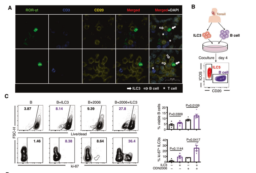
In the latest study, "Reciprocal costimulatory molecules control the activation of mucosal type 3 innate lymphoid cells during engagement with B cells," the authors looked at mucosal type 3 innate lymphocytes (ILC3s) during interactions with B cells How activation is controlled by mutual excitorites. Scientists have unlocked a powerful visual tool that allows us to peer into the "whispers" between immune cells. The key to this is multiplex immunofluorescence staining, which allows us to observe the distribution of multiple biomarkers on the same tissue section at the same time.

Background:
ILC3s are a class of cells in the innate immune system that share a similar phenotype to T helper cells and play a role in mucosal immunity. ICOS (Inducible T-cell costimulator) is a molecule expressed on T cells that is involved in T cell activation and the interaction between T cells and B cells in lymphoid tissues. The role of ICOS in ILC3s and its interaction with the immune microenvironment is unclear.
Objectives:
To explore the expression of ICOS in ILC3s and its role in the interaction between ILC3s and the immune microenvironment; To investigate the specific role of ICOS signaling in the cross-dialogue between ILC3s and B cells.
For this study, the scientists used a multiplex immunofluorescence staining kit from Absin (Cat. No. abs50012), a tiny kit that acts like a magician's wand to make proteins inside cells "glow", revealing their precise location in tissues. The researchers tested CD3, CD20 and RORγt. The results revealed the colocalization of ILC3s and B cells in tonsil tissue, as well as the interactions between them.
Dance steps of experimental techniques
Researchers first undergo tissue sections through a series of treatments, including heating, dewaxing, dehydration, and antigen retrieval, and then label the protein of interest with specific antibodies. These antibodies act as precise navigation systems that target and recognize specific cellular markers. Then, with the help of fluorescent dyes, these markers shine under the microscope, acting as a spotlight on the cell stage, allowing scientists to clearly see the role of each cell and how they interact with each other.
The brilliance of multiplex immunofluorescence staining techniques
This technology not only allows us to see the "conversation" between cells, but also reveals their complex interactions in the tumor microenvironment. It's like being in a choreographed dance, where each cell moves at its own pace, and multiplex immunofluorescence staining allows us to capture these movements and understand their coordination and rhythm.
In summary, through the use of multicolor technology, researchers have drawn important conclusions about the molecular mechanisms of ILC3s and B cell interactions, especially the critical role of ICOS and CD40 signaling in these interactions. These findings help us understand cell-to-cell communication in the mucosal immune system and how these signaling pathways can be modulated to treat related diseases. Multiplex immunofluorescence staining gives us a pair of see-through eyes, allowing us to delve into the microscopic world of cells and explore their secrets. The application of this technology not only pushes the boundaries of scientific research, but also provides a new direction for future medical treatment. Let's hope that these research results can be transformed into powerful weapons against diseases as soon as possible!
Recommended hobbies:
|
Experimental applications |
Catalog number |
trade name |
advantage |
Number of citations |
|
IHC/IF experiments |
abs9756 |
OCT embedding medium |
It has high viscosity and is suitable for embedding fragile tissues in paraffin/frozen sections |
5-star reviews from customers |
|
4% paraformaldehyde (general purpose tissue fixative solution) |
It is used for fixation of tissue and cell specimens |
57 citations |
||
|
Antibody eluent (for mIHC) |
It is used for antibody elution during paraffin, frozen sections, bone tissue sections, cell crawling sections, etc., during TSA staining. |
15 citations |
||
|
abs9299 |
Primary and secondary antibody diluents |
It is used for the dilution and formulation of primary and secondary antibodies in immunohistochemistry, immunofluorescence, ELISA, WB and other immunological experiments |
11 citations |
|
|
abs929 |
Immunohistochemistry pen |
It is used to circle the target area on the slide, which is called the hydrophobic isolation zone |
39 citations |
|
|
abs9235 |
Antifluorescence quenching mountant (with DAPI) |
Mounting reagent for slowing fluorescence quenching |
23 citations |
|
|
abs9248 |
Sodium citrate antigen retrieval solution (50×) |
It is used to remove cross-linking between proteins caused by aldehyde fixation reagents and fully expose epitopes in samples such as paraffin sections |
4 citations |
|
|
abs9342 |
Tris-EDTA antigen retrieval solution (10×, pH 9.0) |
It can be used for antigen retrieval after paraffin sections, frozen sections and other samples are fixed with paraformaldehyde, formaldehyde or other aldehyde reagents |
9 citations |
|
|
Ready-to-use immunohistochemistry secondary antibody kit (pika universal type) |
Kits for IHC experiments |
16 citations |
||
|
Multiplex fluorescence immunohistochemistry (mIHC) |
Four-color multiplex fluorescent immunohistochemical staining kit (pika universal secondary antibody) |
Popular multi-color kit, citing literature impact factor Up to IF: 38.079 |
96 citations |
|
|
Five-color multiplex fluorescent immunohistochemical staining kit (pika universal secondary antibody) |
25 citations |
|||
|
Six-color multiplex fluorescent immunohistochemistry staining kit (pika universal secondary antibody) |
6 citations |
|||
|
Seven-color multiplex fluorescent immunohistochemistry staining kit (plus) (pika universal secondary antibody) |
2 citations |
Absin provides antibodies, proteins, ELISA kits, cell culture, detection kits, and other research reagents. If you have any product needs, please contact us.
|
Absin Bioscience Inc. Email: worldwide@absin.net |
 Follow us on Facebook: Absin Bio Follow us on Facebook: Absin Bio |


