Product Specification
| Host |
Rabbit |
| Antigen |
p63 |
| Synonyms |
Chronic ulcerative stomatitis protein (CUSP), Keratinocyte transcription factor KET, Transformation-related protein 63 (TP63), Tumor protein p73-like (p73L), p40, p51, KET, P63, P73H, P73L, TP73L |
| Immunogen |
Synthetic Peptide |
| Location |
Nucleus |
| Accession |
Q9H3D4 |
| Clone Number |
SDT-054-38 |
| Antibody Type |
Rabbit mAb |
| Application |
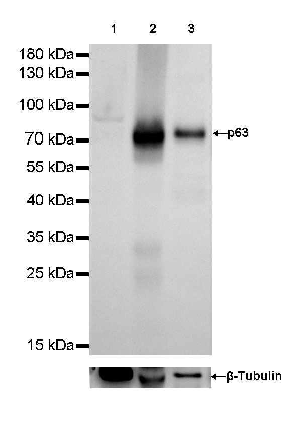
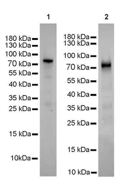
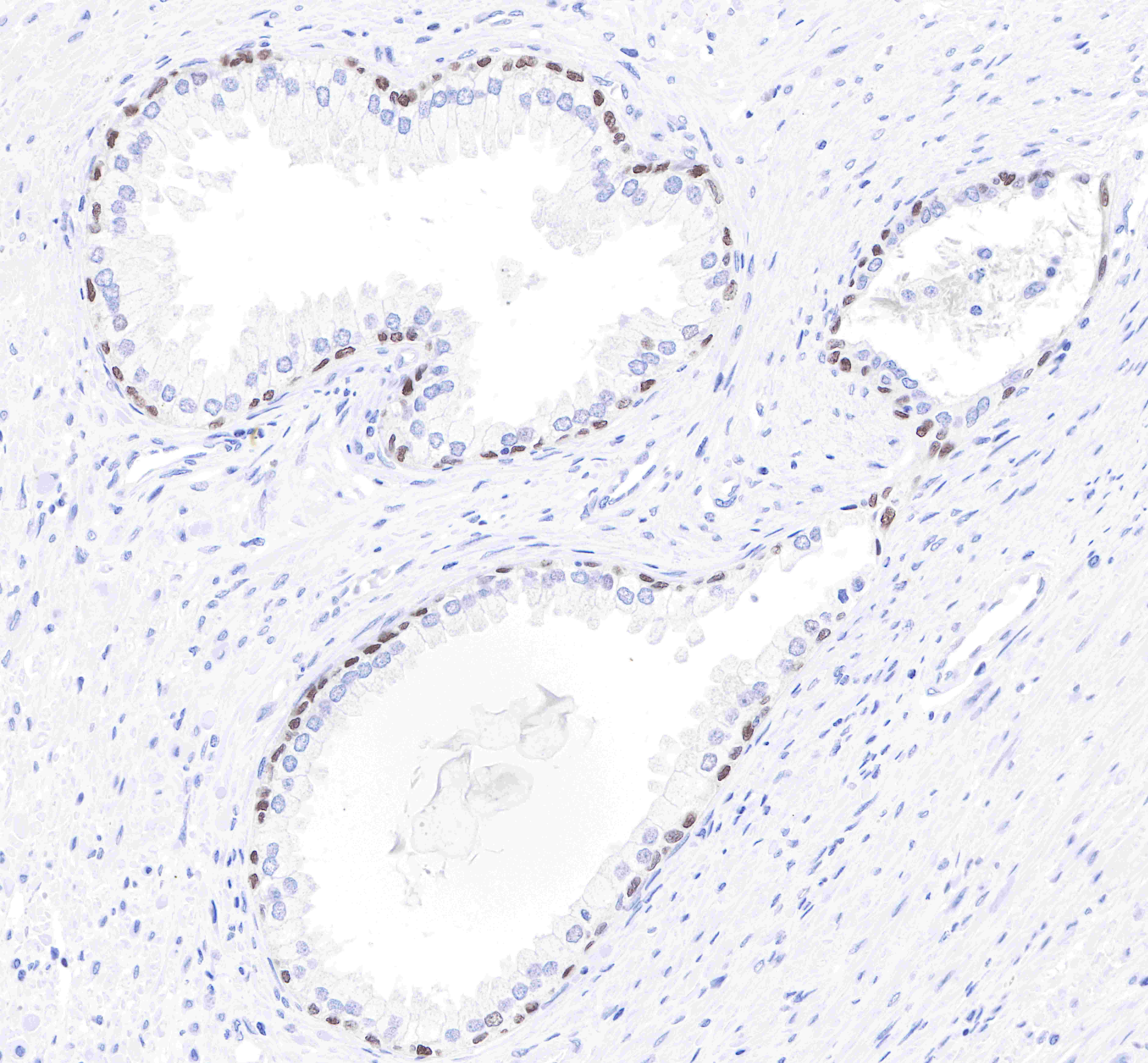
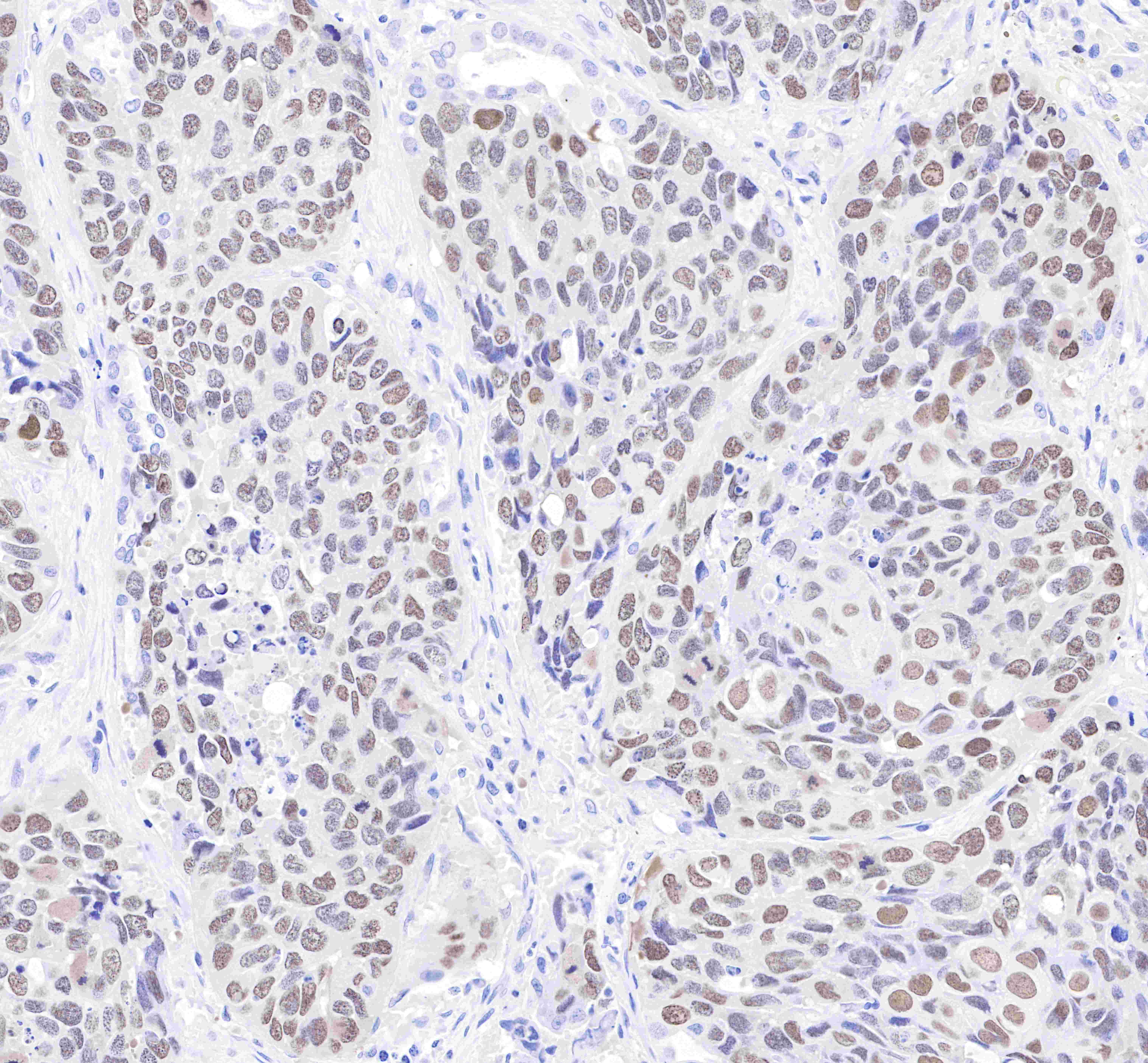
WB, IHC-P |
| Reactivity |
Hu, Ms |
| Purification |
Protein A |
| Concentration |
0.5 mg/ml |
| Conjugation |
Unconjugated |
| Physical Appearance |
Liquid |
| Storage Buffer |
PBS, 40% Glycerol, 0.05%BSA, 0.03% Proclin 300 |
| Stability & Storage |
12 months from date of receipt / reconstitution, -20 °C as supplied |
Dilution
| application |
dilution |
species |
| WB |
1:1000-1:5000 |
|
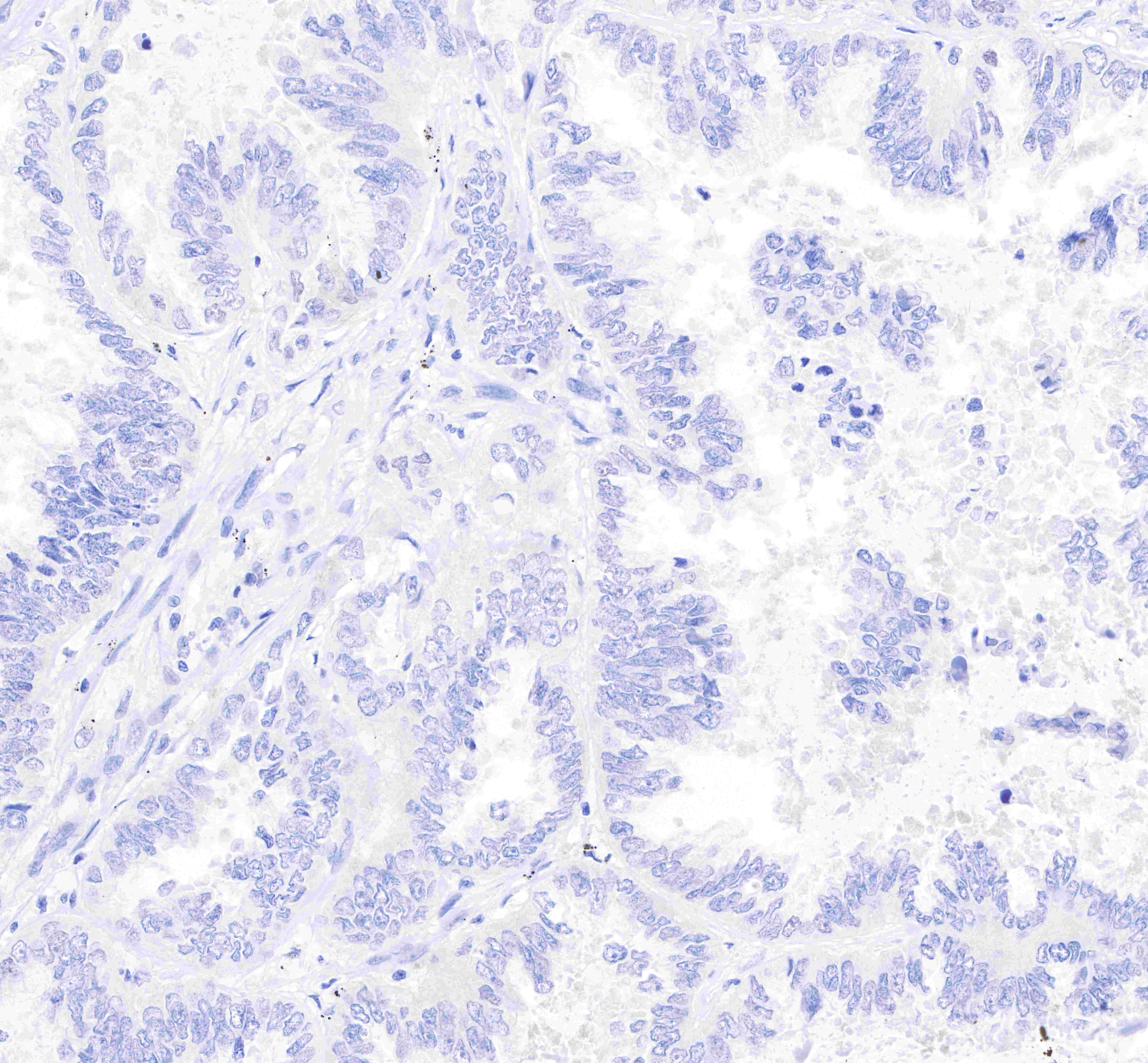
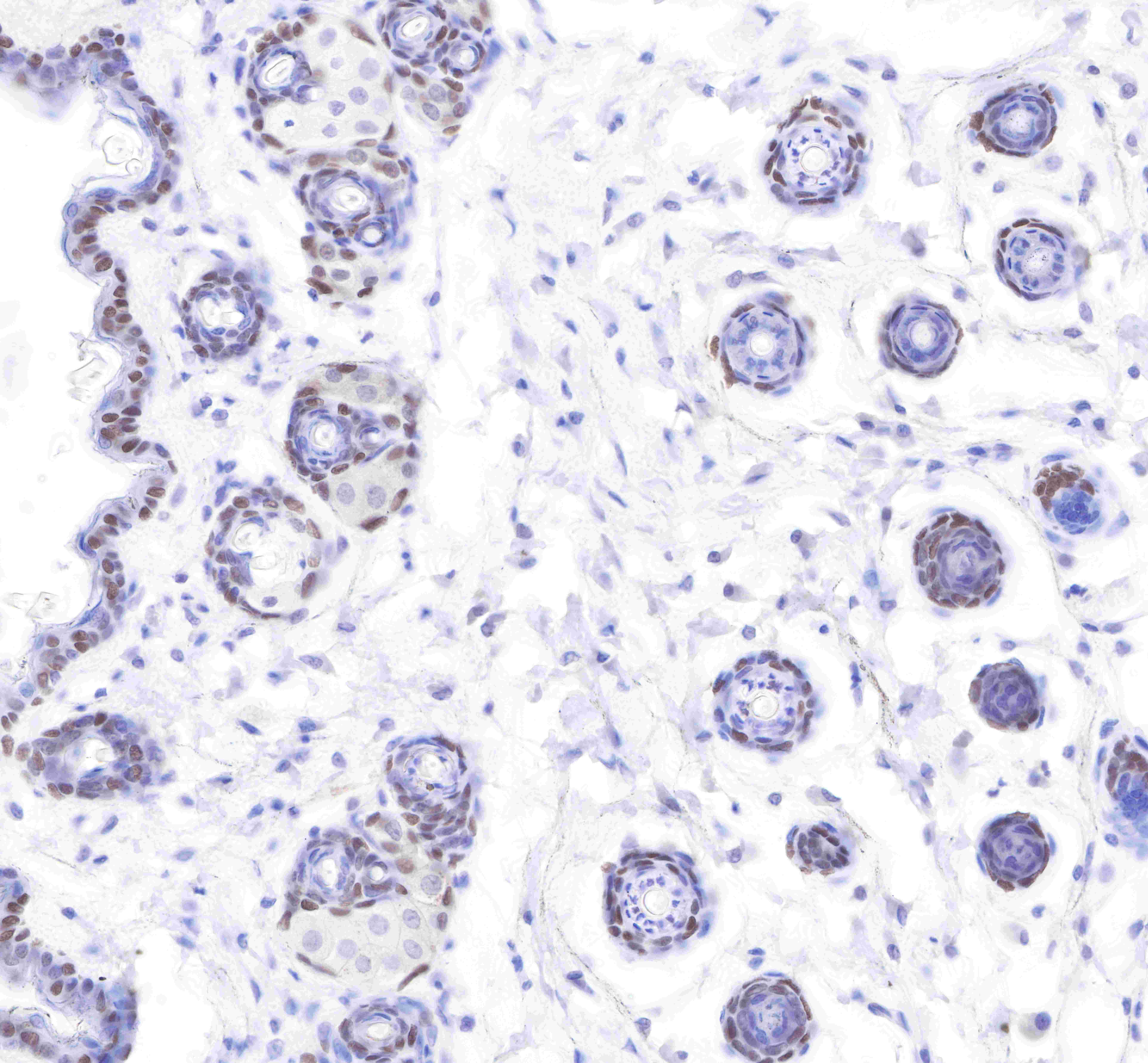
| IHC-P |
1:2000 |
|
Background
Tumor protein p63, typically referred to as p63, also known as transformation-related protein 63 is a protein that in humans is encoded by the TP63 (also known as the p63) gene. Transcription factor p63 is a key regulator of epidermal keratinocyte proliferation and differentiation. Tumor protein p63 is a member of the p53 family of transcription factors. p63 regulates the activity of a multitude of genes involved in growth and development of the ectoderm and derived structures and tissues, such as basal layer keratins and cell cycle control genes. Accordingly, p63 expression is found in basal cell layers of various organs, squamous epithelial cells of many organs and urothelium. p63 (TAp63) is closely related to p40 (ΔNp63) as both proteins represent isoforms of the p63 gene with distinct molecular functions. While “full length” p63 (TAp63) activates p53 target genes such as p21 or BAX, the shorter transcript p40 (ΔNp63) inhibits activation of p53 and “full length” p63. P63 is also helpful in distinguishing poorly differentiated squamous cell carcinoma from small cell carcinoma or adenocarcinoma. P63 should be strongly stained in poorly differentiated squamous cell, but negative in small cell or adenocarcinoma.