WB result of Histone H3 Rabbit mAb
Primary antibody: Histone H3 Rabbit mAb at 1/1000 dilution
Lane 1: HEK293 whole cell lysate 20 µg
Lane 2: HeLa whole cell lysate 20 µg
Secondary antibody: Goat Anti-Rabbit IgG, (H+L), HRP conjugated at 1/10000 dilution
Predicted MW: 15kDa
Observed MW: 17kDa
Exposure time: 3s
Histone H3 Recombinant Rabbit mAb (SDT-266-44)
Histone H3 Recombinant Rabbit mAb (SDT-266-44)
Price:
Regular price
$100 USD
Regular price
Sale price
$100 USD
Unit price
per
For shipping services or bulk orders, you may request a quotation.
Secure checkout with

View full details
Product Details
Product Details
Product Specification
| Host | Rabbit |
| Antigen | Histone H3 |
| Synonyms | Histone H3.1 |
| Immunogen | Synthetic Peptide |
| Location | Nucleus |
| Accession | P68431 |
| Clone Number | SDT-266-44 |
| Antibody Type | Recombinant mAb |
| Isotype | IgG |
| Application | WB, IHC-P, ICC, IP, IF, ICFCM, ChIP |
| Reactivity | Hu, Ms, Rt |
| Positive Sample | HEK293, HeLa, NIH3T3, mouse brain, C6, rat liver, human brain, human spleen, human prostate, human breast cancer, human hepatocellular carcinoma, mouse brain, rat brain |
| Predicted Reactivity | Bv, Dr, C.el, Pl |
| Purification | Protein A |
| Concentration | 0.5 mg/ml |
| Physical Appearance | Liquid |
| Storage Buffer | PBS, 40% Glycerol, 0.05% BSA, 0.03% Proclin 300 |
| Stability & Storage | 12 months from date of receipt / reconstitution, -20 °C as supplied. |
Dilution
| application | dilution | species |
| WB | 1:5000-1:10000 | Hu, Ms, Rt |
| IP | 1:50 | Hu, Ms, Rt |
| IHC-P | 1:4000 | Hu, Ms, Rt |
| ICC | 1:500 | Hu, Ms, Rt |
| IF | 1:500 | Hu, Ms, Rt |
| ICFCM | 1:500 | Hu, Ms, Rt |
| ChIP | 1:20~1:50 | Hu, Ms, Rt |
Background
Histone H3 is a critical component of the nucleosome, which is the basic unit of chromatin. It plays a vital role in the regulation of gene transcription and is subject to various post-translational modifications (PTMs) that affect chromatin structure and gene expression. The H3 family includes several variants, with H3.1, H3.2, and H3.3 being the most well-known. H3.1 and H3.2 are deposited during DNA replication, while H3.3 can be deposited independently of DNA replication and is incorporated throughout the cell cycle2. Post-translational modifications of Histone H3, particularly on the N- and C-terminal tails, can influence the interaction between histones and DNA, as well as with other chromatin factors. These modifications can either promote a relaxed chromatin state that allows gene transcription or lead to a condensed chromatin state that typically suppresses transcription.
Picture
Picture
Western Blot
WB result of Histone H3 Rabbit mAb
Primary antibody: Histone H3 Rabbit mAb at 1/1000 dilution
Lane 1: NIH/3T3 whole cell lysate 20 µg
Lane 2: mouse brain lysate 20 µg
Secondary antibody: Goat Anti-Rabbit IgG, (H+L), HRP conjugated at 1/10000 dilution
Predicted MW: 15kDa
Observed MW: 17kDa
Exposure time: 20s
WB result of Histone H3 Rabbit mAb
Primary antibody: Histone H3 Rabbit mAb at 1/1000 dilution
Lane 1: C6 whole cell lysate 20 µg
Lane 2: rat liver lysate 20 µg
Secondary antibody: Goat Anti-Rabbit IgG, (H+L), HRP conjugated at 1/10000 dilution
Predicted MW: 15kDa
Observed MW: 17kDa
Exposure time: 20s
FC
Flow cytometric analysis of HeLa cells labelling Histone H3 antibody at 1/500 (0.1 μg) dilution/ (red) compared with a Rabbit monoclonal IgG (Black) isotype control and an unlabelled control (cells without incubation with primary antibody and secondary antibody) (Blue). Goat Anti-Rabbit IgG Alexa Fluor® 488 was used as the secondary antibody.
IP
Histone H3 Rabbit mAb at 1/50 dilution (1 µg) immunoprecipitating Histone H3 in 0.4 mg HeLa whole cell lysate.
Western blot was performed on the immunoprecipitate using Histone H3 Rabbit mAb at 1/1000 dilution.
Secondary antibody (HRP) for IP was used at 1/400 dilution.
Lane 1: HeLa whole cell lysate 20 µg (Input)
Lane 2: Histone H3 Rabbit mAb IP in HeLa whole cell lysate
Lane 3: Rabbit monoclonal IgG IP in HeLa whole cell lysate
Predicted MW: 15 kDa
Observed MW: 15 kDa
ChIP
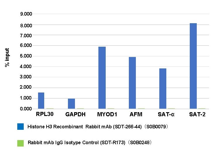
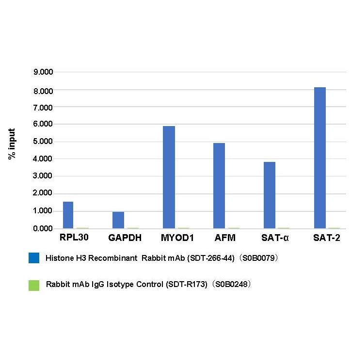
Chromatin immunoprecipitation (ChIP) was performed on HeLa cells cross - linked with 1% formaldehyde for 10 min, then chromatin was fragmented by sonication. Parallel reactions used Histone H3 Recombinant Rabbit mAb (SDT-266-44) and Rabbit mAb IgG Isotype Control (SDT-R173) at 1:50 for immunoprecipitation. Post - immunoprecipitation, both samples were washed, eluted, and cross - links reversed. Purified DNA was analyzed by qPCR.
qPCR (%input: immunoprecipitated DNA/input DNA) showed the enrichment of RPL30, GAPDH, MYOD1, AFM, SAT-α and SAT-2 in Histone H3 Recombinant Rabbit mAb (SDT-266-44)-immunoprecipitated sample.
Immunohistochemistry
IHC shows positive staining in paraffin-embedded human spleen. Anti-Histone H3 antibody was used at 1/4000 dilution, followed by a HRP Polymer for Mouse & Rabbit IgG (ready to use). Counterstained with hematoxylin. Heat mediated antigen retrieval with Tris/EDTA buffer pH9.0 was performed before commencing with IHC staining protocol.
IHC shows positive staining in paraffin-embedded human prostate. Anti-Histone H3 antibody was used at 1/4000 dilution, followed by a HRP Polymer for Mouse & Rabbit IgG (ready to use). Counterstained with hematoxylin. Heat mediated antigen retrieval with Tris/EDTA buffer pH9.0 was performed before commencing with IHC staining protocol.
IHC shows positive staining in paraffin-embedded human breast cancer. Anti-Histone H3 antibody was used at 1/4000 dilution, followed by a HRP Polymer for Mouse & Rabbit IgG (ready to use). Counterstained with hematoxylin. Heat mediated antigen retrieval with Tris/EDTA buffer pH9.0 was performed before commencing with IHC staining protocol.
IHC shows positive staining in paraffin-embedded human hepatocellular carcinoma. Anti-Histone H3 antibody was used at 1/4000 dilution, followed by a HRP Polymer for Mouse & Rabbit IgG (ready to use). Counterstained with hematoxylin. Heat mediated antigen retrieval with Tris/EDTA buffer pH9.0 was performed before commencing with IHC staining protocol.
IHC shows positive staining in paraffin-embedded mouse brain. Anti-Histone H3 antibody was used at 1/4000 dilution, followed by a HRP Polymer for Mouse & Rabbit IgG (ready to use). Counterstained with hematoxylin. Heat mediated antigen retrieval with Tris/EDTA buffer pH9.0 was performed before commencing with IHC staining protocol.
IHC shows positive staining in paraffin-embedded rat brain. Anti-Histone H3 antibody was used at 1/4000 dilution, followed by a HRP Polymer for Mouse & Rabbit IgG (ready to use). Counterstained with hematoxylin. Heat mediated antigen retrieval with Tris/EDTA buffer pH9.0 was performed before commencing with IHC staining protocol.
Immunocytochemistry
ICC shows positive staining in HeLa cells. Anti-Histone H3 antibody was used at 1/500 dilution (Green) and incubated overnight at 4°C. Goat polyclonal Antibody to Rabbit IgG - H&L (Alexa Fluor® 488) was used as secondary antibody at 1/1000 dilution. The cells were fixed with 100% ice-cold methanol and permeabilized with 0.1% PBS-Triton X-100. Nuclei were counterstained with DAPI (Blue). Counterstain with tubulin (red).
Immunofluorescence
IF shows positive staining in paraffin-embedded human breast cancer. Anti-Histone H3 antibody was used at 1/500 dilution (Green) and incubated overnight at 4°C. Goat polyclonal Antibody to Rabbit IgG - H&L (Alexa Fluor® 488) was used as secondary antibody at 1/1000 dilution. Counterstained with DAPI (Blue). Heat mediated antigen retrieval with EDTA buffer pH9.0 was performed before commencing with IF staining protocol.
IF shows positive staining in paraffin-embedded human colon cancer. Anti-Histone H3 antibody was used at 1/500 dilution (Green) and incubated overnight at 4°C. Goat polyclonal Antibody to Rabbit IgG - H&L (Alexa Fluor® 488) was used as secondary antibody at 1/1000 dilution. Counterstained with DAPI (Blue). Heat mediated antigen retrieval with EDTA buffer pH9.0 was performed before commencing with IF staining protocol.
IF shows positive staining in paraffin-embedded human ovarian cancer. Anti-Histone H3 antibody was used at 1/500 dilution (Green) and incubated overnight at 4°C. Goat polyclonal Antibody to Rabbit IgG - H&L (Alexa Fluor® 488) was used as secondary antibody at 1/1000 dilution. Counterstained with DAPI (Blue). Heat mediated antigen retrieval with EDTA buffer pH9.0 was performed before commencing with IF staining protocol.
IF shows positive staining in paraffin-embedded mouse testis. Anti-Histone H3 antibody was used at 1/500 dilution (Green) and incubated overnight at 4°C. Goat polyclonal Antibody to Rabbit IgG - H&L (Alexa Fluor® 488) was used as secondary antibody at 1/1000 dilution. Counterstained with DAPI (Blue). Heat mediated antigen retrieval with EDTA buffer pH9.0 was performed before commencing with IF staining protocol.
IF shows positive staining in paraffin-embedded rat testis. Anti-Histone H3 antibody was used at 1/500 dilution (Green) and incubated overnight at 4°C. Goat polyclonal Antibody to Rabbit IgG - H&L (Alexa Fluor® 488) was used as secondary antibody at 1/1000 dilution. Counterstained with DAPI (Blue). Heat mediated antigen retrieval with EDTA buffer pH9.0 was performed before commencing with IF staining protocol.
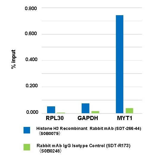
ChIP

Chromatin immunoprecipitation (ChIP) was performed on HeLa cells cross - linked with 1% formaldehyde for 10 min, then chromatin was fragmented by sonication. Parallel reactions used Histone H3 Recombinant Rabbit mAb (SDT-266-44) and Rabbit mAb IgG Isotype Control (SDT-R173) at 1:50 for immunoprecipitation. Post - immunoprecipitation, both samples were washed, eluted, and cross - links reversed. Purified DNA was analyzed by qPCR.
qPCR (%input: immunoprecipitated DNA/input DNA) showed the enrichment of RPL30, GAPDH and MYT1 in Histone H3 Recombinant Rabbit mAb (SDT-266-44)-immunoprecipitated sample.