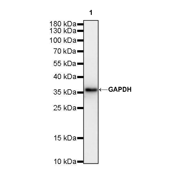
WB result of GAPDH Rabbit mAb
Primary antibody: GAPDH Rabbit mAb at 1/10000 dilution
Lane 1: zebrafish lysate 20 µg
Secondary antibody: Goat Anti-Rabbit IgG, (H+L), HRP conjugated at 1/10000 dilution
Predicted MW: 37 kDa
Observed MW: 37 kDa
GAPDH Recombinant Rabbit mAb (S-240-147)
GAPDH Recombinant Rabbit mAb (S-240-147)
Price:
Regular price
$70 USD
Regular price
Sale price
$70 USD
Unit price
per
For shipping services or bulk orders, you may request a quotation.
Secure checkout with

View full details
Product Details
Product Details
Product Specification
| Host | Rabbit |
| Antigen | GAPDH |
| Synonyms | Glyceraldehyde-3-phosphate dehydrogenase, Peptidyl-cysteine S-nitrosylase GAPDH |
| Immunogen | Synthetic Peptide |
| Location | Cytoplasm, Nucleus, Membrane |
| Accession | P04406、P16858 |
| Clone Number | S-240-147 |
| Antibody Type | Rabbit mAb |
| Application | WB, IHC-P, ICC |
| Reactivity | Hu, Ms, Rt, Zf |
| Predicted Reactivity | Hamster (95%), Mink (95%), Pig (95%), Squirrel (95%),Dog (95%), Cat (95%), Sheep (95%), Quail (95%), Bovine (95%), Chicken (95%) |
| Purification | Protein A |
| Concentration | 1 mg/ml |
| Conjugation | Unconjugated |
| Physical Appearance | Liquid |
| Storage Buffer | PBS, 40% Glycerol, 0.05% BSA, 0.03% Proclin 300 |
| Stability & Storage | 12 months from date of receipt / reconstitution, -20 °C as supplied. |
Dilution
| application | dilution | species |
| WB | 1:10000-1:50000 | |
| IHC | 1:5000 | |
| ICC | 1:50-1:200 |
Background
Glyceraldehyde 3-phosphate dehydrogenase (GAPDH) is an enzyme of about 37kDa that catalyzes the sixth step of glycolysis and thus serves to break down glucose for energy and carbon molecules [PMID: 17072346]. GAPDH comprises a polypeptide chain of 335 amino acids. Structural studies identified two regions, namely the glyceraldehyde-3-phosphate catalytic site and the NAD+ binding site, a primary structure known as the Rossmann fold, which is also required for the activity of other dehydrogenases. Many of these roles are dependent on the ability of GAPDH to bind different macromolecules in the cell.
Picture
Picture
Western Blot
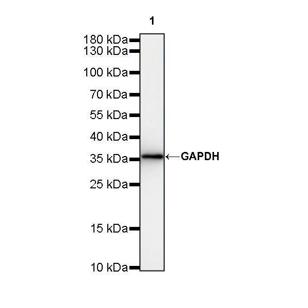
WB result of GAPDH Rabbit mAb
Primary antibody: GAPDH Rabbit mAb at 1/50000 dilution
Lane 1: zebrafish lysate 20 µg
Secondary antibody: Goat Anti-Rabbit IgG, (H+L), HRP conjugated at 1/10000 dilution
Predicted MW: 37 kDa
Observed MW: 37 kDa
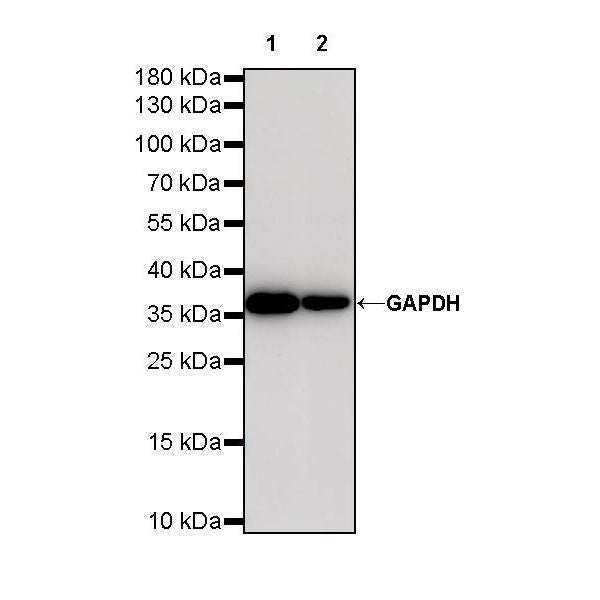
WB result of GAPDH Rabbit mAb Primary antibody: GAPDH Rabbit mAb at 1/10000 dilution Lane 1: HeLa whole cell lysate 20 µg Lane 2: THP-1 whole cell lysate 20 µg Lane 3: MCF7 whole cell lysate 20 µg Lane 4: NIH/3T3 whole cell lysate 20 µg Lane 5: mouse spleen lysate 20 µg Lane 6: C6 whole cell lysate 20 µg Lane 7: rat spleen lysate 20 µg Secondary antibody: Goat Anti-Rabbit IgG, (H+L), HRP conjugated at 1/10000 dilution Predicted MW: 37 kDa Observed MW: 37 kDa
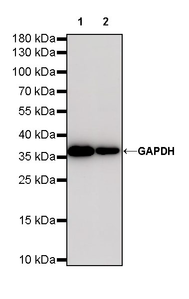
WB result of GAPDH Rabbit mAb Primary antibody: GAPDH Rabbit mAb at 1/50000 dilution Lane 1: HeLa whole cell lysate 20 µg Lane 2: THP-1 whole cell lysate 20 µg Secondary antibody: Goat Anti-Rabbit IgG, (H+L), HRP conjugated at 1/10000 dilution Predicted MW: 37 kDa Observed MW: 37 kDa
Immunohistochemistry
IHC shows positive staining in paraffin-embedded human spleen. Anti-GAPDH antibody was used at 1/5000 dilution, followed by a HRP Polymer for Mouse & Rabbit IgG (ready to use). Counterstained with hematoxylin. Heat mediated antigen retrieval with Tris/EDTA buffer pH9.0 was performed before commencing with IHC staining protocol.
IHC shows positive staining in paraffin-embedded mouse kidney. Anti-GAPDH antibody was used at 1/5000 dilution, followed by a HRP Polymer for Mouse & Rabbit IgG (ready to use). Counterstained with hematoxylin. Heat mediated antigen retrieval with Tris/EDTA buffer pH9.0 was performed before commencing with IHC staining protocol.
IHC shows positive staining in paraffin-embedded rat testis. Anti-GAPDH antibody was used at 1/5000 dilution, followed by a HRP Polymer for Mouse & Rabbit IgG (ready to use). Counterstained with hematoxylin. Heat mediated antigen retrieval with Tris/EDTA buffer pH9.0 was performed before commencing with IHC staining protocol.
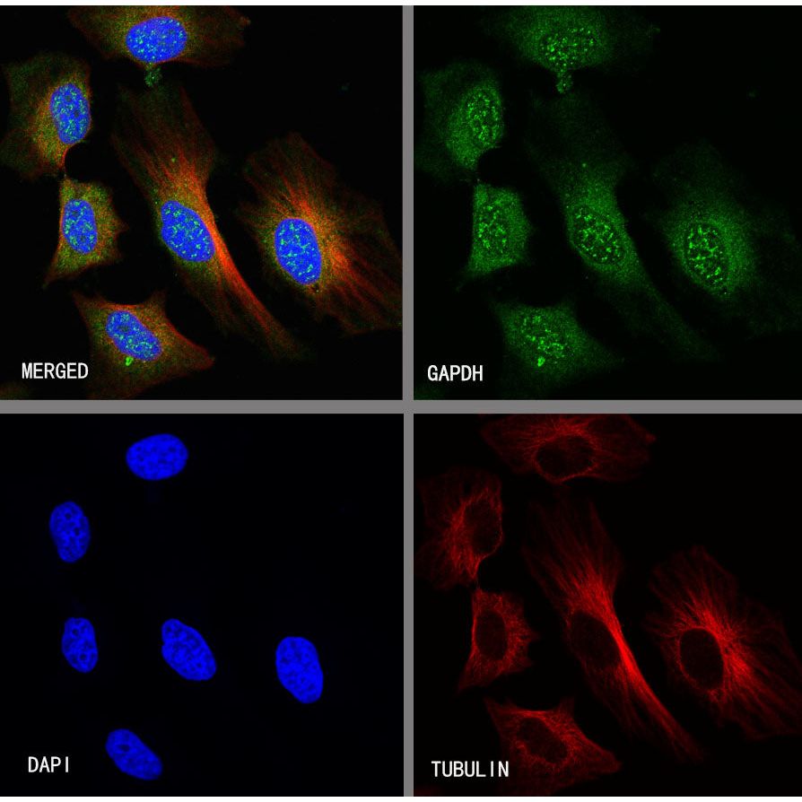
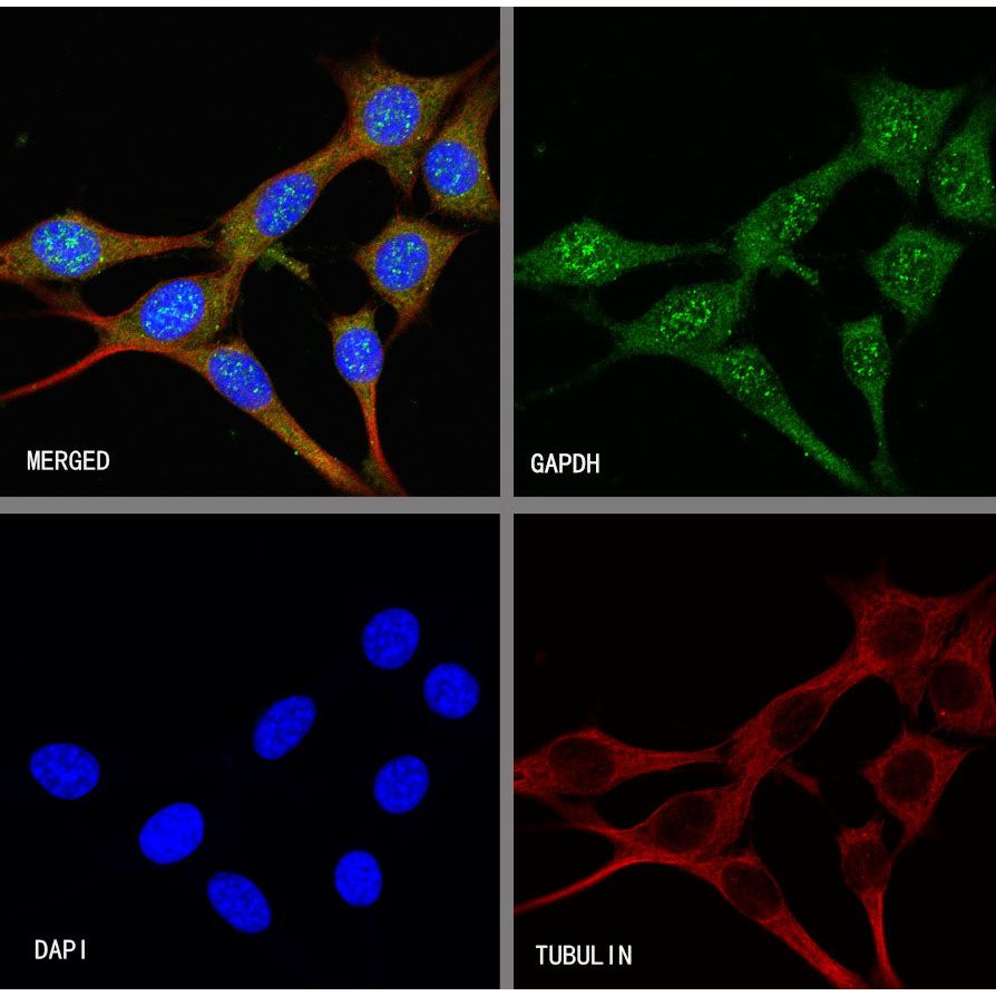
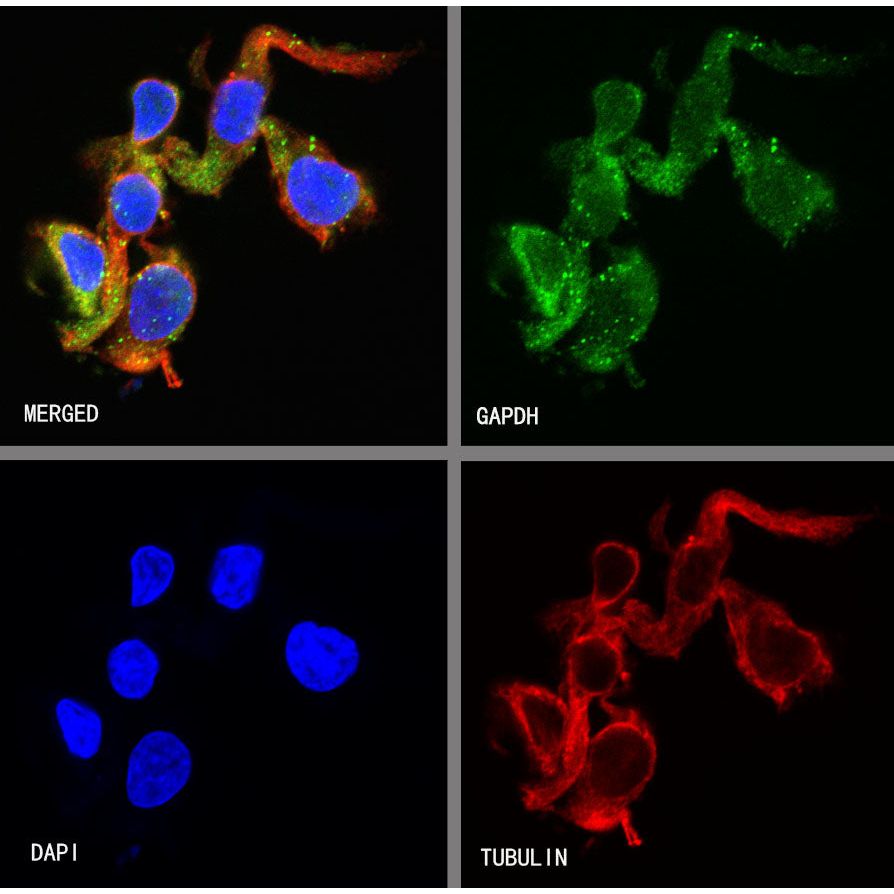
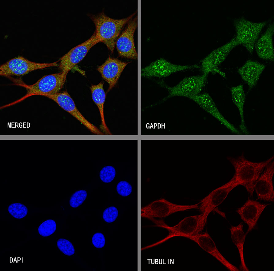
Immunocytochemistry
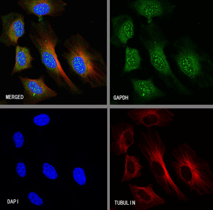
ICC shows positive staining in HeLa cells. Anti-GAPDH antibody was used at 1/50 dilution (Green) and incubated overnight at 4°C. Goat polyclonal Antibody to Rabbit IgG - H&L (Alexa Fluor® 488) was used as secondary antibody at 1/1000 dilution. The cells were fixed with 100% ice-cold methanol and permeabilized with 0.1% PBS-Triton X-100. Nuclei were counterstained with DAPI (Blue).Counterstain with tubulin (Red).
ICC shows positive staining in NIH/3T3 cells. Anti-GAPDH antibody was used at 1/200 dilution (Green) and incubated overnight at 4°C. Goat polyclonal Antibody to Rabbit IgG - H&L (Alexa Fluor® 488) was used as secondary antibody at 1/1000 dilution. The cells were fixed with 100% ice-cold methanol and permeabilized with 0.1% PBS-Triton X-100. Nuclei were counterstained with DAPI (Blue).Counterstain with tubulin (Red).
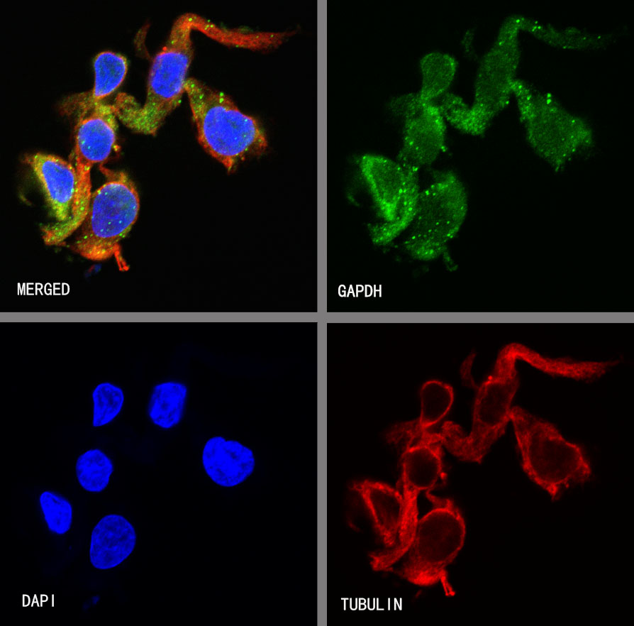
ICC shows positive staining in C6 cells. Anti-GAPDH antibody was used at 1/200 dilution (Green) and incubated overnight at 4°C. Goat polyclonal Antibody to Rabbit IgG - H&L (Alexa Fluor® 488) was used as secondary antibody at 1/1000 dilution. The cells were fixed with 4% PFA and permeabilized with 0.1% PBS-Triton X-100. Nuclei were counterstained with DAPI (Blue).Counterstain with tubulin (Red).