| Usage |
1. Sample collection preparation and preservation
- Serum: Whole blood sample placed at room temperature 2 Hour or 4°C Overnight after 1000×g Centrifugation 20 Minutes, take the supernatant to detect.
The blood collection tubes shall be disposable non-pyrogenic, non-endotoxin tubes.
deposit -20°C Or -80°C Storage, avoid repeated freezing and thawing.
- Plasma: Sample after collection 30 Within minutes 2-8°C 、 1000×g Centrifugation 15 Minutes, take the supernatant to detect.
Anticoagulants recommended EDTA-Na2 , avoid using hemolytic, hyperlipidemic samples.
deposit -20°C Or -80°C Storage, avoid repeated freezing and thawing.
- Tissue homogenate: Take an appropriate amount of tissue block and add it to the pre-cooled PBS ( 0.01M , pH7.0-7.2 ) to remove blood (lysed red blood cells in the homogenate will affect the measurement result), cut the tissue into pieces after weighing, and then mix it with the corresponding volume of PBS (generally according to 1:9 The mass-to-volume ratio, the specific volume can be appropriately adjusted according to the needs of the experiment, and recorded.
It is recommended to PBS Adding a protease inhibitor) into a glass homogenizer and fully grinding on ice;
In order to further lyse tissue cells, the homogenate can be subjected to ultrasonic disruption or repeated freeze-thaw treatment (pay attention to ice bath cooling during ultrasonic disruption, and the repeated freeze-thaw method can be repeated 2 Times).
Finally, the prepared homogenate is mixed in 5000×g Centrifugation 5-10 Minutes, take the supernatant to detect.
- Cell culture supernatant: Take the cell supernatant from 1000×g Centrifugation 20 Minutes, impurities and cell debris were removed.
Take the supernatant to detect and place it in -20°C Or -80°C Store, but repeated freezing and thawing should be avoided.
- Urine: Please collect the first urine in the morning (mid-section urine), or 24 Hourly urine, 2000×g Centrifugation 15 Minutes later the supernatant was collected and the sample was stored at -20°C And repeated freezing and thawing should be avoided.
- Saliva: A sample is collected with a saliva sample collection tube, and then 2-8°C, 1000×g Centrifugation 15 Minutes, take the supernatant to detect, or sub-package -20°C Save.
Avoid repeated freezing and thawing.
- Other biological samples: Please 1000×g Centrifugation 20 Minutes, take the supernatant to detect.
attention :
- The sample should be clear and transparent, and the suspended solids should be removed by centrifugation.
Hemolysis of the sample will affect the results, so hemolyzed samples should not be used.
- After sample collection, if 1 Testing within weeks can be stored at 4°C , if it cannot be detected in time, please pack it according to the one-time usage amount and freeze it in -20°C ( 1 Within months), or -80°C ( 3-6 Test within a month) to avoid repeated freezing and thawing.
Keep the sample at room temperature prior to the experiment.
- If the concentration of the detected substance in your sample is higher than the highest value of the standard, please make an appropriate dilution according to the actual situation (it is recommended to do a pre-experiment first to determine the dilution factor).
Two, Preparation for testing
- Please advance 30 Minutes remove the kit from the refrigerator and equilibrate to room temperature.
- Use double distilled water 25× The concentrated wash liquid is diluted to 1× Working fluid, put back unused 4°C 。
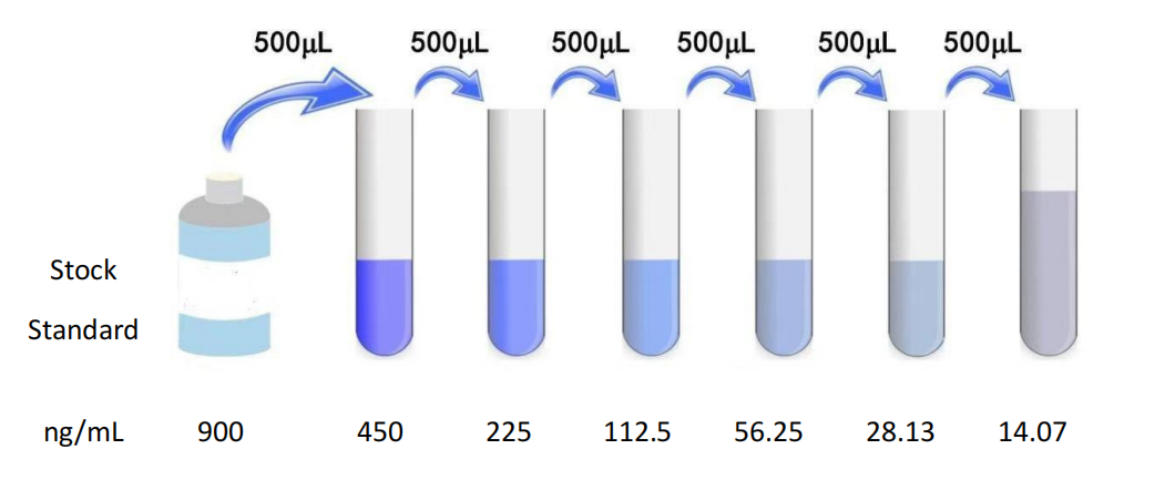
- Standard: Add standard & Sample Universal Diluent 1.0 mL Into the lyophilized standard, screw the tube cap tightly and let stand 10 Minutes, and after it is fully dissolved, gently mix (concentration of 900 ng/mL )。
Thereafter, double dilution is carried out to 900 ng/mL , 450 ng/mL , 225 ng/mL , 112.5 ng/mL , 56.25 ng/mL , 28.13 ng/mL , 14.07 ng/mL 。
Standard dilution ( 0 ng/mL ) is a blank hole.
Configure the standard according to the amount you need for later use.
The configured standards are recommended in 15 Add the sample within minutes, and it is not recommended to leave it for too long.
- Biotin conjugate working solution (1x) : Centrifuge before opening the bottle.
Dilute with biotin conjugate diluent immediately before use, and prepare according to the pre-calculated total amount required for each experiment before dilution ( Per well 50 μL) , the actual preparation should be more prepared 100-200μL 。
Such as 10 μL Biotin conjugate plus 990 μL The proportion of biotin conjugate dilution is formulated, gently mixed, and formulated within one hour before use.
- Streptomycin - Horseradish peroxidase conjugate working solution (1x) : Centrifuge before opening the bottle.
Dilute with enzyme conjugate diluent immediately before use, and prepare according to the pre-calculated total amount required for each experiment before dilution ( Per well 100 μL) , the actual preparation should be more prepared 100-200μL 。
Such as 10 μL Enzyme conjugate plus 990 μL Prepare the proportion of the enzyme conjugate diluent, gently mix well, and prepare within one hour before use.
- TMB Substrate —— Pipette the desired dose of solution and do not pour the residual solution back into the reagent vial again.
attention :
- Please make sure that all components are dissolved and mixed before use of the kit.
If the reconstituted standard is not used, please discard it.
- Concentrated biotin conjugate, concentrated enzyme conjugate is small in volume, may be dispersed in various parts of the tube during transportation, please 1000×g Centrifugation 1 Minutes to allow the liquid of the tube wall or cap to deposit to the bottom of the tube.
Pipette carefully before use 4-5 The solution was mixed once.
Standard, biotin conjugate working solution and enzyme conjugate working solution should be prepared according to the required dosage, and the corresponding diluent should be use to prepare without confusion.
- The concentrated washing liquid taken out of the refrigerator may have crystals, which is a normal phenomenon.
The crystals can be completely dissolved in a water bath or incubator before preparing the washing liquid (the heating temperature should not exceed 40°C )。
The wash liquid should be at room temperature when used.
- Adding samples should be quick, and it is best to control each sample adding within 10 Within minutes, in order to ensure the accuracy of the experiment, it is recommended to use a double hole.
When pipetting reagents, a consistent sequence of addition is maintained from well to well,
This will ensure the same hatch time for all holes.
- During the washing process, the washing liquid remaining in the reaction hole should be patted dry on absorbent paper, and do not put the filter paper directly into the reaction hole to absorb water.
Before reading, pay attention to removing the residual liquid and fingerprints at the bottom, so as not to affect the reading of the microplate reader.
- Color developer TMB Direct exposure to bright light should be avoided during storage and use.
After adding the substrate, pay attention to the color change in the reaction well.
If the gradient is obvious, please terminate the reaction in advance to avoid too dark color affecting the reading of the microplate reader.
- The test tubes and reagents used in the experiment are disposable, and it is strictly forbidden to reuse them, otherwise it will affect the experimental results.
- During the experiment, please wear a laboratory coat and latex gloves for protection, especially when testing blood or other body fluid samples, please follow the national biological laboratory safety protection regulations.
- The kit components of different lot numbers cannot be mixed (except wash solution and reaction stop solution).
- The enzyme labeling strips in the kit are detachable plates, please use them in batches according to the experimental requirements.
Three, Operation steps
- Before the start of the experiment, each reagent should be balanced to room temperature, and all reagents should be configured in advance.
When reagents or samples are diluted, they should be mixed evenly.
Try to avoid foaming when mixing well.
If the sample concentration is too high, dilute with a sample diluent to bring the sample within the range of the kit.
- Sample addition: Standard holes and sample holes to be tested are set respectively.
Add standard or sample to be tested 50 μL , be careful not to have bubbles, add the sample to the bottom of the well of the enzyme labeled plate, try not to touch the well wall, and then add biotin conjugate to each well (1 x) 50 μL Gently shaking and mixing evenly, adding a cover or film to the enzyme label plate, 37°C Incubation 1 Hours.
- To ensure the validity of the experimental results, please use a new standard solution for each experiment.
- Incubation 1 After hours, discard the liquid in the hole, spin dry, and wash the plate 3 Times, add washing solution to each well (1 x) 200 μL , each soak 1-2 Minutes, tumble dry.
- Then add streptomycin to each well -HRP(1 x) 100 μL Gently shaking and mixing evenly, adding a cover or film to the enzyme label plate, 37°C Incubation 1 Hours.
- Discard the liquid in the hole, spin dry, wash the plate 5 Times, add washing solution to each well (1 x) 200 μL , each soak 1-2 Minutes, tumble dry.
- Add each well sequentially TMB Color developer 90 μL , 37°C Color development protected from light 15-20 Minutes (shortened or extended as appropriate according to the actual color development, but not exceeding 30 Minutes).
- Add stop solution to each well sequentially 50 μL , terminate the reaction (blue immediately turns yellow at this time).
The sequence of addition of the stop solution should be the same as that of the substrate solution as possible.
In order to ensure the accuracy of the experimental results, the termination solution should be added as soon as possible after the substrate reaction time expires.
- Use a microplate reader in 450 nm The optical density of each well was measured sequentially by wavelength (OD Value ) 。
After addition of stop solution 5 Test within minutes.
- Samples may need to be diluted.
Please see the sample processing section.
Results Calculation
- Competition Law Standards and Samples OD Values can be directly substituted into the calculation.
If a double hole is set, the average value should be taken for calculation.
- For ease of calculation, although the concentration is an independent variable and OD The value is the dependent variable, and we still use the standard when drawing OD Values as abscissa ( X Axis), the concentration of the standard is the ordinate ( Y Axis).
At the same time, for the intuition of the test results, the figure provides raw data instead of logarithmic values.
Due to the different experimental operating conditions (such as operator, pipetting technology, plate washing technology and temperature conditions, etc.), the standard curve OD Values will vary.
The standard curve provided is for reference only, and the experimenter needs to establish the standard curve according to his own experiment.
Spent sample OD The value can be calculated on the standard curve to calculate the sample concentration and multiply it by the dilution factor, which is the actual concentration of the sample.
It is recommended to use professional curve drawing software such as curve expert 。
Concentration (ng/mL) |
OD |
900 |
0.183 |
450 |
0.385 |
225 |
0.575 |
112.5 |
0.823 |
56.25 |
1.157 |
28.13 |
1.592 |
14.07 |
1.624 |
0 |
2.256 |
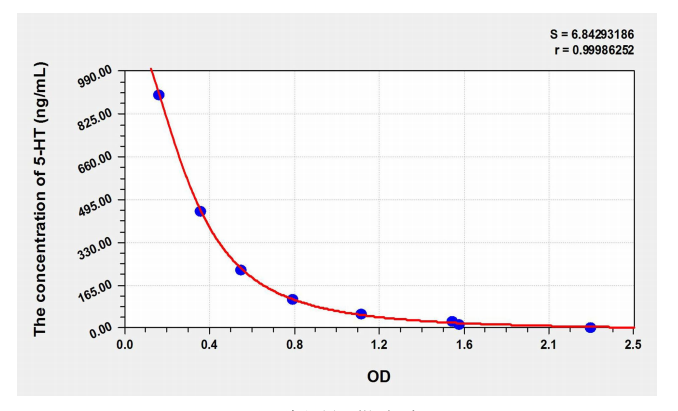
Note: This table is for reference only
Precision
Intraplate precision ( Precision within the assay ):CV%<8%
Three samples with known concentrations were respectively in 1 Test on enzyme label plates 20 Times to evaluate the precision in the assay plate.
Inter-plate precision ( Measure inter-plate precision ):CV%<10%
Three samples with known concentrations were respectively in 3 Tested on different enzyme plates 40 Times to assess the accuracy between the assay plates.
Recovery
Add known concentrations to different samples 5-HT , do the recovery experiment, and get the recovery range and average recovery rate.
Sample Type |
Recovery Range |
Average recovery |
Serum (n=5) |
88-103% |
95% |
EDTA Plasma (n=5) |
95-107% |
101% |
heparin Plasma (n=5) |
80-95% |
93% |
linear
Will be added with 5-HT The samples were diluted separately 2 Times, 4 Times, 8 Times, 16 Double the recovery experiment to obtain the recovery rate range
Sample Type |
1:2 |
1:4 |
1:8 |
1:16 |
Serum (n=5) |
82-93% |
95-104% |
87-98% |
92-99% |
EDTA Plasma (n=5) |
89-99% |
83-96% |
98-104% |
88-101% |
heparin Plasma (n=5) |
87-96% |
87-96% |
96-105% |
95-103% |
|

