Rat Insulin ELISA Kit
Rat Insulin ELISA Kit
Product Details
Product Details
Product Specification
| protein | Insulin | ||||||||||||||||||||||||||||||||||||
| Usage |
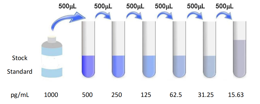
1. Sample collection preparation and preservation 1 Serum: whole blood sample placed at room temperature 2 Hour or 4°C Overnight after 1000×g Centrifugation 20 Minutes, take the supernatant to detect. The blood collection tubes shall be disposable non-pyrogenic, non-endotoxin tubes. deposit -20°C Or -80°C Storage, avoid repeated freezing and thawing. 2 Plasma: the sample after collection 30 Within minutes 2-8°C 、 1000×g Centrifugation 15 Minutes, take the supernatant to detect. Anticoagulants recommended EDTA-Na2 , avoid using hemolytic, hyperlipidemic samples. deposit -20°C Or -80°C Storage, avoid repeated freezing and thawing. 3 , tissue homogenate: take an appropriate amount of tissue block and put it on the pre-cooled PBS ( 0.01M , pH7.0-7.2 ) to remove blood (lysed red blood cells in the homogenate will affect the measurement result), cut the tissue into pieces after weighing, and then mix it with the corresponding volume of PBS (generally according to 1:9 The mass-to-volume ratio, the specific volume can be appropriately adjusted according to the needs of the experiment, and recorded. It is recommended to PBS Adding a protease inhibitor) into a glass homogenizer and fully grinding on ice; In order to further lyse tissue cells, the homogenate can be subjected to ultrasonic disruption or repeated freeze-thaw treatment (pay attention to ice bath cooling during ultrasonic disruption, and the repeated freeze-thaw method can be repeated 2 Times). Finally, the prepared homogenate is mixed in 5000×g Centrifugation 5-10 Minutes, take the supernatant to detect. 4 Cell culture supernatant: the cell supernatant was taken from 1000×g Centrifugation 20 Minutes, impurities and cell debris were removed. Take the supernatant to detect and place it in -20°C Or -80°C Store, but repeated freezing and thawing should be avoided. 5 , urine: Please collect the first urine in the morning (middle urine), or 24 Hourly urine, 2000×g Centrifugation 15 The supernatant was collected after minutes and the sample was saved At -20°C And repeated freezing and thawing should be avoided. 6 Saliva: collecting a sample with a saliva sample collection tube, and then at 2-8°C, 1000×g Centrifugation 15 Minutes, take the supernatant to detect, or sub-package -20°C Save. Avoid repeated freezing and thawing. 7 Other biological samples: please 1000×g Centrifugation 20 Minutes, take the supernatant to detect. attention : 1 The sample should be clear and transparent, and the suspended solids should be removed by centrifugation. Hemolysis of the sample will affect the results, so hemolyzed samples should not be used. 2 , after sample collection, if 1 Testing within weeks can be stored at 4°C , if it cannot be detected in time, please pack it according to the one-time usage amount and freeze it in -20°C ( 1 Within months), or -80°C ( 3-6 Test within a month) to avoid repeated freezing and thawing. Keep the sample at room temperature prior to the experiment. 3 If the concentration of the detected substance in your sample is higher than the highest value of the standard product, please dilute it at an appropriate multiple according to the actual situation (it is recommended to do a pre-experiment first to determine the dilution multiple). Two, Preparation for testing 1 , please advance 30 Minutes remove the kit from the refrigerator and equilibrate to room temperature. 2 , using double-distilled water 25× The concentrated wash liquid is diluted to 1× Working fluid, put back unused 4°C 。 3 Standard: Add standard & Sample Universal Diluent 1 、 0 mL Into the lyophilized standard, screw the tube cap tightly and let stand 10 Minutes, and after it is fully dissolved, gently mix (concentration of 1000 pg/mL )。 Thereafter, double dilution is carried out to 1000 pg/mL , 500 pg/mL , 250 pg/mL , 125 pg/mL , 62.5 pg/mL , 31.25 pg/mL , 15.63 pg/mL Standard dilution ( 0 pg/mL ) is a blank hole. Configure the standard according to the amount you need for later use. The configured standards are recommended in 15 Add the sample within minutes, and it is not recommended to leave it for too long. 4 , biotinylated antibody working solution: calculate the dosage required for the current experiment before the experiment (according to 100 μL/ Hole meter, should be configured more in actual configuration 100-200 μL ), before use 15 Min, concentrated biotinylated antibody was diluted with biotinylated antibody diluent ( 1:100 ) into working concentration, use on the same day. Dilution principle 1 μL Concentrated biotinylated antibody is added to 99 μL In the biotinylated antibody dilution, mix well with a pipette. 5 , enzyme conjugate working solution: calculate the dosage required for the current experiment before the experiment (according to 100 μL/ Hole meter, should be configured more in actual configuration 100-200 μL )。 Before use 15 Minutes, dilute and concentrate with enzyme conjugate diluent HRP Enzyme conjugate ( 1:100 ) into working concentration, use on the same day. Dilution principle 1 μL The concentrated enzyme conjugate is added to 99 μL The enzyme conjugate dilution was mixed well with a pipette. 6 、 TMB Substrate —— Pipette the desired dose of solution and do not pour the residual solution back into the reagent vial again. attention : 1 Before using the kit, please make sure that all components are dissolved and mixed. If the reconstituted standard is not used, please discard it. 2 , concentrated biotinylated antibody, the volume of concentrated enzyme conjugate is small, may be dispersed in various parts of the tube during transportation, please 1000×g Centrifugation 1 Minutes to allow the liquid of the tube wall or cap to deposit to the bottom of the tube. Pipette carefully before use 4-5 The solution was mixed once. Standard, biotinylated antibody working solution and enzyme conjugate working solution should be prepared according to the required dosage, and the corresponding diluent should be use |
||||||||||||||||||||||||||||||||||||
| Species Reactivity | Rat | ||||||||||||||||||||||||||||||||||||
| Theory | This kit adopts the principle of sandwich method. The specific anti-rat INS antibody was coated in a 96-well microplate, and the rat INS standard or sample was added to the microwells, so that the rat INS protein in the standard or the rat INS protein in the sample was bound to the anti-rat INS antibody solid on the microplate, then biotinylated anti-rat INS antibody was added, the unbound biotinylated antibody was washed, HRP-labeled streptavidin was added, and then TMB substrate was added to develop color. TMB is converted to blue under peroxidase catalysis and to final yellow under the action of acid. There was a positive correlation between the depth of color and the rat INS protein in the sample. The absorbance (OD value) was measured with a microplate reader at a wavelength of 450 nm, and the sample concentration was calculated by drawing a standard curve. | ||||||||||||||||||||||||||||||||||||
| Source | Rat | ||||||||||||||||||||||||||||||||||||
| Synonym | Insulin;ins | ||||||||||||||||||||||||||||||||||||
| Detection Type | Recombinant or native rat INS can be detected without cross-reaction with other related proteins | ||||||||||||||||||||||||||||||||||||
| Composition |
|
||||||||||||||||||||||||||||||||||||
| General Notes |
|
||||||||||||||||||||||||||||||||||||
| Storage Temp. | Unopened kit, stored at 4 °C, shelf life 6 months. | ||||||||||||||||||||||||||||||||||||
| Test Range | 15.63-1000 pg/mL; Sensitivity: 4.93 pg/mL |