Product Specification
| Host |
Rabbit |
| Antigen |
CD68 |
| Synonyms |
Gp110, Macrosialin |
| Immunogen |
N/A |
| Location |
Membrane, Lysosome, Endosome |
| Accession |
P34810 |
| Clone Number |
SDT-R025 |
| Antibody Type |
Rabbit mAb |
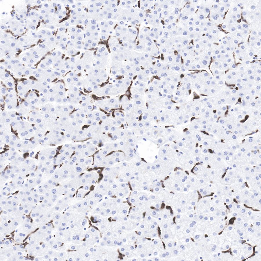
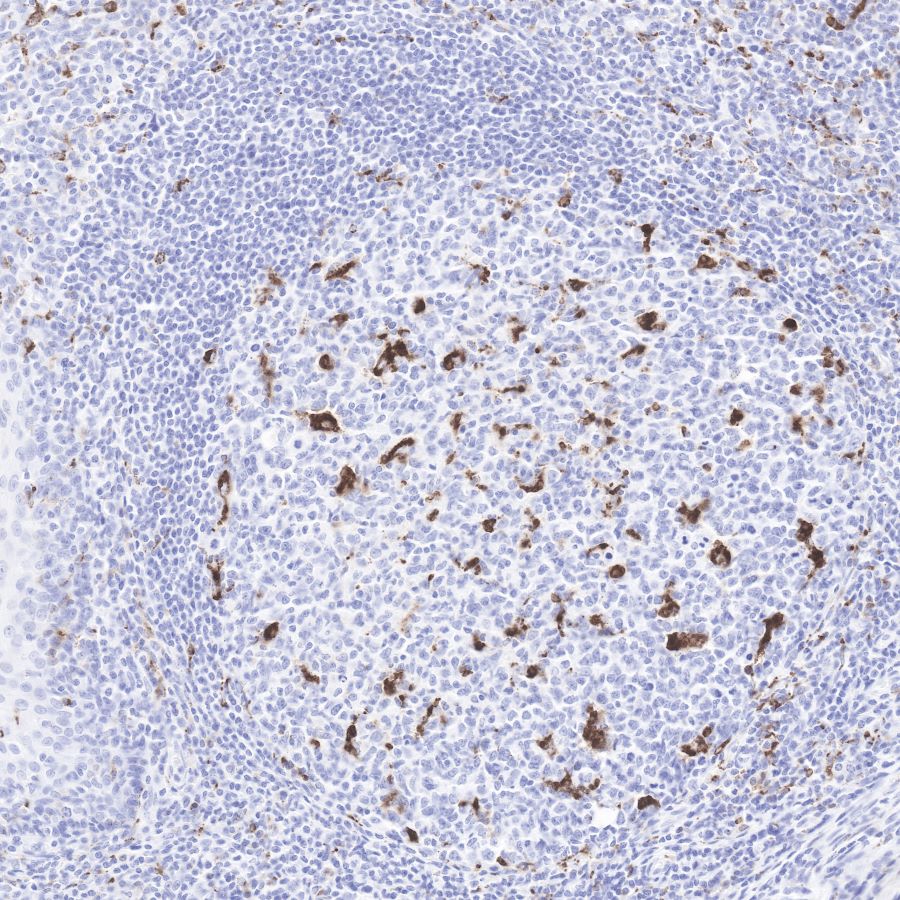
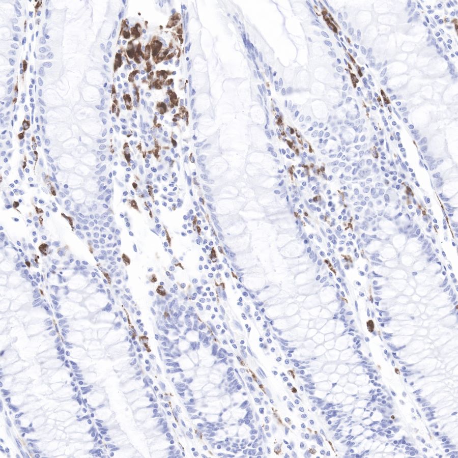
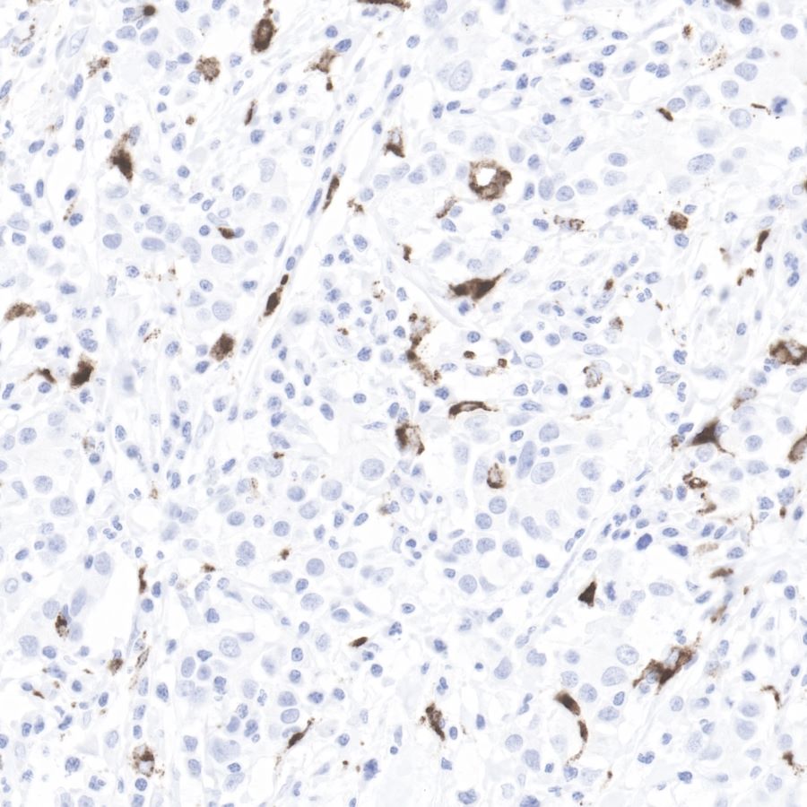
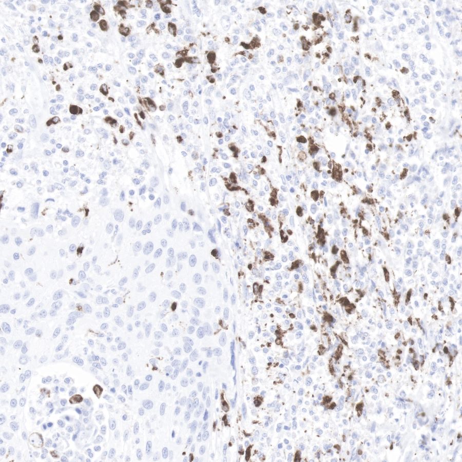
| Application |
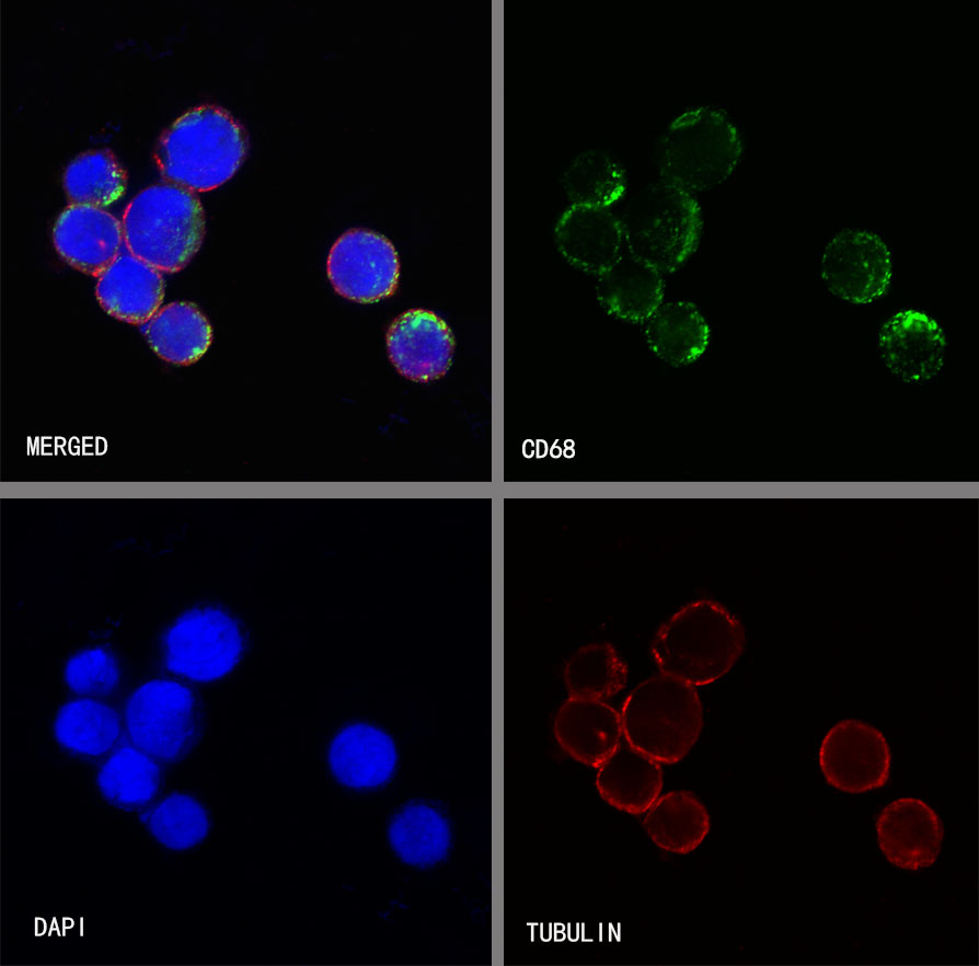
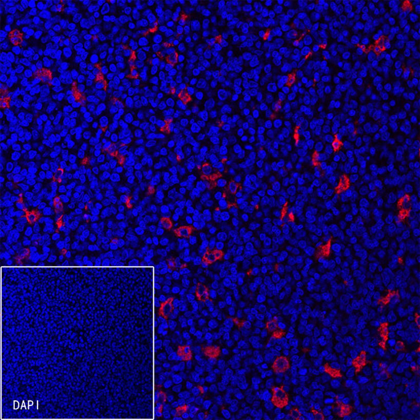
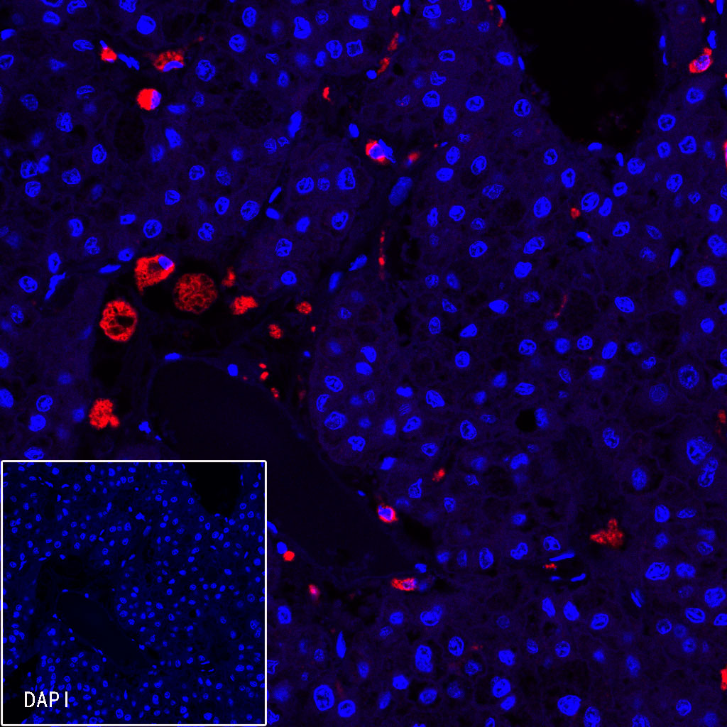
IHC-P, ICC, IF |
| Reactivity |
Hu |
| Purification |
Protein A |
| Concentration |
0.25 mg/ml |
| Physical Appearance |
Liquid |
| Storage Buffer |
PBS, 40% Glycerol, 0.05%BSA, 0.03% Proclin 300 |
| Stability & Storage |
12 months from date of receipt / reconstitution, -20 °C as supplied |
Dilution
| application |
dilution |
species |
| IHC-P |
1:1000 |
|
| ICC |
1:250 |
|
| IF |
1:200 |
|
Background
CD68 (Cluster of Differentiation 68) is a protein highly expressed by cells in the monocyte lineage (e.g., monocytic phagocytes, osteoclasts), by circulating macrophages, and by tissue macrophages. Immunohistochemistry can be used to identify the presence of CD68, which is found in the cytoplasmic granules of a range of different blood cells and myocytes. It is particularly useful as a marker for the various cells of the macrophage lineage, including monocytes, histiocytes, giant cells, Kupffer cells, and osteoclasts. This allows it to be used to distinguish diseases of otherwise similar appearance, such as the monocyte/macrophage and lymphoid forms of leukaemia (the latter being CD68 negative). Its presence in macrophages also makes it useful in diagnosing conditions related to proliferation or abnormality of these cells, such as malignant histiocytosis, histiocytic lymphoma, and Gaucher's disease.