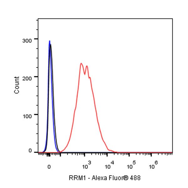
Flow cytometric analysis of 4% PFA fixed 90% methanol permeabilized HeLa (Human cervix adenocarcinoma epithelial cell) cells labelling RRM1 antibody at 1/50 (1 μg) dilution / (red) compared with a Rabbit monoclonal IgG (Black) isotype control and an unlabelled control (cells without incubation with primary antibody and secondary antibody) (Blue). Goat Anti - Rabbit IgG Alexa Fluor® 488 was used as the secondary antibody.
RRM1 Recombinant Rabbit mAb (SDT-R152)
RRM1 Recombinant Rabbit mAb (SDT-R152)
Price:
Regular price
$100 USD
Regular price
Sale price
$100 USD
Unit price
per
For shipping services or bulk orders, you may request a quotation.
Secure checkout with

View full details
Product Details
Product Details
Product Specification
| Host | Rabbit |
| Antigen | RRM1 |
| Synonyms | Ribonucleoside-diphosphate reductase large subunit, Ribonucleoside-diphosphate reductase subunit M1, Ribonucleotide reductase large subunit |
| Immunogen | N/A |
| Location | Cytoplasm |
| Accession | P23921 |
| Clone Number | SDT-R152 |
| Antibody Type | Recombinant mAb |
| Application | IHC-P, ICC, ICFCM |
| Reactivity | Hu |
| Purification | Protein A |
| Concentration | 0.5 mg/ml |
| Physical Appearance | Liquid |
| Storage Buffer | PBS, 40% Glycerol, 0.05% BSA, 0.03% Proclin 300 |
| Stability & Storage | 12 months from date of receipt / reconstitution, -20 °C as supplied |
Dilution
| application | dilution | species |
| IHC-P | 1:100 | |
| ICC | 1:500 | |
| ICFCM | 1:50 |
Background
The large subunit of human ribonucleotide reductase, RRM1, is involved in the regulation of cell proliferation, cell migration, tumour and metastasis development, and the synthesis of deoxyribonucleotides for DNA synthesis. It is also a cellular target for the chemotherapeutic agent, gemcitabine. RRM1 has been studied in a large number of patients with different types of cancer, such as non-small-cell lung cancer, pancreatic cancer, breast cancer, and biliary tract cancer, to establish its prognostic or predictive value when patients were treated with gemcitabine, and mRNA expression and genetic variants as determined by genotyping have in some cases been associated with clinical outcome of patients with cancer [PMID: 21163702].
Picture
Picture
FC
Immunohistochemistry

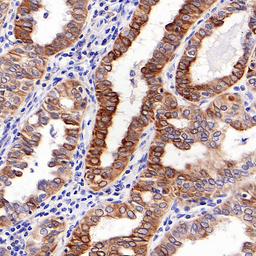
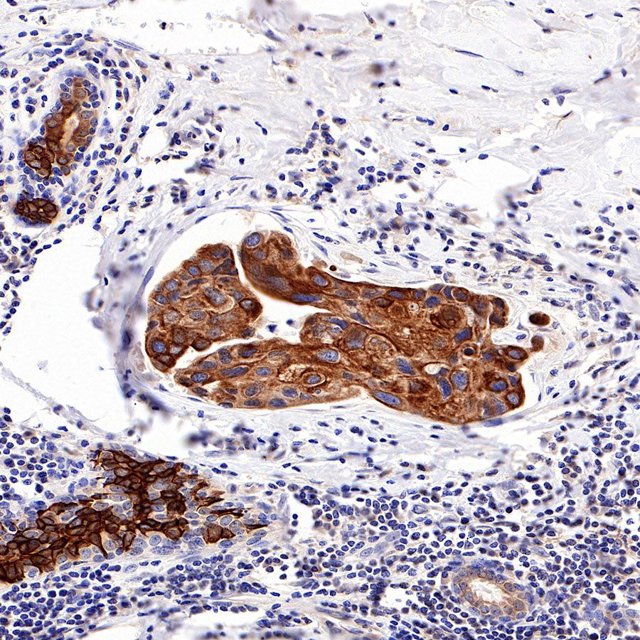
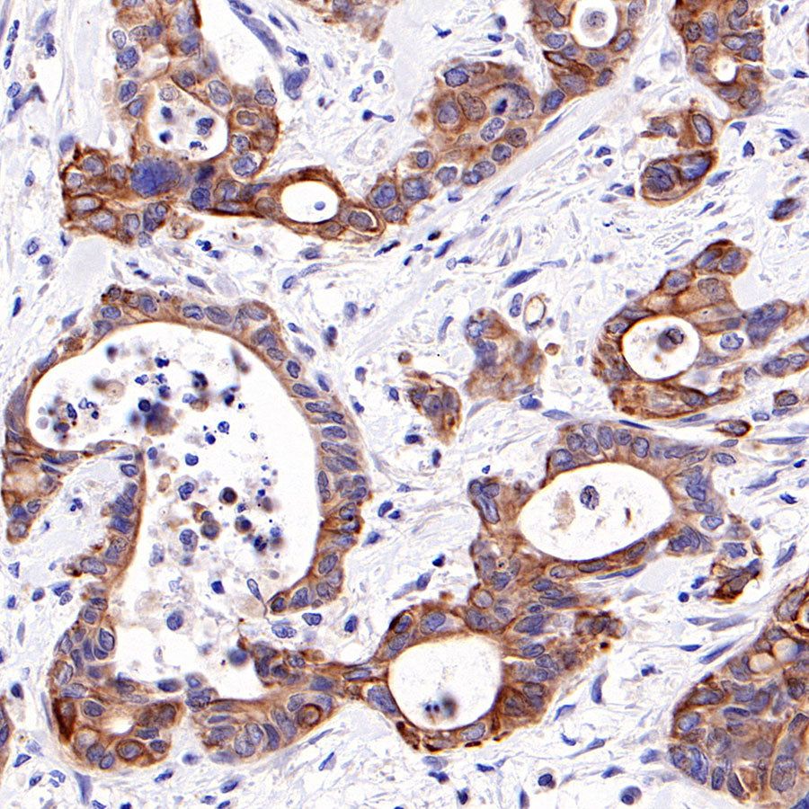
IHC shows positive staining in paraffin-embedded human thyroid cancer. Anti-RRM1 antibody was used at 1/100 dilution, followed by a HRP Polymer for Mouse & Rabbit IgG (ready to use). Counterstained with hematoxylin. Heat mediated antigen retrieval with Tris/EDTA buffer pH9.0 was performed before commencing with IHC staining protocol.
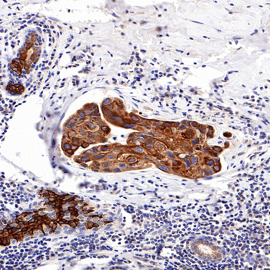
IHC shows positive staining in paraffin-embedded human breast cancer. Anti-RRM1 antibody was used at 1/100 dilution, followed by a HRP Polymer for Mouse & Rabbit IgG (ready to use). Counterstained with hematoxylin. Heat mediated antigen retrieval with Tris/EDTA buffer pH9.0 was performed before commencing with IHC staining protocol.

IHC shows positive staining in paraffin-embedded human pancreatic cancer. Anti-RRM1 antibody was used at 1/100 dilution, followed by a HRP Polymer for Mouse & Rabbit IgG (ready to use). Counterstained with hematoxylin. Heat mediated antigen retrieval with Tris/EDTA buffer pH9.0 was performed before commencing with IHC staining protocol.
Immunocytochemistry
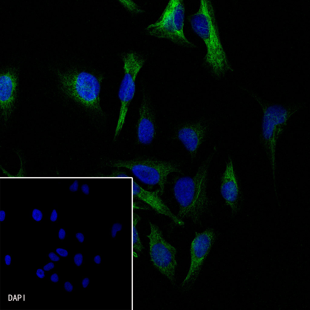
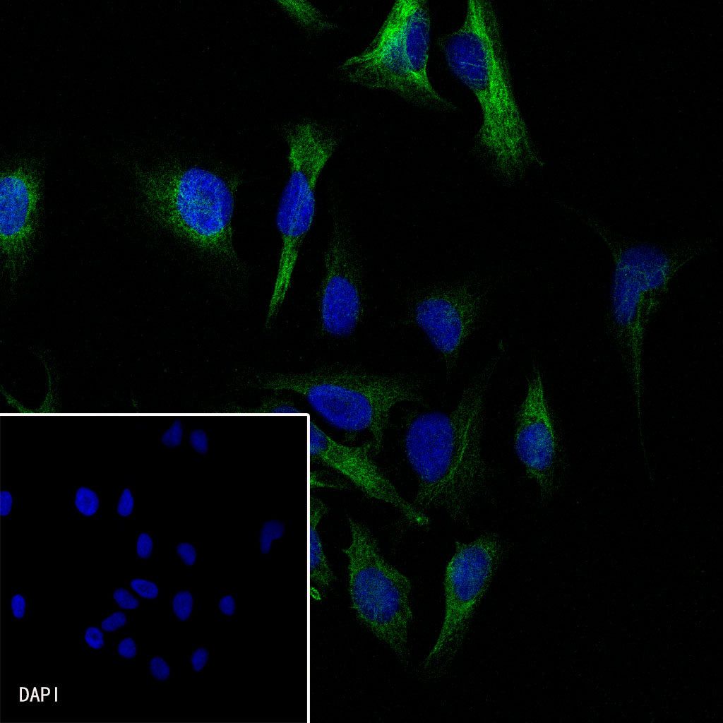
ICC shows positive staining in HeLa cells. Anti-RRM1 antibody was used at 1/500 dilution (Green) and incubated overnight at 4°C. Goat polyclonal Antibody to Rabbit IgG - H&L (Alexa Fluor® 488) was used as secondary antibody at 1/1000 dilution. The cells were fixed with 100% ice-cold methanol and permeabilized with 0.1% PBS-Triton X-100. Nuclei were counterstained with DAPI (Blue).