GST-π Recombinant Rabbit mAb (SDT-R054)
GST-π Recombinant Rabbit mAb (SDT-R054)
Price:
Regular price
$100 USD
Regular price
Sale price
$100 USD
Unit price
per
For shipping services or bulk orders, you may request a quotation.
Secure checkout with

View full details
Product Details
Product Details
Product Specification
| Host | Rabbit |
| Antigen | GSTP-1 |
| Synonyms | Glutathione S-Transferase Pi, GST-pi, GST class-pi, GSTP1-1 |
| Immunogen | N/A |
| Location | Cytoplasm, Nucleus |
| Accession | P09211 |
| Clone Number | SDT-R054 |
| Antibody Type | Rabbit mAb |
| Application | WB, IHC-P, ICC |
| Reactivity | Hu, Ms, Rt |
| Purification | Protein A |
| Concentration | 0.25 mg/ml |
| Physical Appearance | Liquid |
| Storage Buffer | PBS, 40% Glycerol, 0.05%BSA, 0.03% Proclin 300 |
| Stability & Storage | 12 months from date of receipt / reconstitution, -20 °C as supplied |
Dilution
| application | dilution | species |
| WB | 1:500-1:2500 | null |
| IHC-P | 1:2000 | null |
| ICC | 1:50 | null |
Background
GST-π is abundantly expressed in some mammalian tissues, particularly those associated with malignancies. While the enzyme can catalyze thioether bond formation between some electrophilic chemicals and GSH, novel non-detoxification functions are now ascribed to it.
Picture
Picture
Western Blot
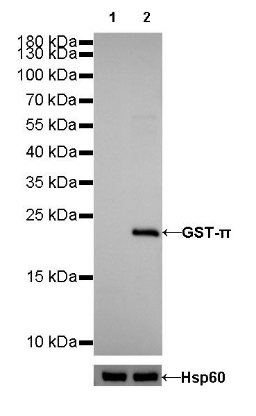
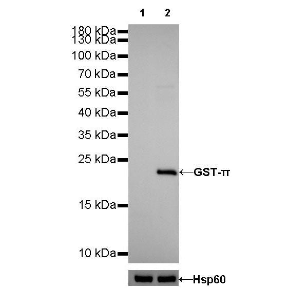
WB result of GST-π Rabbit mAb Primary antibody: GST-π Rabbit mAb at 1/2500 dilution Lane 1: LNCaP whole cell lysate 20 µg Lane 2: K-562 whole cell lysate 20 µg Negative control: LNCaP whole cell lysate Secondary antibody: Goat Anti-Rabbit IgG, (H+L), HRP conjugated at 1/10000 dilution Predicted MW: 23 kDa Observed MW: 23 kDa
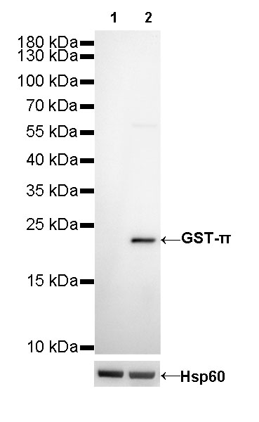
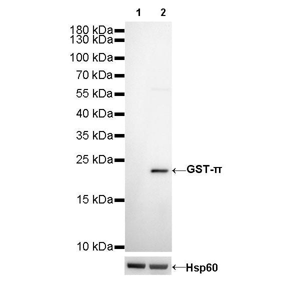
WB result of GST-π Rabbit mAb Primary antibody: GST-π Rabbit mAb at 1/2500 dilution Lane 1: LNCaP whole cell lysate 20 µg Lane 2: Jurkat whole cell lysate 20 µg Negative control: LNCaP whole cell lysate Secondary antibody: Goat Anti-Rabbit IgG, (H+L), HRP conjugated at 1/10000 dilution Predicted MW: 23 kDa Observed MW: 23 kDa
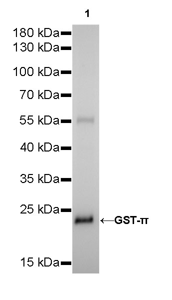
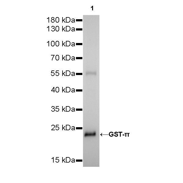
WB result of GST-π Rabbit mAb Primary antibody: GST-π Rabbit mAb at 1/500 dilution Lane 1: mouse liver lysate 20 µg Secondary antibody: Goat Anti-Rabbit IgG, (H+L), HRP conjugated at 1/10000 dilution Predicted MW: 23 kDa Observed MW: 23 kDa
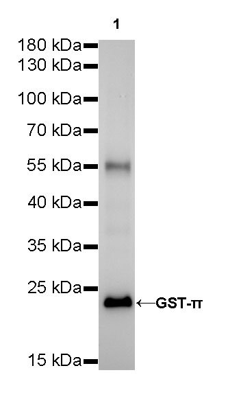
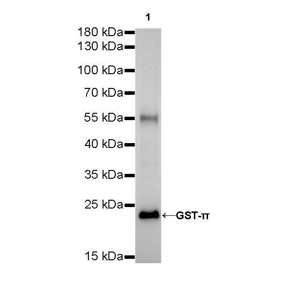
WB result of GST-π Rabbit mAb Primary antibody: GST-π Rabbit mAb at 1/500 dilution Lane 1: rat kidney lysate 20 µg Secondary antibody: Goat Anti-Rabbit IgG, (H+L), HRP conjugated at 1/10000 dilution Predicted MW: 23 kDa Observed MW: 23 kDa
Immunohistochemistry
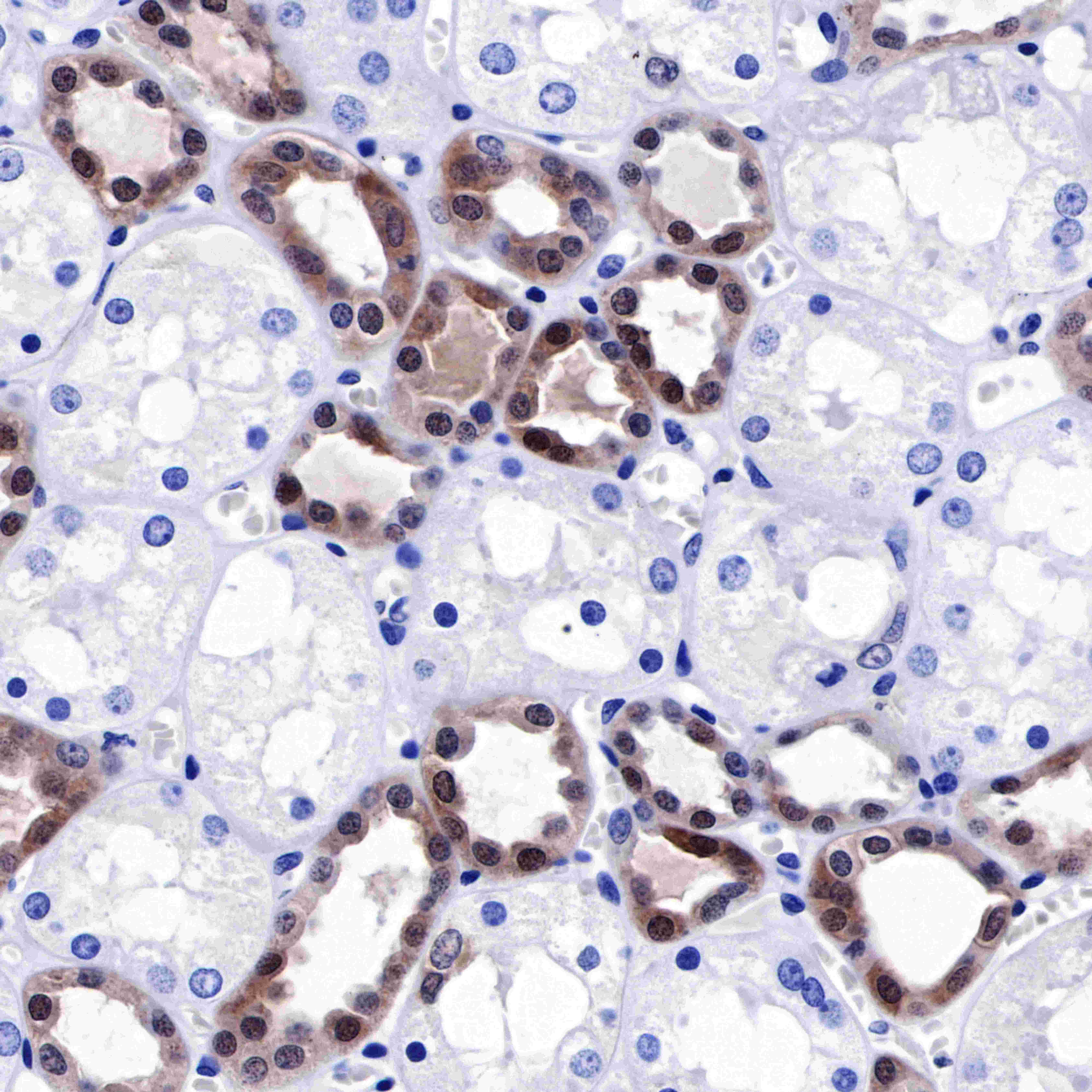
IHC shows positive staining in paraffin-embedded human kidney. Anti-GST-π antibody was used at 1/2000 dilution, followed by a HRP Polymer for Mouse & Rabbit IgG (ready to use). Counterstained with hematoxylin. Heat mediated antigen retrieval with Tris/EDTA buffer pH9.0 was performed before commencing with IHC staining protocol.
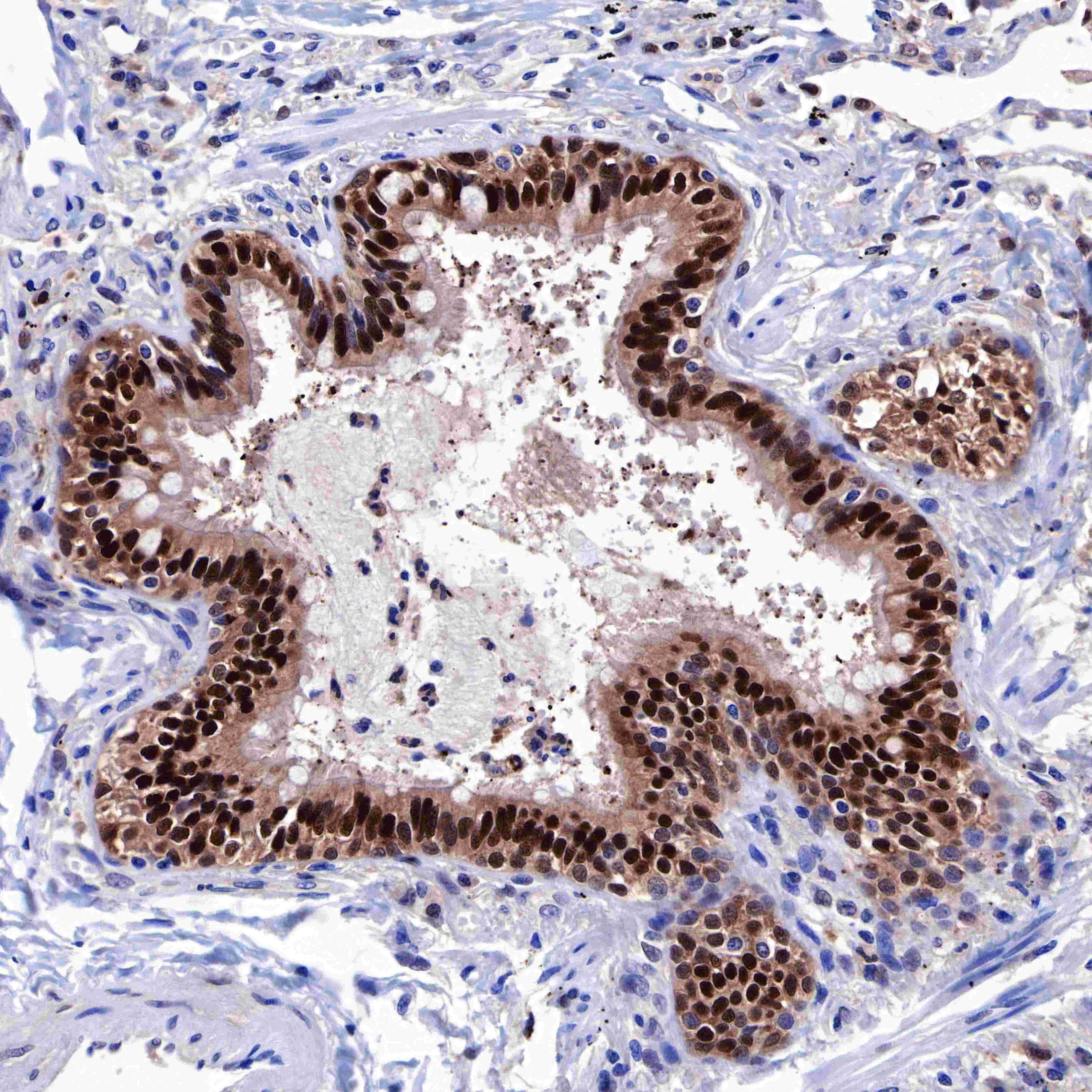
IHC shows positive staining in paraffin-embedded human lung. Anti-GST-π antibody was used at 1/2000 dilution, followed by a HRP Polymer for Mouse & Rabbit IgG (ready to use). Counterstained with hematoxylin. Heat mediated antigen retrieval with Tris/EDTA buffer pH9.0 was performed before commencing with IHC staining protocol.
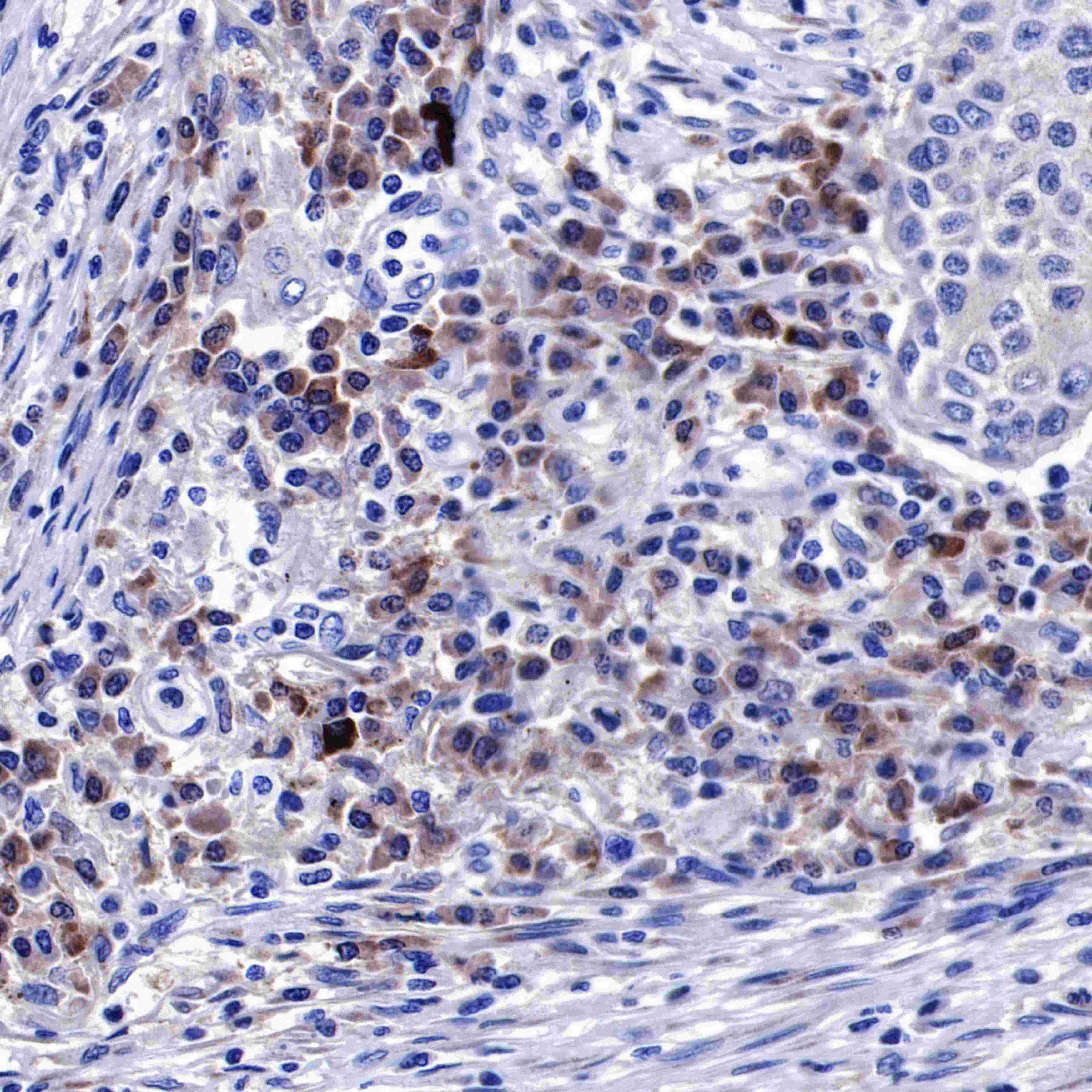
IHC shows positive staining in paraffin-embedded human lung squamous cell carcinoma. Anti-GST-π antibody was used at 1/2000 dilution, followed by a HRP Polymer for Mouse & Rabbit IgG (ready to use). Counterstained with hematoxylin. Heat mediated antigen retrieval with Tris/EDTA buffer pH9.0 was performed before commencing with IHC staining protocol.
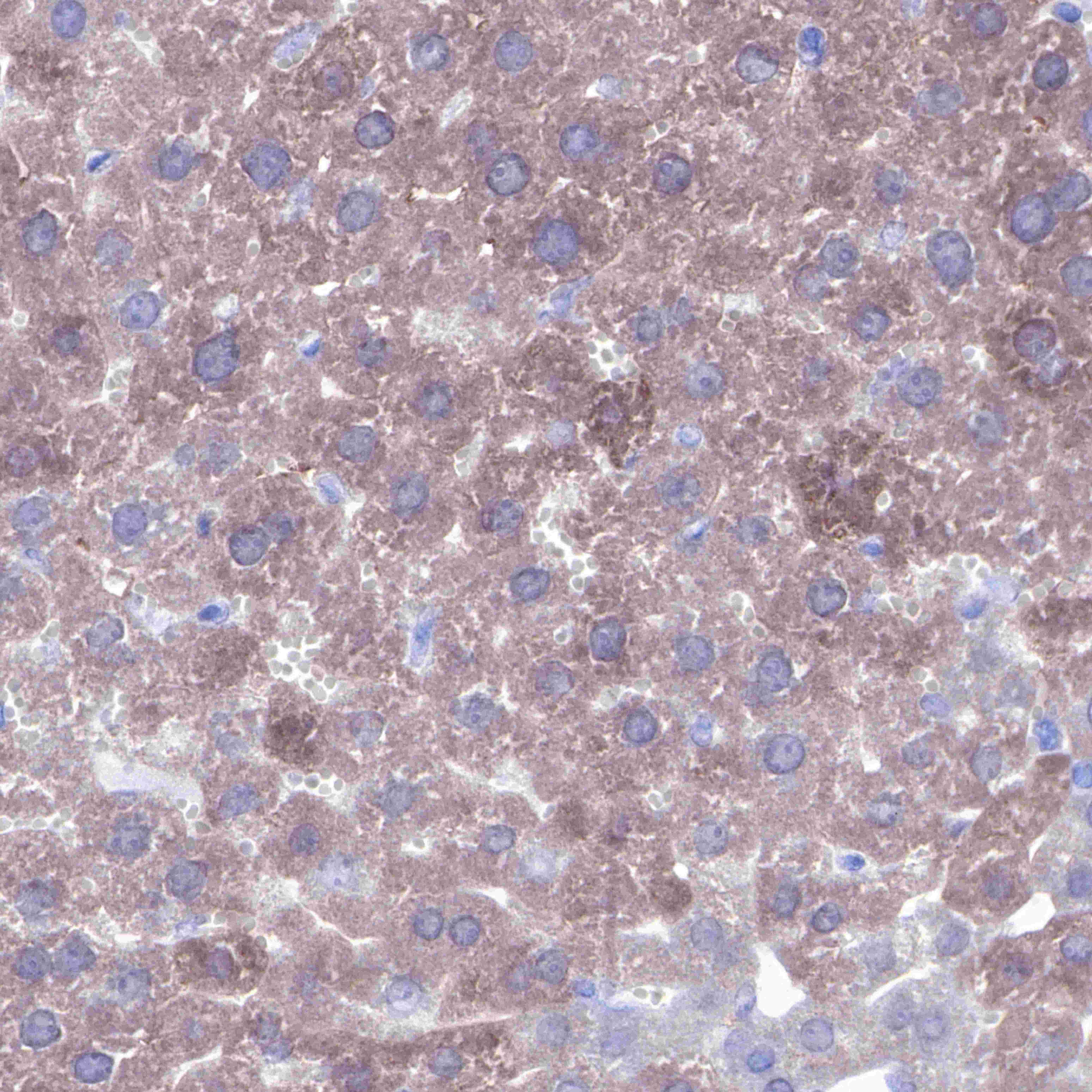
IHC shows positive staining in paraffin-embedded mouse liver. Anti-GST-π antibody was used at 1/2000 dilution, followed by a HRP Polymer for Mouse & Rabbit IgG (ready to use). Counterstained with hematoxylin. Heat mediated antigen retrieval with Tris/EDTA buffer pH9.0 was performed before commencing with IHC staining protocol.
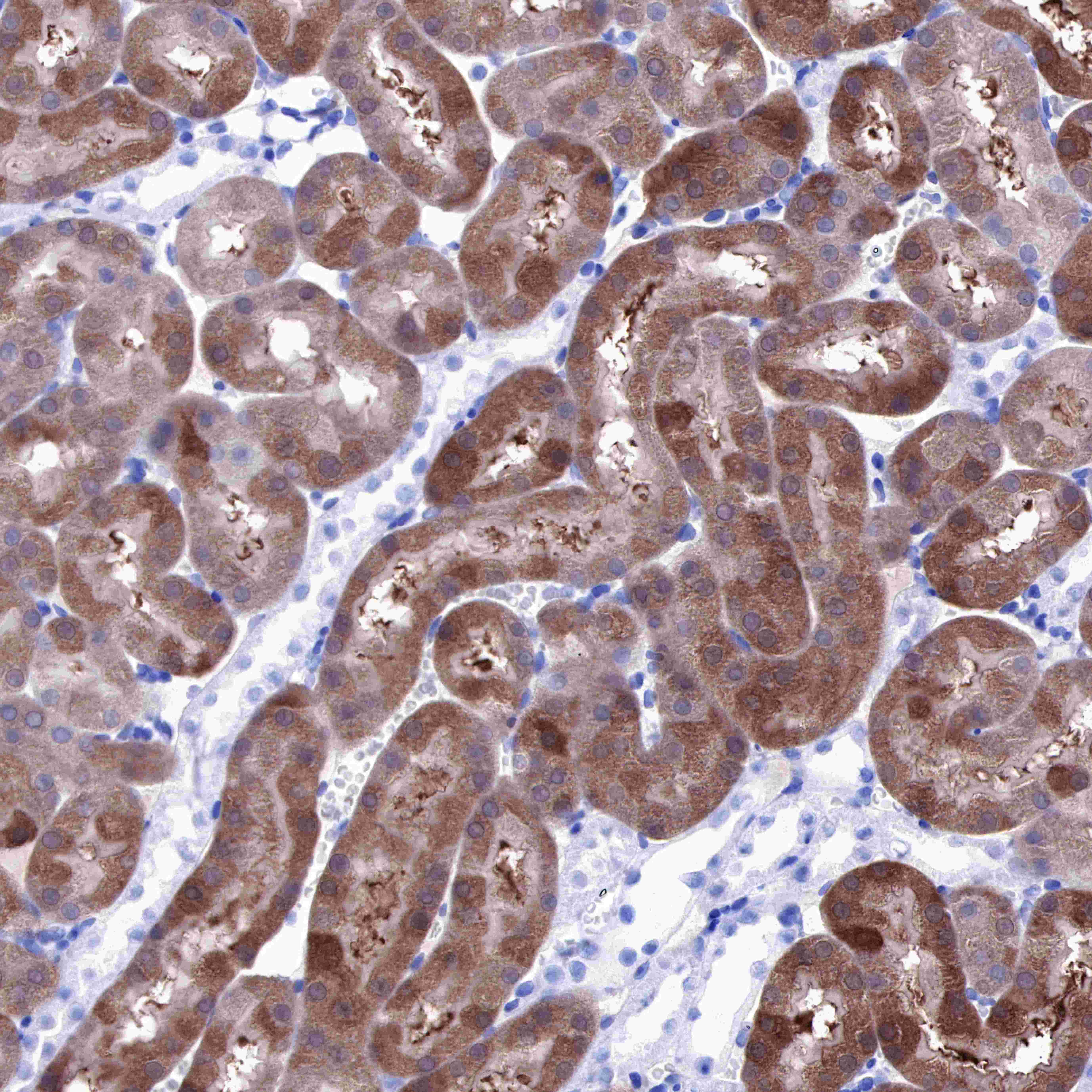
IHC shows positive staining in paraffin-embedded rat kidney. Anti-GST-π antibody was used at 1/2000 dilution, followed by a HRP Polymer for Mouse & Rabbit IgG (ready to use). Counterstained with hematoxylin. Heat mediated antigen retrieval with Tris/EDTA buffer pH9.0 was performed before commencing with IHC staining protocol.
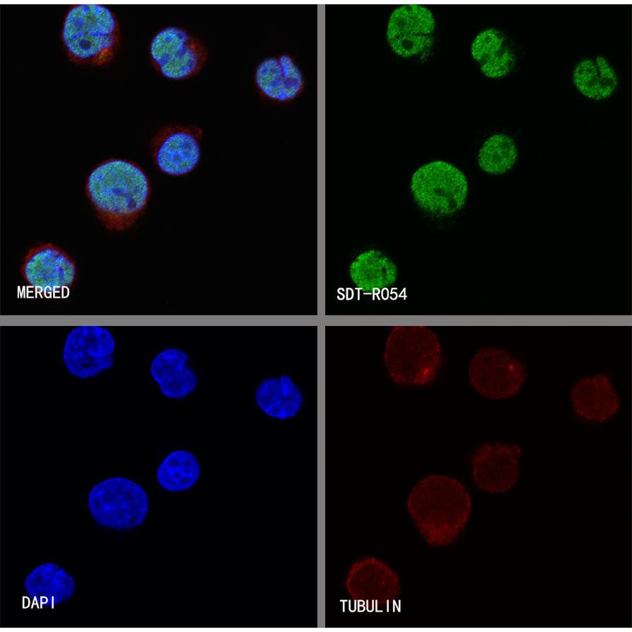
Immunocytochemistry
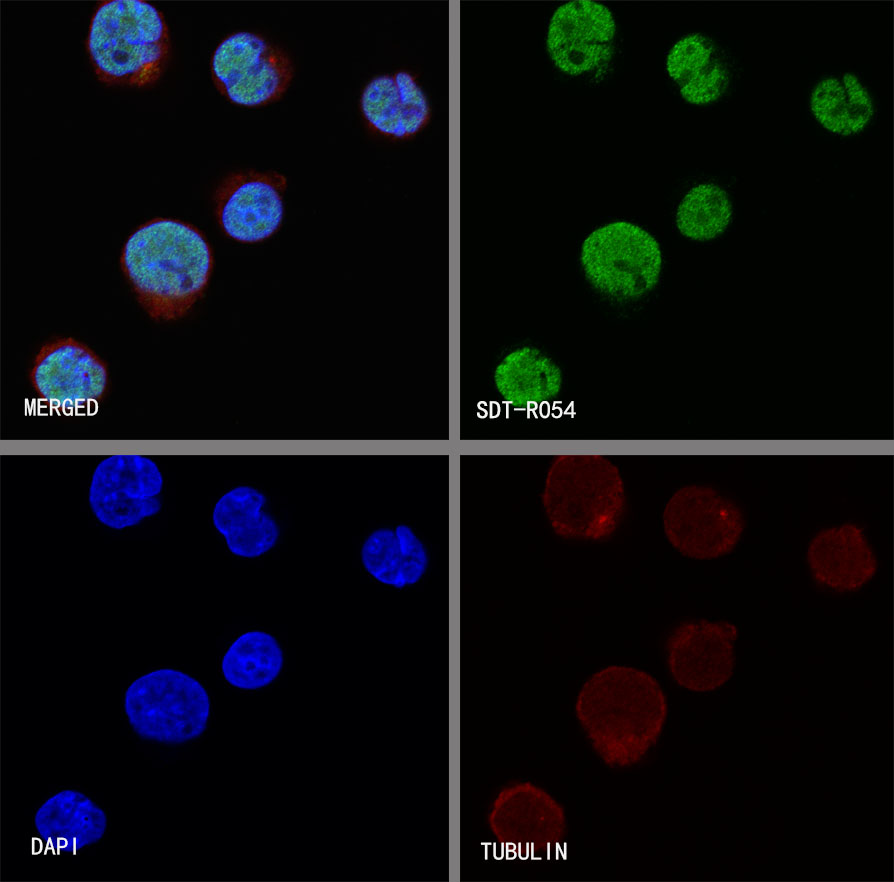
ICC shows positive staining in K562 cells. Anti-GST-π antibody was used at 1/50 dilution (Green) and incubated overnight at 4°C. Goat polyclonal Antibody to Rabbit IgG - H&L (Alexa Fluor® 488) was used as secondary antibody at 1/1000 dilution. The cells were fixed with 4% PFA and permeabilized with 0.1% PBS-Triton X-100. Nuclei were counterstained with DAPI (Blue). Counterstain with tubulin (red).