Product Specification
| Synonyms |
Mycoplasma luciferase detection kits |
| Stability & Storage |
Dry ice transport. -20℃ storage is protected from light, and the validity period is 12 months
|
Background
The UA-Glo Mycoplasma Detection Kit is used for the qualitative detection of mycoplasma contamination in cell cultures. Over 95% of mycoplasma contamination in cell cultures is caused by Mycoplasma fermentans, M. orale, M. pirum, M. hyorhinis, M. hominis, M. salivarius, M. arginine, and Acholeplasma laidlawii. Live mycoplasmas produce kinases that are not expressed by eukaryotic cells, which can convert ADP into ATP. The Mycoplasma Detection Kit provides ADP and other substrates required for mycoplasma kinases to convert ADP into ATP, and then detects the generated ATP via a luciferase luminescence method to confirm whether there is mycoplasma contamination in the cell culture. This kit can quickly detect the eight aforementioned species as well as many other common live mycoplasma strains.
Components
|
Size
|
component |
|
10T
|
Mycoplasma luciferase detection reagent 0.5ml
Mycoplasma luciferase detection reagent 0.1mL
|
|
25T
|
Mycoplasma luciferase detection reagent 1.25ml
Mycoplasma luciferase detection reagent 0.25mL
|
|
50T
|
Mycoplasma luciferase detection reagent 2.5ml
Mycoplasma luciferase detection reagent 0.5mL
|
Protocol
1. Sample Preparation for Cell Culture Medium to be Tested
-
Take 1 mL of cell culture medium from cells in the logarithmic growth phase.
-
Centrifuge the cell culture medium at room temperature (200 g) for 5 minutes. Transfer approximately 800 μL of the supernatant to a new centrifuge tube, making sure not to include any cell debris at the bottom.
-
Use the prepared cell culture medium for the mycoplasma detection with luciferase assay.
2. Luciferase Mycoplasma Detection
-
Add 50 μL of the cell culture medium sample to a white opaque 96-well plate.
-
Equilibrate the luciferase mycoplasma detection reagent and substrate to room temperature (22°C-25°C), and mix gently.
-
Add 50 μL of the luciferase mycoplasma detection reagent to the 50 μL of the cell culture medium sample.
-
Mix by gentle shaking for 10 seconds, incubate at room temperature for 5 minutes, and then read the fluorescence value (Reading 1).
-
Add 10 μL of the luciferase mycoplasma detection substrate.
-
Mix by gentle shaking for 10 seconds, incubate at room temperature for 10 minutes, and then read the fluorescence value (Reading 2).
3. Results
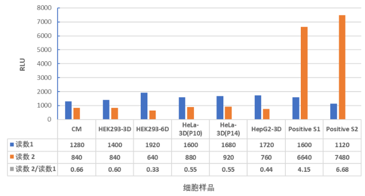
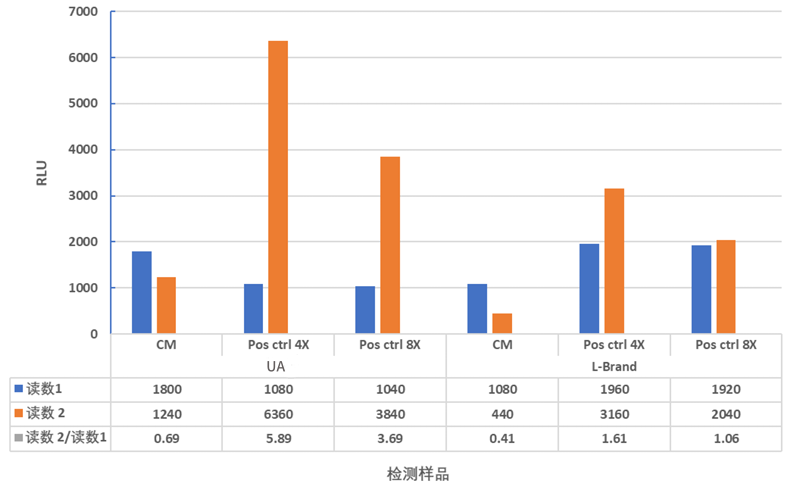
The ratio of Reading 2 to Reading 1 is used to determine if mycoplasma contamination is present.
|
The ratio of reading 2 to reading 1
|
Results Explanation and Recommendations
|
|
< 1
|
No Mycoplasma contamination
|
|
1-1.2
|
Possible Mycoplasma contamination. The cells were isolated and tested again after culture for 24-48hr.
|
|
> 1.2
|
Mycoplasma contamination
|
Guidelines
-
Aliquot Storage: Aliquot and store the reagents according to the instructions at -20°C to ensure reagent stability.
-
Freeze-Thaw Stability: Reagents stored at -20°C, protected from light, can be freeze-thawed twice without affecting the test results.
-
Sample Collection: Collect samples from cells in the logarithmic growth phase for testing. Newly passaged cells should be tested 24-48 hours after subculture.
-
Luciferase Reaction Sensitivity: The luciferase reaction is sensitive to temperature changes. Reagents and samples should be equilibrated to room temperature (22°C-25°C), and the temperature should remain constant (±1°C) throughout the test.
-
For Research Use Only: This product is intended for research purposes only.