SOX9 Recombinant Rabbit mAb (SDT-019-29)
SOX9 Recombinant Rabbit mAb (SDT-019-29)
Product Details
Product Details
Product Specification
| Host | Rabbit |
| Antigen | SOX9 |
| Synonyms | Transcription factor SOX-9 |
| Immunogen | Recombinant Protein |
| Location | Nucleus |
| Accession | P48436 |
| Clone Number | SDT-019-29 |
| Antibody Type | Recombinant mAb |
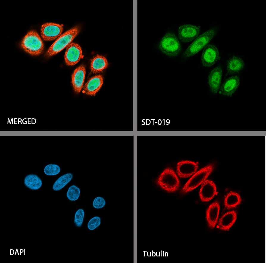
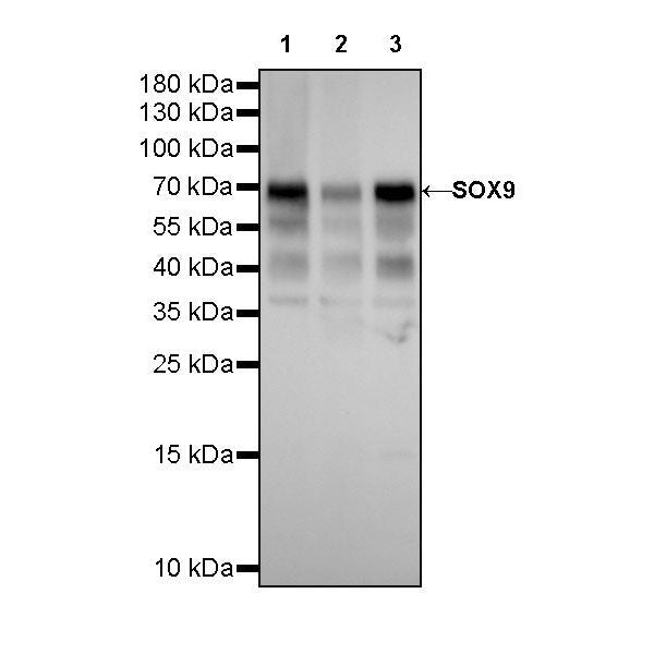
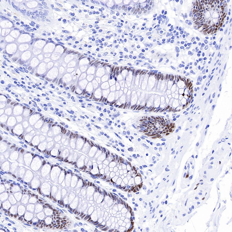
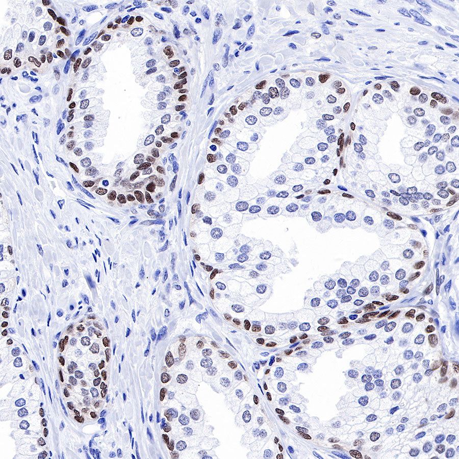
| Application | WB, IHC-P, ICC, ICFCM |
| Reactivity | Hu |
| Predicted Reactivity | Pr, Cz, Pg |
| Purification | Protein A |
| Concentration | 0.25 mg/ml |
| Physical Appearance | Liquid |
| Storage Buffer | PBS, 40% Glycerol, 0.05% BSA, 0.03% Proclin 300 |
| Stability & Storage | 12 months from date of receipt / reconstitution, -20 °C as supplied |
Dilution
| application | dilution | species |
| WB | 1:500 | |
| IHC-P | 1:1000 | |
| ICC | 1:250 | |
| ICFCM | 1:250 |
Background
SOX9 is a pivotal transcription factor in developing and adult cartilage. Its gene is expressed from the multipotent skeletal progenitor stage and is active throughout chondrocyte differentiation [PMID: 27128146]. The SOX9 transcription factor controls the differentiation of many cell types among vertebrates. The SOX9 gene locus is large and complex and contains various tissue-specific enhancers. Individual enhancers direct specific expression of SOX9 in chondrocytes, Sertoli cells and cranial neural crest cells. Human SOX9 mutations can lead to either the complete Campomelic Dysplasia syndrome, or isolated clinical features, depending upon whether the mutation occurs in the coding region or in enhancer regions. Chromatin Immunoprecipitation has helped to define SOX9 control of target gene expression at the genome wide level in hair follicle stem cells and in chondrocytes where SOX9 binds at super-enhancers. SOX9 binding proximal to promoters controls basal cell activity whereas cell type specificity is directed from distal enhancers [PMID: 28323209].
Picture
Picture
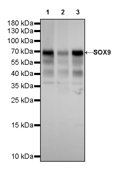
Western Blot
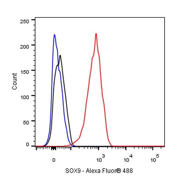
FC
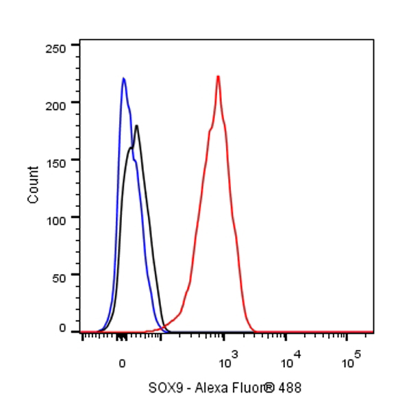
Flow cytometric analysis of 4% PFA fixed 90% methanol permeabilized HeLa (Human cervix adenocarcinoma epithelial cell) cells labelling SOX9 antibody at 1/250 (0.1 μg) dilution / (red) compared with a Rabbit monoclonal IgG (Black) isotype control and an unlabelled control (cells without incubation with primary antibody and secondary antibody) (Blue). Goat Anti - Rabbit IgG Alexa Fluor® 488 was used as the secondary antibody.
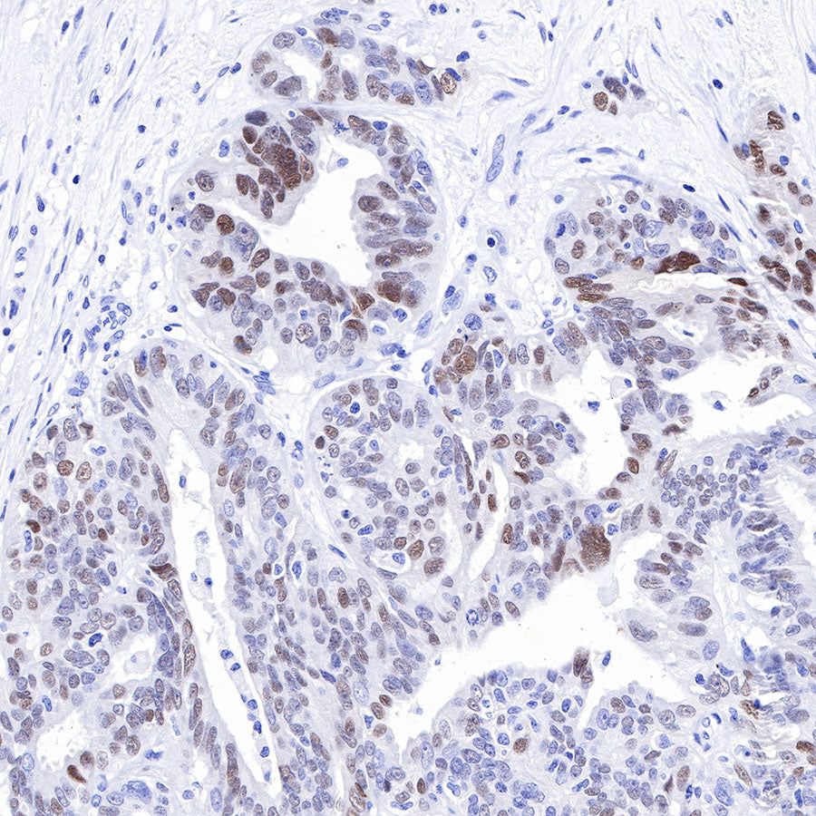
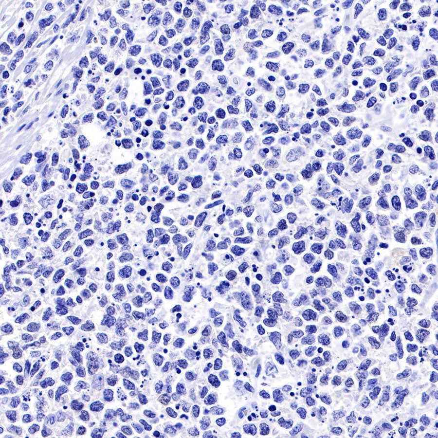
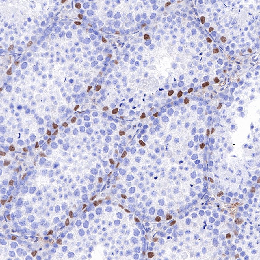
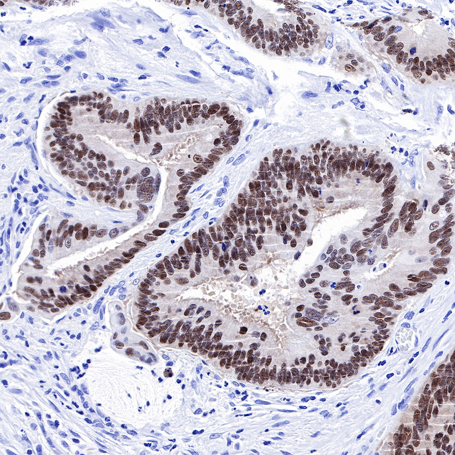
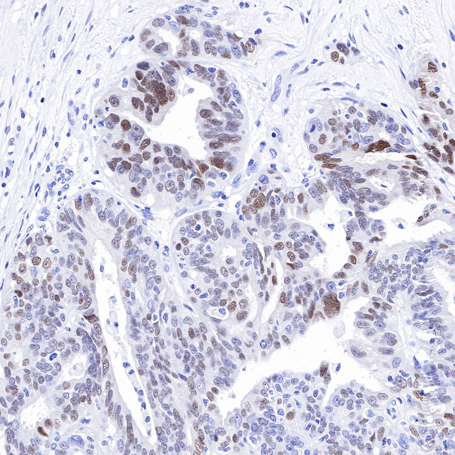
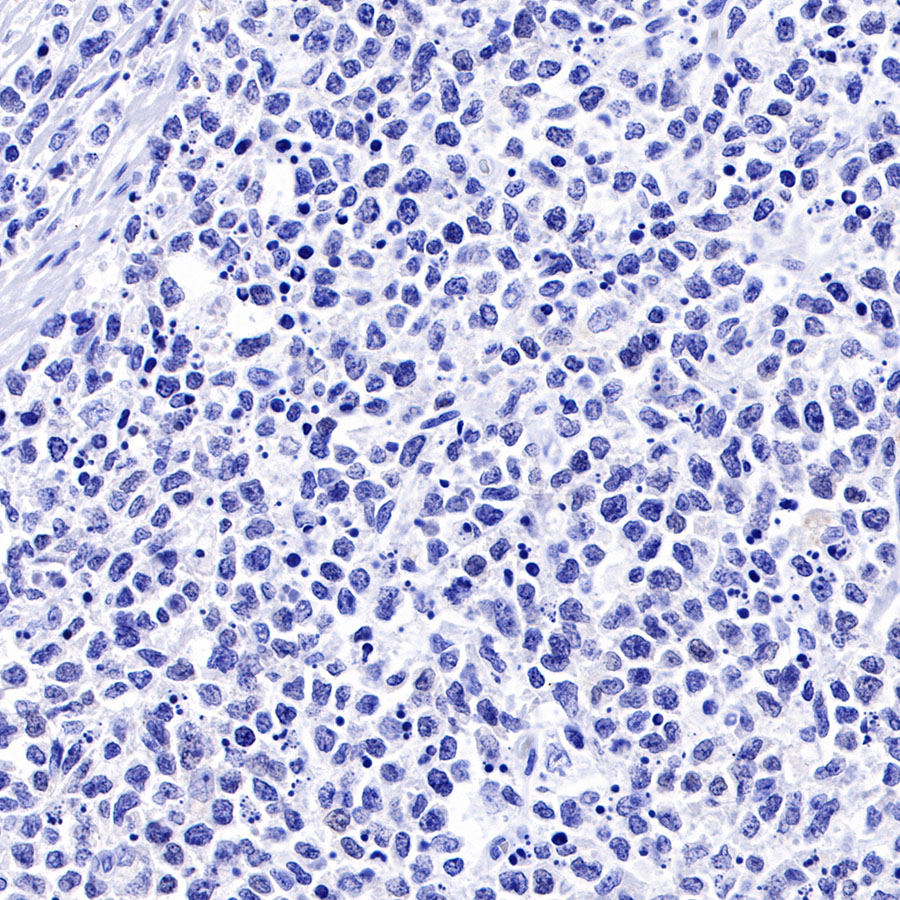
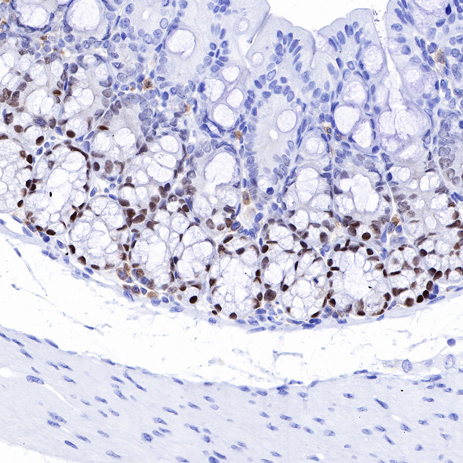
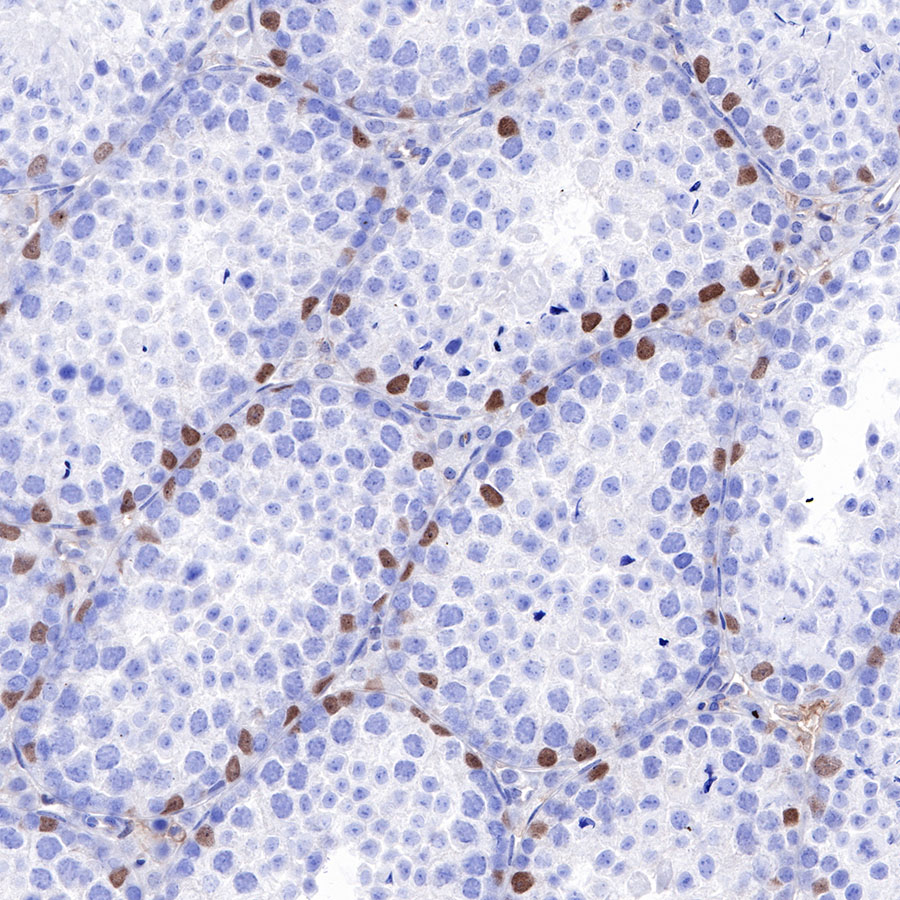
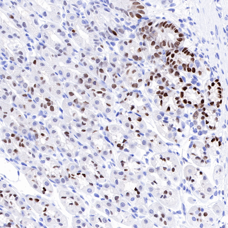
Immunohistochemistry


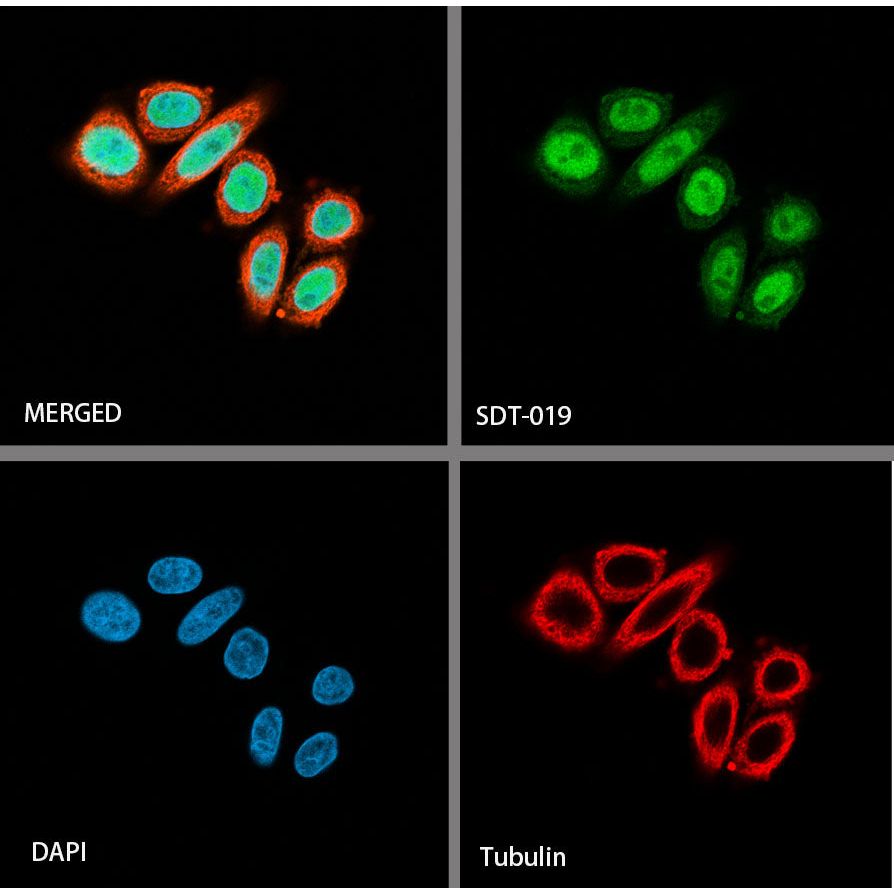
Immunocytochemistry